. 2022 Jun 28;13(3):e0025822.
doi: 10.1128/mbio.00258-22. Epub 2022 May 31.

InvL, an Invasin-Like Adhesin, Is a Type II Secretion System Substrate Required for Acinetobacter baumannii Uropathogenesis

Affiliations

InvL, an Invasin-Like Adhesin, Is a Type II Secretion System Substrate Required for Acinetobacter baumannii Uropathogenesis

Clay D Jackson-Litteken et al. mBio. .

Abstract

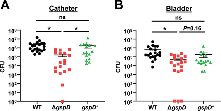
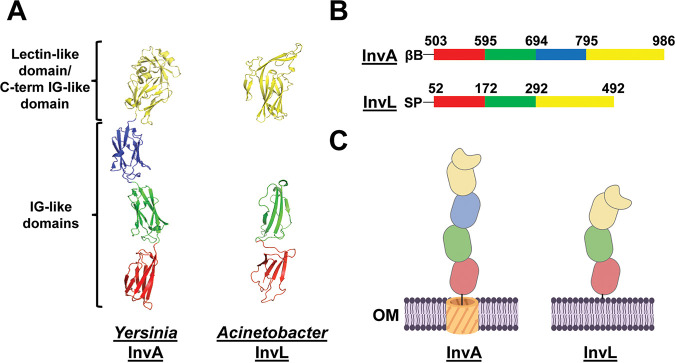
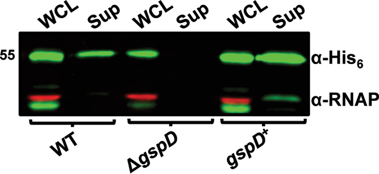
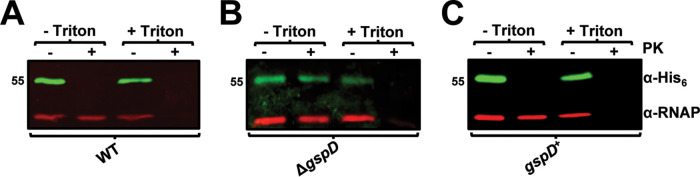
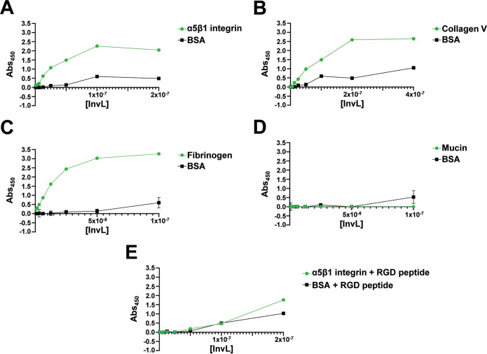
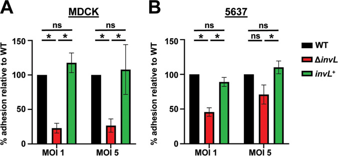
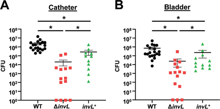
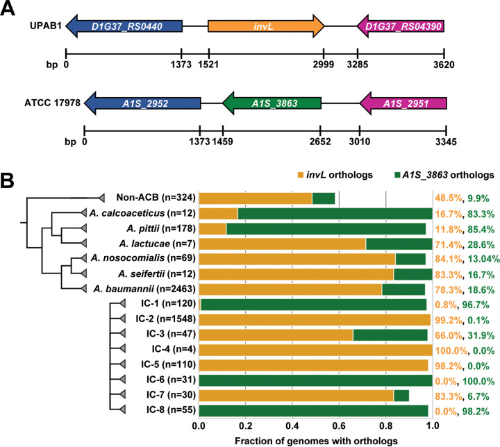
Acinetobacter baumannii is an opportunistic pathogen of growing concern, as isolates are commonly multidrug resistant. While A. baumannii is most frequently associated with pulmonary infections, a significant proportion of clinical isolates come from urinary sources, highlighting its uropathogenic potential. The type II secretion system (T2SS) of commonly used model Acinetobacter strains is important for virulence in various animal models, but the potential role of the T2SS in urinary tract infection (UTI) remains unknown. Here, we used a catheter-associated UTI (CAUTI) model to demonstrate that a modern urinary isolate, UPAB1, requires the T2SS for full virulence. A proteomic screen to identify putative UPAB1 T2SS effectors revealed an uncharacterized lipoprotein with structural similarity to the intimin-invasin family, which serve as type V secretion system (T5SS) adhesins required for the pathogenesis of several bacteria. This protein, designated InvL, lacked the β-barrel domain associated with T5SSs but was confirmed to require the T2SS for both surface localization and secretion. This makes InvL the first identified T2SS effector belonging to the intimin-invasin family. InvL was confirmed to be an adhesin, as the protein bound to extracellular matrix components and mediated adhesion to urinary tract cell lines in vitro. Additionally, the invL mutant was attenuated in the CAUTI model, indicating a role in Acinetobacter uropathogenesis. Finally, bioinformatic analyses revealed that InvL is present in nearly all clinical isolates belonging to international clone 2, a lineage of significant clinical importance. In all, we conclude that the T2SS substrate InvL is an adhesin required for A. baumannii uropathogenesis. IMPORTANCE While pathogenic Acinetobacter can cause various infections, we recently found that 20% of clinical isolates come from urinary sources. Despite the clinical relevance of Acinetobacter as a uropathogen, few virulence factors involved in urinary tract colonization have been defined. Here, we identify a novel type II secretion system effector, InvL, which is required for full uropathogenesis by a modern urinary isolate. Although InvL has predicted structural similarity to the intimin-invasin family of autotransporter adhesins, InvL is predicted to be anchored to the membrane as a lipoprotein. Similar to other invasin homologs, however, we demonstrate that InvL is a bona fide adhesin capable of binding extracellular matrix components and mediating adhesion to urinary tract cell lines. In all, this work establishes InvL as an adhesin important for Acinetobacter's urinary tract virulence and represents the first report of a type II secretion system effector belonging to the intimin-invasin family.

Keywords: Acinetobacter; adhesin; infection; invasin; pathogenesis; type II secretion system; urinary tract infection; virulence.


Conflict of interest statement

The authors declare no conflict of interest.

Figures

FIG 1
FIG 1
The T2SS is required for full virulence in a murine CAUTI model. Mice were implanted with a catheter followed by transurethral inoculation with UPAB1 WT, ΔgspD, or gspD+ strains. At 24 h postinfection, mice were sacrificed, and bacterial burdens on the catheter (A) and in the bladder (B) were quantified. Shown are results from at least three pooled experiments. Each data point represents an individual mouse, the horizontal line represents the mean, and the standard error of the mean (SEM) is indicated by error bars. *, P < 0.05; two-tailed Mann-Whitney U test; ns, not significant.
FIG 2
FIG 2
InvL has predicted structural similarity to InvA of Yersinia. (A) The crystal structure of the passenger domain of Yersinia InvA (PDB entry 1CWV) is shown on the left, and the AlphaFold2 predicted structure of the analogous region of InvL is pictured on the right. (B) Amino acids corresponding to subdomains of the passenger domain of InvA and the predicted homologous region of InvL. (C) Graphic depiction of the structure of InvA and the predicted structure of InvL; created with BioRender.com. Red, green, and blue colors denote individual IG-like domains, and yellow denotes the C-terminal IG-like domain in intimate contact with the lectin-like domain. βB, β-barrel domain; SP, signal peptide; OM, outer membrane.
FIG 3
FIG 3
The T2SS is required for InvL secretion. Whole-cell lysate (WCL) and supernatant (Sup) fractions from WT, ΔgspD, and gspD+ UPAB1 cultures harboring pBAV-Apr::invL-his6 were probed for InvL (α-His6) and RNAP by immunoblotting. Numbers to the left indicate molecular weight in kDa. At least two biological replicates were performed, yielding similar results, and a representative blot from one replicate is shown.
FIG 4
FIG 4
The T2SS is required for InvL surface localization. WT (A), ΔgspD (B), and gspD+ (C) UPAB1 cells harboring pBAV-Apr::invL-his6 were treated with proteinase K (PK; +) or left untreated (−) in the presence or absence of Triton X-100. Cells were then probed for InvL (α-His6) and RNAP by immunoblotting. Numbers to the left indicate molecular weight in kDa. At least two biological replicates were performed, yielding similar results, and a representative blot from one replicate is shown.
FIG 5
FIG 5
InvL binds to multiple ECM components. ELISAs were performed to assess binding of InvL to α5β1 integrin (A), collagen V (B), fibrinogen (C), mucin (D), or α5β1 integrin in the presence of RGD-containing peptide (E). BSA served as a negative binding control. Shown are the results from two biological replicates, and SEM is indicated by error bars.
FIG 6
FIG 6
InvL facilitates adhesion to epithelial cells in vitro. UPAB1 WT, ΔinvL, and invL+ strains were used in adhesion assays with MDCK (A) and 5637 (B) epithelial cells. The mean from six replicates is shown, and error bars represent the SEM. *, P < 0.05; one-way ANOVA, Tukey’s test for multiple comparisons.
FIG 7
FIG 7
InvL is required for full virulence in a murine CAUTI model. Mice were implanted with a catheter followed by transurethral inoculation with UPAB1 WT, ΔinvL, or invL+ strains. At 24 h postinfection, mice were sacrificed and bacterial burden on the catheter (A) and in the bladder (B) were quantified. Shown are results from at least three pooled experiments. Each data point represents an individual mouse, the horizontal line represents the mean, and the SEM is indicated by error bars. *, P < 0.05; two-tailed Mann-Whitney U test.
FIG 8
FIG 8
InvL is encoded by ACB complex strains and has high prevalence in IC-2 clones. (A) InvL is encoded at a common locus in ACB complex strains. Orthologs of InvL (top) and orthologs of A1S_3863 (bottom) are encoded at a common locus between genes encoding a dihydrolipoyl dehydrogenase (D1G37_RS0440 in UPAB1 and A1S_2952 in ATCC 17978; blue) and a TIGR01244 family phosphatase (D1G37_RS04390 in UPAB1 and A1S_2951 in ATCC 17978; purple). (B) Distribution of invL and A1S_3863 in Acinetobacter. The fractions of genomes encoding InvL and A1S_3863 are reported by ACB species and international clone number. Percentage of genomes containing each ortholog by species and international clone number are indicated to the right.

References

    1. Vijayakumar S, Biswas I, Veeraraghavan B. 2019. Accurate identification of clinically important Acinetobacter spp.: an update. Future Sci OA 5:FSO395. doi: 10.2144/fsoa-2018-0127. - DOI - PMC - PubMed
    1. Cerqueira GM, Peleg AY. 2011. Insights into Acinetobacter baumannii pathogenicity. IUBMB Life 63:1055–1060. doi: 10.1002/iub.533. - DOI - PubMed
    1. Sarshar M, Behzadi P, Scribano D, Palamara AT, Ambrosi C. 2021. Acinetobacter baumannii: an ancient commensal with weapons of a pathogen. Pathogens 10:387. doi: 10.3390/pathogens10040387. - DOI - PMC - PubMed
    1. Dexter C, Murray GL, Paulsen IT, Peleg AY. 2015. Community-acquired Acinetobacter baumannii: clinical characteristics, epidemiology and pathogenesis. Expert Rev Anti Infect Ther 13:567–573. doi: 10.1586/14787210.2015.1025055. - DOI - PubMed
    1. Cisneros JM, Rodriguez-Baño J. 2002. Nosocomial bacteremia due to Acinetobacter baumannii: epidemiology, clinical features and treatment. Clin Microbiol Infect 8:687–693. doi: 10.1046/j.1469-0691.2002.00487.x. - DOI - PubMed

Publication types

MeSH terms

Substances

LinkOut - more resources