. 2022 Jun 22;96(12):e0016821.
doi: 10.1128/jvi.00168-21. Epub 2022 May 31.

Race against Time between the Virus and Host: Actin-Assisted Rapid Biogenesis of Replication Organelles is Used by TBSV to Limit the Recruitment of Cellular Restriction Factors

Affiliations

Race against Time between the Virus and Host: Actin-Assisted Rapid Biogenesis of Replication Organelles is Used by TBSV to Limit the Recruitment of Cellular Restriction Factors

Melissa Molho et al. J Virol. .

Abstract

Positive-strand RNA viruses build large viral replication organelles (VROs) with the help of coopted host factors. Previous works on tomato bushy stunt virus (TBSV) showed that the p33 replication protein subverts the actin cytoskeleton by sequestering the actin depolymerization factor, cofilin, to reduce actin filament disassembly and stabilize the actin filaments. Then, TBSV utilizes the stable actin filaments as "trafficking highways" to deliver proviral host factors into the protective VROs. In this work, we show that the cellular intrinsic restriction factors (CIRFs) also use the actin network to reach VROs and inhibit viral replication. Disruption of the actin filaments by expression of the Legionella RavK protease inhibited the recruitment of plant CIRFs, including the CypA-like Roc1 and Roc2 cyclophilins, and the antiviral DDX17-like RH30 DEAD box helicase into VROs. Conversely, temperature-sensitive actin and cofilin mutant yeasts with stabilized actin filaments reduced the levels of copurified CIRFs, including cyclophilins Cpr1, CypA, Cyp40-like Cpr7, cochaperones Sgt2, the Hop-like Sti1, and the RH30 helicase in viral replicase preparations. Dependence of the recruitment of both proviral and antiviral host factors into VROs on the actin network suggests that there is a race going on between TBSV and its host to exploit the actin network and ultimately to gain the upper hand during infection. We propose that, in the highly susceptible plants, tombusviruses efficiently subvert the actin network for rapid delivery of proviral host factors into VROs and ultimately overcome host restriction factors via winning the recruitment race and overwhelming cellular defenses. IMPORTANCE Replication of positive-strand RNA viruses is affected by the recruitment of host components, which provide either proviral or antiviral functions during virus invasion of infected cells. The delivery of these host factors into the viral replication organelles (VROs), which represent the sites of viral RNA replication, depends on the cellular actin network. Using TBSV, we uncover a race between the virus and its host with the actin network as the central player. We find that in susceptible plants, tombusviruses exploit the actin network for rapid delivery of proviral host factors into VROs and ultimately overcome host restriction factors. In summary, this work demonstrates that the actin network plays a major role in determining the outcome of viral infections in plants.

Keywords: DEAD box RNA helicase; cochaperone; cyclophilin; host factor; plant; replication; restriction factor; tomato bushy stunt virus; viral replicase; virus-host interaction; yeast.


Conflict of interest statement

The authors declare no conflict of interest.

Figures

FIG 1
FIG 1
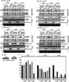
Temperature-sensitive actin and cofilin mutant yeasts affect the recruitment of cyclophilins into TBSV VRC. (A) The amounts of copurified yeast Cpr1 cyclophilin in the p33/p92 replicase preparations were reduced in act1-121ts mutant yeast cultured at the semipermissive temperature (32°C). The Flag-tagged p33 and p92pol viral replication proteins and the viral (+)repRNA together with His6-tagged Cpr1 were expressed in WT (BY4741) and act1-121ts mutant yeasts, followed by Flag affinity purification from the detergent-solubilized membrane fraction of yeasts. Expression of the His6-tagged p33 and p92pol viral replication proteins were used as a control. (Top row) Western blot analysis shows the copurified levels of His6-Cpr1 in WT and act1-121ts. Cpr1 protein was detected with anti-His antibody. (Second row) Western blot of the purified viral Flag-p33 and Flag-p92pol detected with anti-FLAG antibody. (Third row) Western blot analysis of the His6-Cpr1 levels in the total protein extracts from yeasts. (Fourth row) Western blot analysis of the His6-p33 protein levels in the total protein extract. (Fifth row) Coomassie blue-stained SDS-PAGE of the total protein levels. (B) The copurified levels of Cpr1 were reduced in cof1-8ts mutant yeasts cultured at the semipermissive temperature. See further details in panel A. (C to D) The copurified levels of CypA were reduced in actin and cofilin mutant yeasts cultured at the semipermissive temperature. Flag-p33 and Flag p92pol were coexpressed with His6-tagged CypA. See further details in panel A. (E) Each experiment was repeated three times, and the bar plot graphic represents the average levels of co-purified Cpr1 and CypA in the viral replicase preparations in WT, act1-121ts, and cof1-8ts. Error bars represent the standard error of the mean (SEM).
FIG 2
FIG 2
Temperature-sensitive actin and cofilin mutant yeasts affect the recruitment of Cyp40-like Cpr7 and its Cyp and TPR domains into TBSV VRC. (A) The amounts of copurified yeast Cpr7 cyclophilin in the p33/p92 replicase preparations were reduced in act-121ts mutant yeast cultured at the semipermissive temperature (32°C). The Flag-tagged p33 and p92pol viral replication proteins and the viral (+)repRNA together with His6-tagged Cpr7 were expressed in WT (BY4741) and act1-121ts mutant yeasts, followed by Flag affinity purification from the detergent-solubilized membrane fraction of yeasts. Expression of the His6-tagged p33 and p92pol viral replication proteins were used as a control. (Top row) Western blot analysis shows the copurified levels of His6-Cpr7 in WT and act-121ts yeasts detected with anti-His antibody. (Second row) Western blot of the purified viral Flag-p33 and Flag-p92pol detected with anti-FLAG antibody. (Third row) Western blot analysis of the His6-Cpr7 levels in the total protein extracts from yeasts. (Fourth row) Western blot analysis of the His6-p33 protein levels in the total protein extract. (Fifth row) Coomassie blue-stained SDS-PAGE of the total protein levels. (B) The copurified levels of Cpr7 were reduced in cof1-8ts mutant yeasts cultured at semipermissive temperature. See further details in panel A. (C to D) The copurified levels of His6-TPRcpr7 (only the TPR domain of Cpr7 was expressed) were reduced in actin and cofilin mutant yeasts cultured at the semipermissive temperature. Flag-p33 and Flag p92pol were coexpressed with His6-TPRcpr7. See further details in panel A. (E) The domain structure of the Cyp40-like Cpr7. (F) Each experiment was repeated three times, and the bar plot graphic represents the average levels of copurified full-length Cpr7, TPRcpr7, and CYPcpr7, respectively, in the viral replicase preparations in WT, act1-121ts, and cof1-8ts. Error bars represent the standard error.
FIG 3
FIG 3
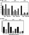
The recruitment of the Sgt2 cochaperone and the RH30 helicase into TBSV VRC depends on the actin network. (A and B) Graphic representation of the average levels of copurified full-length Sgt2, TPRsgt2, and RH30 DEAD box helicase, respectively, in the viral replicase preparations in WT, act1-121ts, and cof1-8ts. The experiments were performed as described in the legend to Fig. 1. Each experiment was repeated three times. Error bars represent the standard error.
FIG 4
FIG 4
The recruitment of the anti-CIRV-specific Hop1-like Sti1 cochaperone into CIRV VRC depends on the actin network. (A and B) The amounts of copurified yeast Sti1 cochaperone in the CIRV p36/p95pol replicase preparations were reduced in act-121ts and cof1-8ts mutant yeasts, respectively, cultured at the semipermissive temperature (32°C). The Flag-tagged p36 and p95pol CIRV replication proteins and the viral (+)repRNA together with His6-tagged Sti1 were expressed in WT (BY4741) and mutant yeasts, followed by Flag affinity purification from the detergent-solubilized membrane fraction of yeasts. See further details in the legend to Fig. 1. (C) Graphic representation of the average levels of copurified full-length Sti1 in the CIRV replicase preparations in WT, act1-121ts, and cof1-8ts yeasts. Each experiment was repeated three times. Error bars represent the standard error.
FIG 5
FIG 5
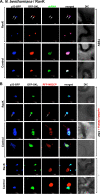
Transient expression of the Legionella RavK effector affects the architecture of the actin network and TBSV VROs in GFP-mTalin N. benthamiana transgenic plants. (A, top row) Transgenic N. benthamiana plants expressing GFP-mTalin actin-binding protein and coexpressing p33-BFP and RFP-SKL peroxisomal luminal marker (to visualize TBSV VROs). (Second row) GFP-mTalin N. benthamiana plants expressing p33-BFP, RFP-SKL, and the RavK effector were visualized via confocal microscopy. The plants were infected with TBSV. All of the plants were inoculated with TBSV 16 h after agroinfiltration. Plant samples were analyzed using confocal microscopy 36 h postinfection. (B) Merged three-dimensional (3D) image of plant leaves expressing the tagged-proteins as shown. See details in panel A. (C) Control transgenic N. benthamiana plants expressing GFP-mTalin and coexpressing RFP-SKL without (top) or with (bottom) expressing RavK effector. The plants were mock-inoculated. The scale bar is 10 μm.
FIG 6
FIG 6
Transient expression of the Legionella RavK effector inhibits the production of dsRNA replication intermediate and (+)ssRNA progeny in TBSV VROs in N. benthamiana plants. (A) Reduced production of the viral double-stranded RNA replication intermediate in N. benthamiana leaves infected with TBSV and expressing RavK effector. The TBSV dsRNA was detected via a dsRNA detector assay based on dsRNA binding-dependent fluorescence complementation. (Top) Viral dsRNA is poorly visualized within the VRO, which is marked by p33-BFP and RFP-SKL. (Bottom) TBSV dsRNA is detected in VROs in the absence of RavK expression in the control samples. Expression of the above proteins from 35S promoter was done after coagroinfiltration into N. benthamiana leaves. RavK effector was expressed in N. benthamiana leaves, and 16 h later plant leaves were inoculated with TBSV. Plant samples were collected 1.5 days postinfection (dpi). Scale bars represent 10 μm. Each experiment was repeated three times. (B) Reduced production of the viral (+)RNA products in N. benthamiana leaves expressing TBSV repRNA carrying MS2 hairpins and the RFP-MS2CP sensor. The plants were infected with CNV and expressed RavK effector as shown. The TBSV (+)RNA was detected via an RFP-MS2CP using confocal microscopy. Bottom images show the control images of plants with or without RavK expression and in the absence of TBSV repRNA(+)MS2hp when RFP-MS2-CP is targeted to the nucleus in the absence of cytosolic targets. Scale bars represent 10 μm. Each experiment was repeated three times.
FIG 7
FIG 7
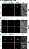
Disruption of the plant actin filaments by RavK expression inhibits the recruitment of plant cyclophilin restrictions factors into VROs during TBSV replication. (A, top row) Bimolecular fluorescence complementation (BiFC) analysis shows the interaction between the plant cyclophilin nYFP-Roc1 and TBSV replication protein p33-cYFP within VROs marked by RFP-SKL in control N. benthamiana. (Second row) On the contrary, BiFC analysis of plants also expressing RavK indicates the lack of interaction between nYFP-Roc1 and p33-cYFP within VROs. Plants were inoculated with TBSV 16 h after agroinfiltration, followed by BiFC 36 hpi. (B) Negative control experiments were performed as in panel A. (C) BiFC analysis shows the interaction between the plant cyclophilin nYFP-Roc2 and TBSV replication protein p33-cYFP within VROs marked by RFP-SKL in N. benthamiana not expressing and expressing RavK, respectively. Experiments were performed as in panel A. (D) Negative control experiments were performed as in panel A. (E to F) BiFC analysis shows the interaction between the plant cyclophilin nYFP-Roc1 or nYFP-Roc2 and the CIRV replication protein p36-cYFP within VROs marked by RFP-Tim21 in N. benthamiana not expressing and expressing RavK, respectively. Experiments were performed as in panel A, except the plant samples were visualized with confocal microscope 50 h after infection.
FIG 8
FIG 8
Inhibition of the recruitment of the antiviral RH30 DEAD box helicase into VROs via disruption of the plant actin network by RavK expression during TBSV replication. (A, top row) BiFC analysis shows the interaction between the plant nYFP-RH30 helicase and TBSV p33-cYFP replication protein within VROs marked by RFP-SKL in control N. benthamiana. (Second row) Comparable BiFC analysis of plants also expressing RavK indicates the lack of interaction between nYFP-RH30 and p33-cYFP within VROs. Plants were inoculated with TBSV 16 h after agroinfiltration followed by BiFC 36 hpi. (B) Negative control experiments were performed as in panel A. (C) BiFC analysis shows the interaction between the nYFP-RH30 helicase and the CIRV p36-cYFP within VROs marked by RFP-Tim21 in N. benthamiana not expressing and expressing RavK, respectively. Experiments were performed as in panel A, except the plant samples were visualized with confocal microscope 50 h after infection. Each experiment was repeated three times.
FIG 9
FIG 9
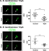
Inhibition of the recruitment of the antiviral CIRFs into VROs via stabilization of the plant actin filaments by VipA expression during tombusvirus replication in N. benthamiana. (A, top row) BiFC analysis shows the interaction between the plant nYFP-Roc1 cyclophilin and TBSV p33-cYFP replication protein within VROs in control N. benthamiana. (Second row) Comparable BiFC analysis of plants also expressing VipA effector indicates the reduced interaction between nYFP-Roc1 and p33-cYFP within VROs. Plants were inoculated with CNV via agroinfiltration, followed by BiFC 48 hpi. Arrows point at the VROs. The BiFC signal intensity was measured by image J. The same confocal laser microscopy setting was used in these experiments. Scale bars represent 20 μm. Each experiment was repeated three times. (B, top row) BiFC analysis shows the interaction between the plant nYFP-Roc2 cyclophilin and TBSV p33-cYFP replication protein within VROs in control N. benthamiana. (Second row) Comparable BiFC analysis of plants also expressing VipA effector indicates the reduced interaction between nYFP-Roc2 and p33-cYFP within VROs. See further details in panel A.
FIG 10
FIG 10
A model on the role of the actin network in the race to deliver restriction factors into tombusvirus VROs. (A) Disruption of the actin filaments (by RavK expression) leads to poor recruitment of both selected proviral and restriction factors into VROs. This leads to low level of replication and inhibition of VRO formation due to the unsatisfactory amounts of recruited proviral host factors. (B) During normal replication, for example in WT yeast or N. benthamiana plants, tombusviruses stabilize the actin filaments via sequestering cofilins with p33 replication proteins. Then, the stable actin filaments are utilized to deliver proviral host factors and also restriction factors into VROs. This is a race driven by p33 among the host factors: in these susceptible hosts, tombusviruses are capable of delivering the proviral factors more efficiently to VROs than the combined inhibitory effects provided by the recruited host restriction factors. This results in robust TBSV replication in these hosts. (C) When the host has a stabilized actin network, for example due to given mutations in actin or cofilin or expression of VipA actin nucleator, then tombusviruses easily outcompete the restriction factors via enhanced recruitment of the p33 proviral host factor complexes into VROs. Altogether, this is a race for/against VRO biogenesis between proviral and antiviral factors depending on the availability of these cellular factors and the ability of tombusviruses to subvert the host actin network in the infected hosts.

References

    1. Wu X, Valli A, Garcia JA, Zhou X, Cheng X. 2019. The tug-of-war between plants and viruses: great progress and many remaining questions. Viruses 11:203. 10.3390/v11030203. - DOI - PMC - PubMed
    1. Zorzatto C, Machado JP, Lopes KV, Nascimento KJ, Pereira WA, Brustolini OJ, Reis PA, Calil IP, Deguchi M, Sachetto-Martins G, Gouveia BC, Loriato VA, Silva MA, Silva FF, Santos AA, Chory J, Fontes EP. 2015. NIK1-mediated translation suppression functions as a plant antiviral immunity mechanism. Nature 520:679–682. 10.1038/nature14171. - DOI - PMC - PubMed
    1. Nicaise V. 2014. Crop immunity against viruses: outcomes and future challenges. Front Plant Sci 5:660. 10.3389/fpls.2014.00660. - DOI - PMC - PubMed
    1. Cillo F, Palukaitis P. 2014. Transgenic resistance. Adv Virus Res 90:35–146. 10.1016/B978-0-12-801246-8.00002-0. - DOI - PubMed
    1. Carr JP, Lewsey MG, Palukaitis P. 2010. Signaling in induced resistance. Adv Virus Res 76:57–121. 10.1016/S0065-3527(10)76003-6. - DOI - PubMed

Publication types

MeSH terms

LinkOut - more resources