

Clinical applicability of quantitative atrophy measures on MRI in patients suspected of Alzheimer's disease
- PMID: 35639148
- PMCID: PMC9668763
- DOI: 10.1007/s00330-021-08503-7
Clinical applicability of quantitative atrophy measures on MRI in patients suspected of Alzheimer's disease
Abstract
Objectives: Neurodegeneration in suspected Alzheimer's disease can be determined using visual rating or quantitative volumetric assessments. We examined the feasibility of volumetric measurements of gray matter (GMV) and hippocampal volume (HCV) and compared their diagnostic performance with visual rating scales in academic and non-academic memory clinics.
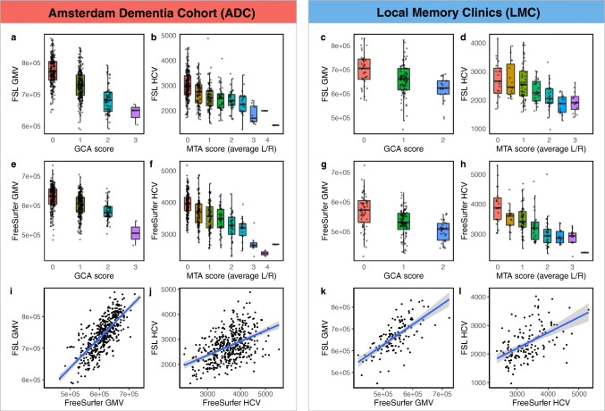
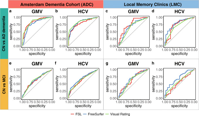
Materials and methods: We included 231 patients attending local memory clinics (LMC) in the Netherlands and 501 of the academic Amsterdam Dementia Cohort (ADC). MRI scans were acquired using local protocols, including a T1-weighted sequence. Quantification of GMV and HCV was performed using FSL and FreeSurfer. Medial temporal atrophy and global atrophy were assessed with visual rating scales. ROC curves were derived to determine which measure discriminated best between cognitively normal (CN), mild cognitive impairment (MCI), and Alzheimer's dementia (AD).
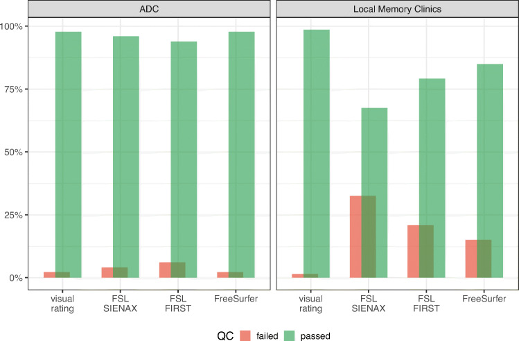
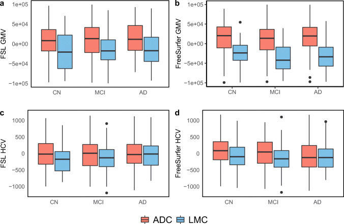
Results: Patients attending LMC (age 70.9 ± 8.9 years; 47% females; 19% CN; 34% MCI; 47% AD) were older, had more cerebrovascular pathology, and had lower GMV and HCV compared to those of the ADC (age 64.9 ± 8.2 years; 42% females; 35% CN, 43% MCI, 22% AD). While visual ratings were feasible in > 95% of scans in both cohorts, quantification was achieved in 94-98% of ADC, but only 68-85% of LMC scans, depending on the software. Visual ratings and volumetric outcomes performed similarly in discriminating CN vs AD in both cohorts.
Conclusion: In clinical settings, quantification of GM and hippocampal atrophy currently fails in up to one-third of scans, probably due to lack of standardized acquisition protocols. Diagnostic accuracy is similar for volumetric measures and visual rating scales, making the latter suited for clinical practice. In a real-life clinical setting, volumetric assessment of MRI scans in dementia patients may require acquisition protocol optimization and does not outperform visual rating scales.
Key points: • In a real-life clinical setting, the diagnostic performance of visual rating scales is similar to that of automatic volumetric quantification and may be sufficient to distinguish Alzheimer's disease groups. • Volumetric assessment of gray matter and hippocampal volumes from MRI scans of patients attending non-academic memory clinics fails in up to 32% of cases. • Clinical MR acquisition protocols should be optimized to improve the output of quantitative software for segmentation of Alzheimer's disease-specific outcomes.
Keywords: Alzheimer’s disease; Gray matter volume (GMV); Hippocampal volume (HCV); Magnetic resonance imaging (MRI); Visual rating scales.
© 2022. The Author(s).
Conflict of interest statement
Prof. Frederik Barkhof received payment and honoraria from Bayer, Biogen, TEVA, Merck-Serono, Novartis, Roche, IXICO Ltd, GeNeuro, and Combinostics for consulting; research support via grants from EU/EFPIA Innovative Medicines Initiative Joint Undertaking (AMYPAD consortium), EuroPOND (H2020), UK MS Society, Dutch MS Society, PICTURE.
(IMDI NWO), NIHR UCLH Biomedical Research Centre (BRC), ECTRIMS-MAGNIMS.
Research programs of Prof. Wiesje van der Flier have been funded by ZonMW, NWO, EU-FP7, EU-JPND, Alzheimer Nederland, CardioVascular Onderzoek Nederland, Health~Holland, Topsector Life Sciences & Health, stichting Dioraphte, Gieskes-Strijbis fonds, stichting Equilibrio, Pasman stichting, Biogen MA Inc, Boehringer Ingelheim, Life-MI, AVID, Roche BV, Fujifilm, Combinostics. WF holds the Pasman chair. WF has performed contract research for Biogen MA Inc and Boehringer Ingelheim. WF has been an invited speaker at Boehringer Ingelheim and Biogen MA Inc. All funding is paid to her institution. Other authors declare that they have no competing interests.
Figures
References
MeSH terms
Grants and funding
LinkOut - more resources
Full Text Sources
Medical
