. 2022 Jul;8(4):1694-1708.
doi: 10.1002/vms3.834. Epub 2022 May 31.

CT anatomy of the head in the Ile de France sheep

Affiliations

CT anatomy of the head in the Ile de France sheep

Majid Masoudifard et al. Vet Med Sci. 2022 Jul.

Abstract

Background: CT scan images provide accurate anatomical data from different areas of the body that can be used to diagnose diseases.

Objectives: The present work aimed to describe the normal anatomical structures of the Ile de France sheep head and its morphometric and volumetric properties using computed tomography (CT) and stereological methods.

Methods: Five adult Ile de France sheep heads, which were of mature age (above 10 months), were included in this study. The different cavities of the head, including the nasal cavity, paranasal sinuses, oral cavity, orbital cavity and vestibulocochlear system, were evaluated using CT scans, cross, sagittal and coronal sections.

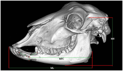
Results: The mean length, height and width of the skull were 25.3 ± 1.02, 9.8 ± 0.93 and 12.3 ± 0.91 cm, respectively. The results showed that the nasal cavity is divided into three regions. Vestibular, respiratory and olfactory regions. The paranasal sinuses are composed of maxillary, frontal, palatine, sphenoid, lacrimal and ethmoidal that were identified and named in the CT scan images and their corresponding anatomical cross-sections. The total volume of the head, nasal cavity and oral cavity was estimated to be 2998 ± 202.00, 303 ± 31.33 and 229.3 ± 10.61 cm3 , respectively.

Conclusions: The frontal sinus in the Ile de France sheep was limited to the frontal bone without extending into the parietal, temporal, or occipital bones, similar to Saanen goat. This study provided a comprehensive atlas of Ile de France sheep anatomy to internal medicine veterinarians and surgeons.

Keywords: Ile de France sheep; anatomy; computed tomography; morphometric study; volumetric study.


Conflict of interest statement

The authors declare that they have no conflict of interest.

Figures

FIGURE 1
FIGURE 1
2D CT scan images (transverse view–bone window) and cross‐sectional images in the Ile de France sheep. (a, b) At level of incisive teeth and (c, d) at level of inter‐alveolar space. (1) Dorsal nasal meatus; (2) straight fold of dorsal nasal concha; (3) alar fold of ventral nasal meatus; (4) middle nasal meatus; (5) ventral nasal meatus; (6) nasal septum; (7) nasal process of incisive bone; (8) hard palate; (9) vomeronasal organ; (10) body of tongue; (11) mandible
FIGURE 2
FIGURE 2
2D CT scan images (transverse view–bone window) and cross‐sectional images in the Ile de France sheep. (a, b) At level of 1 cm rostral to the first pre‐molar tooth and (c, d) at level of first pre‐molar tooth. (1) Nasal bone; (2) dorsal nasal concha; (3) dorsal conchal sinus; (4) dorsal spiral lamella of ventral nasal concha; (5) ventral nasal concha; (6) ventral spiral lamella of ventral nasal concha; (7) basal fold of ventral nasal concha; (8) nasal septum; (9) nasal process of incisive bone; (10) infra‐orbital canal; (11) palatine process of maxilla; (12) palatine sinus; (13) hard palate; (14) first pre‐molar tooth; (15) body of tongue; (16) mandible; (17) vomeronasal organ
FIGURE 3
FIGURE 3
2D CT scan images (transverse view–bone window) and cross‐sectional images in the Ile de France sheep. (a, b) At level of third pre‐molar tooth and (c, d) at level of first molar tooth. (1) Nasal bone; (2) frontal bone; (3) dorsal nasal concha; (4) dorsal conchal sinus; (5) lacrimal sinus; (6) dorsal spiral lamella of ventral nasal concha; (7) ventral nasal concha; (8) nasolacrimal duct; (9) middle nasal sinus; (10) nasal septum; (11) dorsal compartment of maxillary sinus; (12) maxillary sinus; (13) palatine sinus; (14) infra‐orbital canal; (15) palatine process of maxillary sinus; (16) hard palate; (17) second molar tooth; (18) third pre‐molar tooth; (19) body of tongue; (20) mandible; (21) ventral spiral lamella of ventral nasal concha
FIGURE 4
FIGURE 4
2D CT scan images (transverse view–bone window) and cross‐sectional images in the Ile de France sheep. (a, b) At level of third molar tooth and (c, f) At level of eye ball centre. (1) Frontal bone; (2) inter‐frontal septum; (3) medial compartment of rostral frontal sinus; (4) intermediate compartment of rostral frontal sinus; (5) lateral compartment of rostral frontal sinus; (6) rostral part of frontal sinus; (7) supra‐orbital canal; (8) brain; (9) eye ball (retina and choroid); (10) periorbital fat; (11) lens of eye; (12) zygomatic arch; (13) maxillary sinus; (14) ethmoidal labyrinth; (15) sclera; (16) nasal septum; (17) choanae; (18) third molar tooth; (19) soft palate; (20) larynx; (21) root of last molar tooth; (22) hyoid bone; (23) mandible
FIGURE 5
FIGURE 5
2D CT scan images (transverse view–bone window) and cross‐sectional images in the Ile de France sheep. (a, b) At level of temporo‐mandibular joint (TMJ). (1) Parietal bone, (2) brain, (3) zygomatic arch, (4) temporo‐mandibular joint, (5) basilar part of occipital bone; (6) optic chiasma; (7) larynx; (8) hyoid bone
FIGURE 6
FIGURE 6
2D CT scan images (longitudinal view–bone window) and cross‐sectional images in the Ile de France sheep. (a, b) At level temporo‐mandibular joint (TMJ) and (c, d) At level of at level of medulla oblongata. (1) Frontal sinus; (2) maxillary sinus; (3) internal acoustic meatus; (4) temporo‐mandibular joint; (5) third molar tooth of upper jaw; (6) second molar tooth of upper jaw; (7) first molar tooth of upper jaw; (8) brain; (9) olfactory bulb; (10) hypophyseal fossa; (11) inter‐thalamic adhesion; (12) cerebellum; (13) medulla oblongata; (14) basisphenoid bone; (15) ethmoidal labyrinth; (16) dorsal nasal concha and its sinus; (17) middle nasal concha and its sinus; (18) ventral nasal concha and its sinus; (19) ventral nasal meatus; (20) palatine process of incisive bone; (21) hard palate; (22) incisor's tooth; (23) palatine process of incisive bone; (24) body of tongue; (25) epiglottis; (26) Soft palate; (27) Mandible
FIGURE 7
FIGURE 7
3D reconstruction (osseous‐shaped‐vp) in the Ile de France sheep. (a) Left median section, (b) caudal view. Morphometric parameters of the head. Skull length (SL), skull height (SH), skull width (SW), cranial length (CL), cranial width (CW), facial length (FL)
FIGURE 8
FIGURE 8
3D reconstruction (osseous‐shaped‐vp) lateral view in the Ile de France sheep. morphometric parameters of the Mandible. Mandibular height (MH), mandibular length (ML), distance from the mental foramen to the most lateral incisive tooth (MFID), distance from the mental foramen to the cranial margin of the first pre‐molar tooth (MFPD), distance from the mental foramen to the ventral margin of the mandibular body (MFVD), distance from the caudal margin of the mandibular ramus to the mental foramen (MFC)
FIGURE 9
FIGURE 9
3D reconstruction (osseous‐shaped‐vp) lateral view in the Ile de France sheep. morphometric parameters of the hyoid bone. Stylohyoid length (S), epihyoid length (E), ceratohyoid length (C), lingual process of the basihyoid bone length (LB), thyrohyoid length (T)

References

    1. Alsafy, M. A. , El‐Gendy, S. A. , & Abumandour, M. M. (2014). Computed tomography and gross anatomical studies on the head of one‐humped camel (Camelus dromedarius). Anatomical Record (Hoboken, N.J.: 2007), 297(4), 630–642. - PubMed
    1. Alsafy, M. A. , El‐Gendy, S. A. , & El Sharaby, A. A. (2013). Anatomic reference for computed tomography of paranasal sinuses and their communication in the Egyptian buffalo (Bubalus bubalis). Anatomia, Histologia, Embryologia, 42(3), 220–231. - PubMed
    1. Arencibia, A. , Vázquez, J. M. , Rivero, M. , Latorre, R. , Sandoval, J. A. , Vilar, J. M. , & Ramírez, J. A. (2000). Computed tomography of normal cranioencephalic structures in two horses. Anatomia, Histologia, Embryologia, 29(5), 295–299. - PubMed
    1. Awaad, A. S. , Abdel Maksoud, M. , & Fathy, M. Z. (2019). Surgical anatomy of the nasal and paranasal sinuses in Egyptian native sheep (Ovis aries) using computed tomography and cross sectioning. Anatomia, Histologia, Embryologia, 48(4), 279–289. - PubMed
    1. Badlangana, N. L. , Adams, J. W. , & Manger, P. R. (2011). A comparative assessment of the size of the frontal air sinus in the giraffe (Giraffa camelopardalis). Anatomical Record (Hoboken, N.J.: 2007), 294(6), 931–940. - PubMed

LinkOut - more resources