. 2022 Jun 21;48(4):728-737.
doi: 10.1093/schbul/sbac050.

Schizophrenia in Translation: Why the Eye?

Affiliations

Schizophrenia in Translation: Why the Eye?

Steven M Silverstein et al. Schizophr Bull. .

Abstract

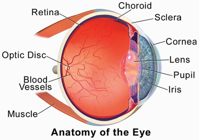
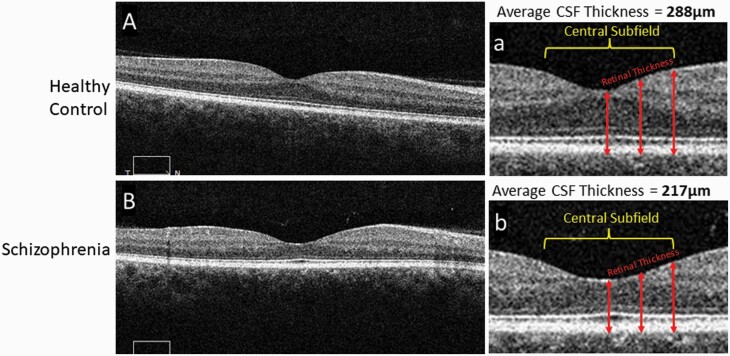
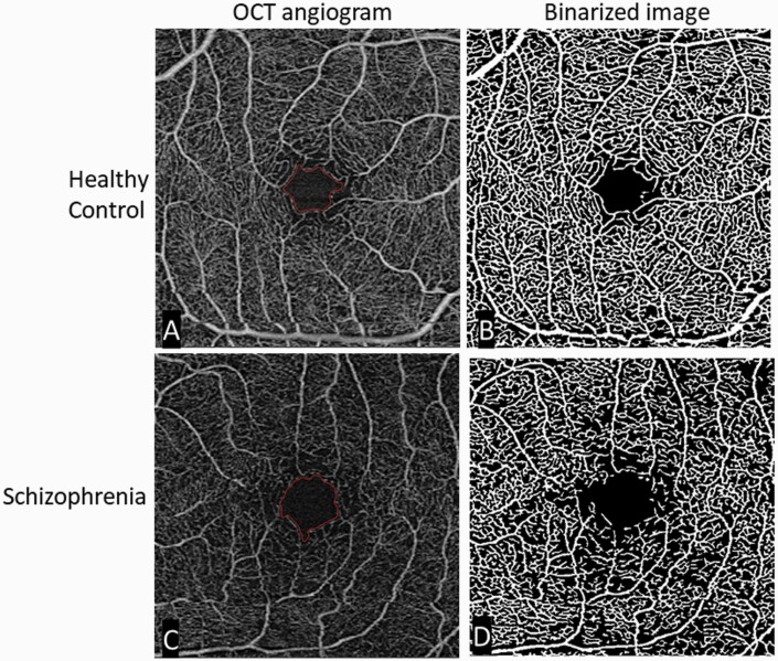
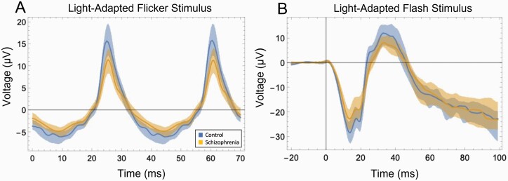
Schizophrenia is increasingly recognized as a systemic disease, characterized by dysregulation in multiple physiological systems (eg, neural, cardiovascular, endocrine). Many of these changes are observed as early as the first psychotic episode, and in people at high risk for the disorder. Expanding the search for biomarkers of schizophrenia beyond genes, blood, and brain may allow for inexpensive, noninvasive, and objective markers of diagnosis, phenotype, treatment response, and prognosis. Several anatomic and physiologic aspects of the eye have shown promise as biomarkers of brain health in a range of neurological disorders, and of heart, kidney, endocrine, and other impairments in other medical conditions. In schizophrenia, thinning and volume loss in retinal neural layers have been observed, and are associated with illness progression, brain volume loss, and cognitive impairment. Retinal microvascular changes have also been observed. Abnormal pupil responses and corneal nerve disintegration are related to aspects of brain function and structure in schizophrenia. In addition, studying the eye can inform about emerging cardiovascular, neuroinflammatory, and metabolic diseases in people with early psychosis, and about the causes of several of the visual changes observed in the disorder. Application of the methods of oculomics, or eye-based biomarkers of non-ophthalmological pathology, to the treatment and study of schizophrenia has the potential to provide tools for patient monitoring and data-driven prediction, as well as for clarifying pathophysiology and course of illness. Given their demonstrated utility in neuropsychiatry, we recommend greater adoption of these tools for schizophrenia research and patient care.

Keywords: ERG; OCT; brain; cornea; eye; imaging; oculomics; pupil; retina; schizophrenia.


Figures

Fig. 1.
Fig. 1.
Depiction of the components of the eye.Figure reproduced from Blausen.com staff. Medical gallery of Blausen Medical 2014. WikiJ Med. 2014;1(2). doi:10.15347/wjm/2014.010. ISSN 2002-4436, via a creative commons license (CC BY 3.0).
Fig. 2.
Fig. 2.
Comparison of macular thickness, from OCT images, between a healthy control and subject with schizophrenia.Representative 6 mm OCT scans of the retina in both a healthy control (A) and subject with schizophrenia (B), along with enlarged images of the central portion of the scans (a, b) respectively. Alternating darker and lighter bands seen in the scans represent unique layers in the retina. The central subfield is a 1 mm central section of the scan. Retinal thickness at different points in the scan are measured from the superficial internal limiting membrane to the deep retinal pigment epithelium (arrows). The thickness at multiple corresponding points are appreciably thinner in the schizophrenia scan. The average central subfield thickness is calculated on the OCT device by averaging the retinal thickness at many points in a central circular area of retinal tissue measuring 1 mm in diameter. In these images the thickness of the macula central subfield is 217 microns for the person with schizophrenia and 288 microns for the control subject. OCT, optical coherence tomography.
Fig. 3.
Fig. 3.
Comparison of retinal microvasculature, from OCTA images, between a healthy control and a subject with schizophrenia.Original and binarized OCT angiograms of a patient with schizophrenia and a healthy control. A standard OCT angiogram of a healthy eye is seen in (A). Vessels are seen in white, with larger arterioles and smaller capillaries represented in the 3 × 3 mm2 window. The foveal avascular zone (the most central region of the macula, where blood vessels are not present) is delineated in red. (B): A binarized version of the same angiogram, allowing for easier qualitative and quantitative assessment of vascular density. The bottom row shows similar images from a patient with schizophrenia. Compared to the healthy control, there is an enlargement of the foveal avascular zone (typically reflecting loss of microvasculature in the surrounding area) and a decrease in vessel density (C) most easily appreciated in the binarized image (D). OCT, optical coherence tomography; OCTA, OCT angiography; Figure reproduced from Green KM, Choi JJ, Ramchandran RS, Silverstein SM. OCT and OCT angiography offer new insights and opportunities in schizophrenia research and treatment. Front Digit Health. 2022;4:836851. doi:10.3389/fdgth.2022.836851 via a creative commons license (CC BY 4.0).
Fig. 4.
Fig. 4.
Schizophrenia-control differences in strength of retinal cell firing as indicated by ERG.(A) Data from a light-adapted flicker stimulus ERG test, depicting cone photoreceptor response. The stimulus was white light flickering at 28.3 Hz, at an intensity of 85 troland seconds. The difference between the schizophrenia group (tan) and control group (blue) was large: t(48) = −3.58, P = .001, d = 1.01. Shaded areas depict the 25th–75th percentile interquartile range. Substantial non-overlap between groups can be observed. (B) Data from a light-adapted flash stimulus ERG test. The stimulus was light flashing at 1 Hz, at an intensity of 100 troland seconds. The differences between the schizophrenia group (tan) and control group (blue) were large: For the initial negative peak (a-wave), reflecting photoreceptor activity, t(48) = 2.86, P = .006, d = .81. For the subsequent positive peak (b-wave), reflecting bipolar cell and glial cell activity, t(48) = 2.92, P = .005, d = .83. Substantial non-overlap between groups can be observed here as well. Data in both cases from Demmin DL, Davis Q, Roché M, Silverstein SM. Electroretinographic anomalies in schizophrenia. J Abnorm Psychol. 2018;127(4):417–428. doi:10.1037/abn0000347. PMID: 29745706. ERG, electroretinography.

References

    1. Alonso Y, Valiente-Palleja A, Verge B, Vilella E, Martorell L. High frequency of clinical conditions commonly associated with mitochondrial disorders in schizophrenia. Acta Neuropsychiatr. 2020;32(5):265–269. - PubMed
    1. Perry BI, Bowker N, Burgess S, et al. . Evidence for shared genetic aetiology between schizophrenia, cardiometabolic, and inflammation-related traits: genetic correlation and colocalization analyses. Schizophr Bull Open. 2022;3(1):sgac001. - PMC - PubMed
    1. Little JD. Schizophrenia: a multi-system disorder? Aust N Z J Psychiatry. 2015;49(4):390. - PubMed
    1. Malaspina D. Looking schizophrenia in the eye. Am J Psychiatry. 2013;170(12):1382–1384. - PubMed
    1. Mitchell AJ, Dinan TG. Schizophrenia: a multisystem disease? J Psychopharmacol. 2010;24(suppl 4):5–7. - PMC - PubMed

Publication types