

Effect of orthodontic forces on root length of immature mandibular second premolars: a split-mouth randomized clinical trial
- PMID: 35640080
- PMCID: PMC8576854
- DOI: 10.1590/2177-6709.26.5.e2119355.oar
Effect of orthodontic forces on root length of immature mandibular second premolars: a split-mouth randomized clinical trial
Abstract
Objective: To assess the effect of orthodontic forces on changes in root length of immature mandibular second premolars.
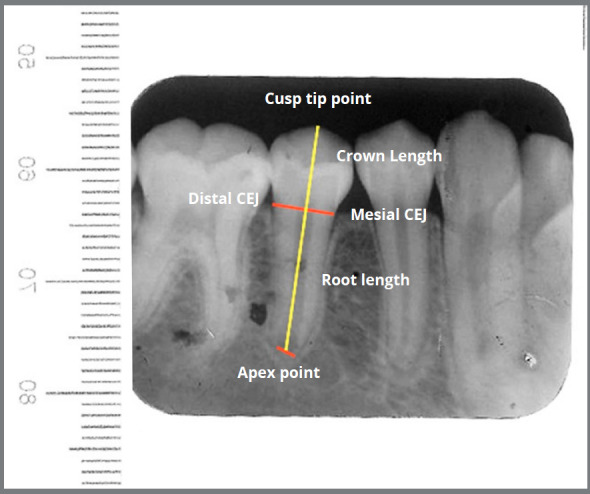
Methods: Sixty-four mandibular second premolars (MSP) with immature apices (left and right sides) of 32 patients aged between 10 and 13 years were evaluated. Orthodontic treatment was started after obtaining periapical radiographs (T1) from the MSPs of each patient. Brackets were bonded, except the ones of MSPs (left or right by random as control MSP, and the other side as test MSP). After 9-12 months, a second periapical radiograph (T2) was obtained from the MSPs of each patient. Then, brackets were bonded to the control MSPs, which were not bonded before. After 18 ± 3 months, a third periapical radiograph (T3) was obtained. Changes in root length were evaluated by using a new formula. The test and control MSPs at T1, T2 and T3 were compared using repeated measures ANOVA and parametric tests. P-value smaller than 0.05 was statistically significant.
Results: There was no significant difference between the test and control groups in the mean root length of MSP at T1 (p= 0.48) and T3 (p= 0.078). The root length at T2 (p= 0.001) was significantly different between test and control MSPs, and the test group showed longer root length than the control group.
Conclusions: Orthodontic force applied for leveling and alignment of immature MSPs may not have destructive effects on the roots, and may accelerates root formation in short-term. Normal root length was achieved at the end of root development.
Objetivo:: Avaliar o efeito das forças ortodônticas nas mudanças do comprimento radicular de segundos pré-molares inferiores com raízes incompletas.
Métodos:: Foram avaliados 64 segundos pré-molares inferiores (SPI) com raízes incompletas (lados direito e esquerdo da mandíbula) de 32 pacientes com idades entre 10 e 13 anos. O tratamento ortodôntico teve início após a obtenção de radiografias periapicais (T1) dos SPIs de cada paciente. Foram colados braquetes em todos os dentes, com exceção dos SPIs (do lado esquerdo ou direito, de forma aleatória, como grupo controle; e o SPI contralateral como grupo teste). Após 9 a 12 meses, uma segunda radiografia periapical (T2) foi obtida dos SPIs de cada paciente. Então, braquetes foram colados nos SPIs do grupo controle, que não haviam sido colados anteriormente. Após 18 ± 3 meses, uma terceira radiografia periapical (T3) foi obtida. As mudanças no comprimento radicular foram avaliadas por meio de uma nova fórmula. Os SPIs teste e controle foram comparados em T1, T2 e T3 usando ANOVA para medidas repetidas e testes paramétricos. Valores de p< 0,05 foram considerados estatisticamente significativos.
Resultados:: Não foi encontrada diferença significativa no comprimento radicular médio dos SPIs entre os grupos teste e controle em T1 (p= 0,48) e T3(p= 0,078). O comprimento radicular dos SPIs em T2 (p= 0,001) foi significativamente diferente entre os lados teste e controle, sendo que o grupo teste apresentou maior comprimento radicular do que o grupo controle.
Conclusões:: As forças ortodônticas aplicadas para o alinhamento e nivelamento de SPIs com raízes incompletas podem não ter efeitos danosos nas raízes, podendo, inclusive, acelerar a formação radicular em curto prazo. O comprimento radicular normal foi alcançado ao fim da formação radicular.
Conflict of interest statement
The authors report no commercial, proprietary or financial interest in the products or companies described in this article.
Figures


References
-
- Mavragani M, Vergari A, Selliseth NJ, Bøe OE, Wisth PL. A radiographic comparison of apical root resorption after orthodontic treatment with a standard edgewise and a straight-wire edgewise technique. Eur J Orthod. 2000;22(6):665–674. - PubMed
-
- Hendrix I, Carels C, Kuijpers-Jagtman AM, Van 'T Hof M. A radiographic study of posterior apical root resorption in orthodontic patients. Am J Orthod Dentofacial Orthop. 1994;105(4):345–349. - PubMed
-
- Brezniak N, Wasserstein A. Root resorption after orthodontic treatment Part 2. Literature review. Am J Orthod Dentofacial Orthop. 1993;103(2):138–146. - PubMed
-
- Mavragani M, Bøe OE, Wisth PJ, Selvig KA. Changes in root length during orthodontic treatment advantages for immature teeth. Eur J Orthod. 2002;24(1):91–97. - PubMed
Publication types
MeSH terms
LinkOut - more resources
Full Text Sources
Research Materials
