. 2022 Jun 30;145(6):2077-2091.
doi: 10.1093/brain/awab456.

Heterozygous PRKN mutations are common but do not increase the risk of Parkinson's disease

Affiliations

Heterozygous PRKN mutations are common but do not increase the risk of Parkinson's disease

William Zhu et al. Brain. .

Abstract

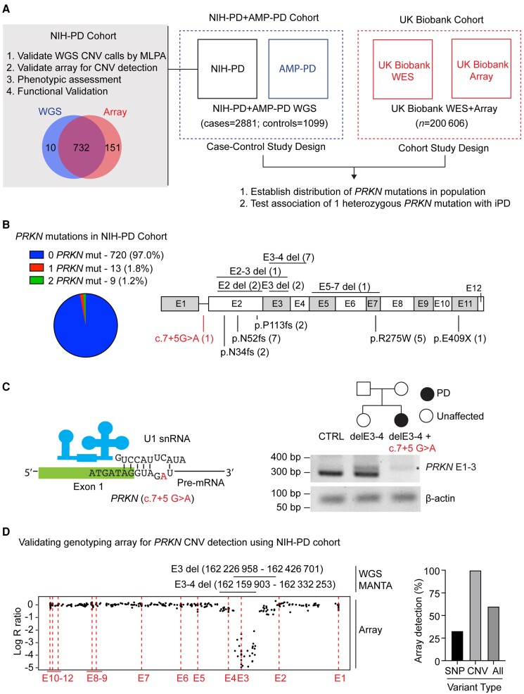
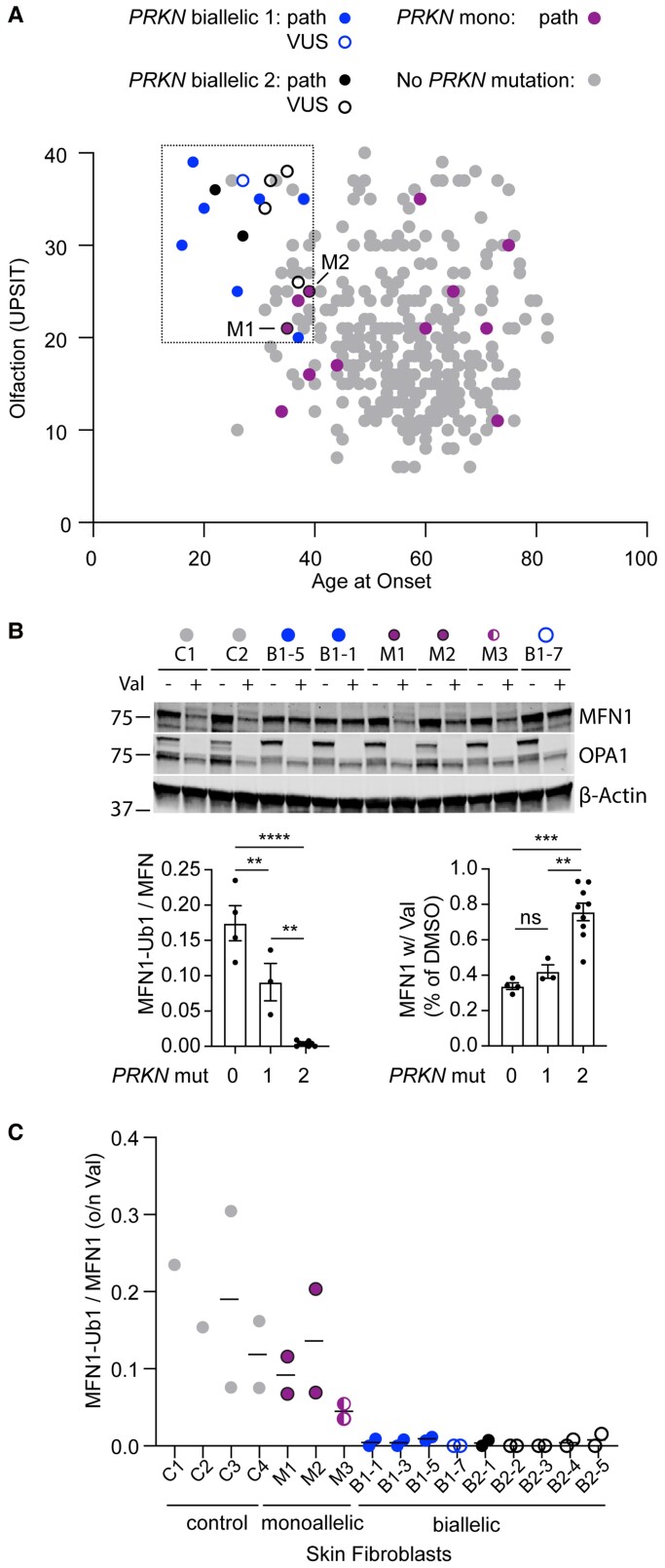
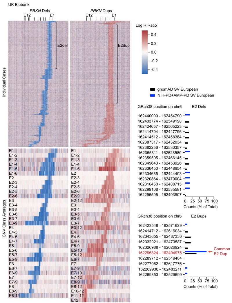
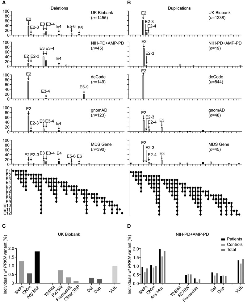
PRKN mutations are the most common recessive cause of Parkinson's disease and are a promising target for gene and cell replacement therapies. Identification of biallelic PRKN patients at the population scale, however, remains a challenge, as roughly half are copy number variants and many single nucleotide polymorphisms are of unclear significance. Additionally, the true prevalence and disease risk associated with heterozygous PRKN mutations is unclear, as a comprehensive assessment of PRKN mutations has not been performed at a population scale. To address these challenges, we evaluated PRKN mutations in two cohorts with near complete genotyping of both single nucleotide polymorphisms and copy number variants: the NIH-PD + AMP-PD cohort, the largest Parkinson's disease case-control cohort with whole genome sequencing data from 4094 participants, and the UK Biobank, the largest cohort study with whole exome sequencing and genotyping array data from 200 606 participants. Using the NIH-PD participants, who were genotyped using whole genome sequencing, genotyping array, and multi-plex ligation-dependent probe amplification, we validated genotyping array for the detection of copy number variants. Additionally, in the NIH-PD cohort, functional assays of patient fibroblasts resolved variants of unclear significance in biallelic carriers and suggested that cryptic loss of function variants in monoallelic carriers are not a substantial confounder for association studies. In the UK Biobank, we identified 2692 PRKN copy number variants from genotyping array data from nearly half a million participants (the largest collection to date). Deletions or duplications involving exon 2 accounted for roughly half of all copy number variants and the vast majority (88%) involved exons 2, 3, or 4. In the UK Biobank, we found a pathogenic PRKN mutation in 1.8% of participants and two mutations in ∼1/7800 participants. Those with one PRKN pathogenic variant were as likely as non-carriers to have Parkinson's disease [odds ratio = 0.91 (0.58-1.38), P-value 0.76] or a parent with Parkinson's disease [odds ratio = 1.12 (0.94-1.31), P-value = 0.19]. Similarly, those in the NIH-PD + AMP + PD cohort with one PRKN pathogenic variant were as likely as non-carriers to have Parkinson's disease [odds ratio = 1.29 (0.74-2.38), P-value = 0.43]. Together our results demonstrate that heterozygous pathogenic PRKN mutations are common in the population but do not increase the risk of Parkinson's disease.

Keywords: PARK2; early onset Parkinson’s disease; mitophagy; parkin; young onset Parkinson’s disease.


Figures

Figure 1
Figure 1
Study design and investigation of PRKN variants in the NIH-PD cohort. (A) PRKN variants were investigated in two large cohorts, the US based NIH-PD + AMP-PD cohort and the UK Biobank cohort. More extensive investigation was undertaken for NIH-PD participants, which cross-validated genotyping methods used in the two cohorts and provided context for the overall study through phenotypic and functional analysis of PRKN mutation carriers. (B) Proportion of PRKN variants detected in the NIH-PD cohort by whole genome sequencing is shown in the pie chart (left). The position of pathogenic PRKN variants (black) and a novel variant of unclear significance (VUS) (c.7+5G>A) detected by whole genome sequencing are shown in schematic of the PRKN gene (right). CNVs are shown on the top and SNPs on the bottom. (C) Schematic of the exon 1–intron 1 junction is shown with the position of the intronic substitution c.7+5G>A in red (left). This is predicted to disrupt a base pairing with the U1 small nuclear RNA of the spliceosome. Pedigree of family with this mutation appears on the top right. RT-PCR of RNA isolated from fibroblast cell lines from a control (CTRL), the unaffected sister of the proband with a single exon 3–4 deletion, and the proband with an exon 3–4 deletion and c.7+5G>A variants is depicted on the bottom right. The c.7+5G>A mutation disrupted the PCR product from primers in exons 1 and 3. An aberrant product (asterisk) was seen in both exon 3–4 deletion carriers. β-Actin served as a loading control. (D) Correspondence between CNV calls by WGS and by genotyping array is shown for a representative case with compound heterozygous PRKN deletions (left). The x-axis represents the relative genomic position. The position of the exons is indicated by the dotted lines. Log R ratio is shown on the y-axis. A value of −0.5 corresponds to a single deletion and a lower value is expected for two deletions. The relative position of the deletions called from WGS is shown above. Log R ratio is around −0.5 for probes in the non-overlapping regions of the deletions and <−2 for probes in the overlapping region. The graph on the right represents the proportion of PRKN variants (either SNPs or CNVs) detected by genotyping array.
Figure 2
Figure 2
Biallelic and monoallelic PRKN mutation carriers can be distinguished by their phenotype and functional assays in their fibroblasts. (A) Scatterplot depicts scores from smell identification test (UPSIT, y-axis) and age-at-onset (x-axis) for genotyped Parkinson disease patients in the NIH-PD cohort with no PRKN mutations (grey), one PRKN mutation (magenta), or two PRKN mutations (blue). A second group of patients with two PRKN mutations (black) was identified outside of the consecutive series. Those with a novel variant or VUS are shown as open symbols. Two patients with one PRKN mutation and whose fibroblasts were evaluated in C have a black border. (B and C) Patient fibroblasts were treated with vehicle (DMSO) or 10 mM of valinomycin (val) overnight (o/n), separated on SDS-PAGE gels, and immunoblotted for the PARKIN substrate MFN1, as shown in representative blot. The ratio of MFN-Ub1 (corresponding to the slower migrating band) to MFN1 was calculated for each cell line treated with val. Each cell line was assayed in two biological replicates with exception of lines C1 and C2, which were assayed in one replicate each. All cell lines from PRKN carriers were from individuals with Parkinson’s disease, except for M3, who was the unaffected sibling of B1-7. Graphs of the summary data are shown on the bottom. (C) Individual data for experiment described in B.
Figure 3
Figure 3
Identification of PRKN CNVs in UK Biobank from genotyping array data. Heat maps represent Log R ratio across the PRKN locus (on the x-axis). Deletions (dels) are shown on the left and duplications (dups) on the right. The top heat maps depict all individual cases (on the y-axis) in which a single CNV was detected. The locations of exon 2 deletions and exon 2 duplications are shown. Heat maps (bottom) represent the average Log R ratios for each class of CNV. Graphs (right) show the distributions of exon 2 deletions (top) and exon 2 duplications (bottom) for the non-Finnish European population in the gnomAD structural variant database and the NIH-PD + AMP-PD cohort. A common exon 2 duplication detected in European populations is indicated by the arrow.
Figure 4
Figure 4
Distribution of PRKN CNVs in UK Biobank and other studies. (A and B) Graphs depict distributions of PRKN deletions (A) and duplications (B) reported here for the UK Biobank and NIH-PD + AMP-PD cohorts versus those reported in the deCode study, the gnomAD structural variant database, and the MDS gene database. Major peaks identified in the UK Biobank are indicated by black arrows. Grey arrows signify major peaks (>15% of deletions or duplications, respectively) that were detected in one of the other databases and were rare in the UK Biobank (<1%). (C and D) Graphs represents PRKN variants detected in the UK Biobank for participants with both WES and genotyping array data (C) and PRKN variants detected in the NIH-PD + AMP-PD cohort by WGS (D).

References

    1. Marras C, Beck JC, Bower JH, et al. Prevalence of Parkinson’s disease across North America. NPJ Parkinsons Dis. 2018;4:21. - PMC - PubMed
    1. Blauwendraat C, Nalls MA, Singleton AB. The genetic architecture of Parkinson’s disease. Lancet Neurol. 2020;19(2):170–178. - PMC - PubMed
    1. Kitada T, Asakawa S, Hattori N, et al. Mutations in the parkin gene cause autosomal recessive juvenile parkinsonism. Nature. 1998;392(6676):605–608. - PubMed
    1. Mochizuki H. Parkin gene therapy. Parkinsonism Relat Disord. 2009;15:S43–S45. - PubMed
    1. Kunath T, Natalwala A, Chan C, et al. Are PARKIN patients ideal candidates for dopaminergic cell replacement therapies? Eur J Neurosci. 2019;49(4):453–462. - PMC - PubMed

Publication types

MeSH terms