. 2022 Jun 10;50(10):5974-5987.
doi: 10.1093/nar/gkac433.

Structural basis of transcription activation by Rob, a pleiotropic AraC/XylS family regulator

Affiliations

Structural basis of transcription activation by Rob, a pleiotropic AraC/XylS family regulator

Jing Shi et al. Nucleic Acids Res. .

Abstract

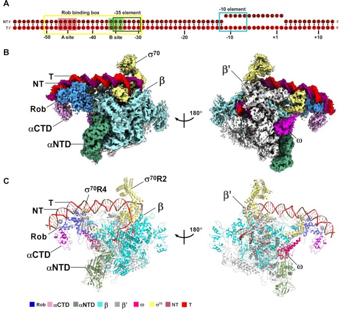
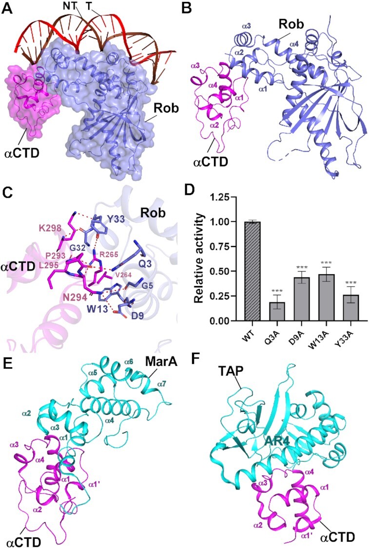
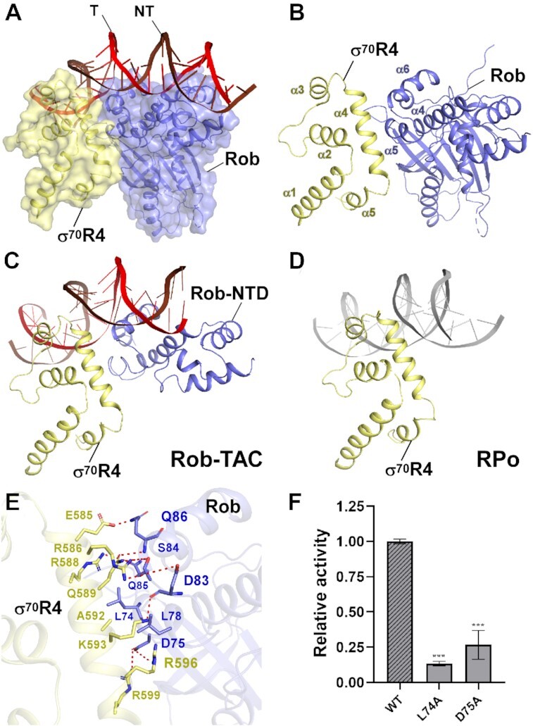
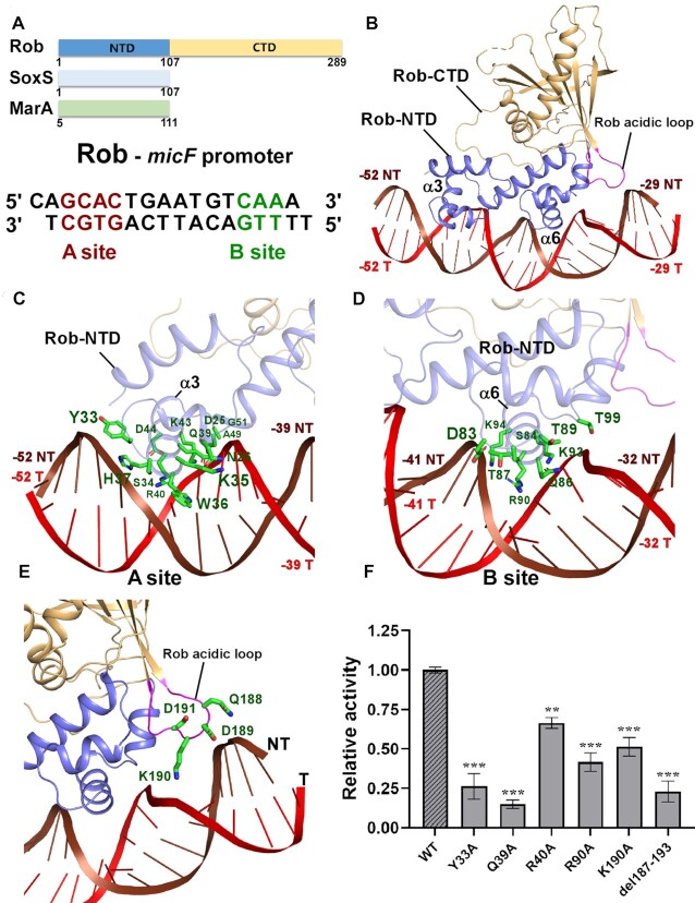
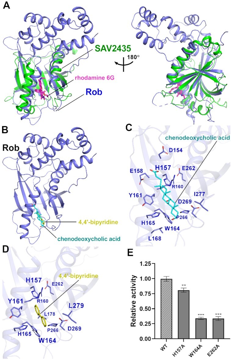
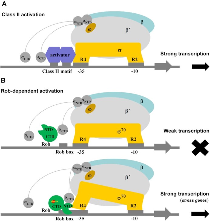
Rob, which serves as a paradigm of the large AraC/XylS family transcription activators, regulates diverse subsets of genes involved in multidrug resistance and stress response. However, the underlying mechanism of how it engages bacterial RNA polymerase and promoter DNA to finely respond to environmental stimuli is still elusive. Here, we present two cryo-EM structures of Rob-dependent transcription activation complex (Rob-TAC) comprising of Escherichia coli RNA polymerase (RNAP), Rob-regulated promoter and Rob in alternative conformations. The structures show that a single Rob engages RNAP by interacting with RNAP αCTD and σ70R4, revealing their generally important regulatory roles. Notably, by occluding σ70R4 from binding to -35 element, Rob specifically binds to the conserved Rob binding box through its consensus HTH motifs, and retains DNA bending by aid of the accessory acidic loop. More strikingly, our ligand docking and biochemical analysis demonstrate that the large Rob C-terminal domain (Rob CTD) shares great structural similarity with the global Gyrl-like domains in effector binding and allosteric regulation, and coordinately promotes formation of competent Rob-TAC. Altogether, our structural and biochemical data highlight the detailed molecular mechanism of Rob-dependent transcription activation, and provide favorable evidences for understanding the physiological roles of the other AraC/XylS-family transcription factors.


Figures

Figure 1.
Figure 1.
The overall structure of E. coli Rob-TAC. (A) DNA scaffold used in structure determination of E. coli Rob-TACII. (B, C) Two views of the cryo-EM density map (B) and structure model (C) of E. coli Rob-TACII. The EM density maps and cartoon representations of Rob-TACII are colored as indicated in the color key. NT, non-template-strand promoter DNA; T, template-strand promoter DNA.
Figure 2.
Figure 2.
The interactions between Rob and RNAP αCTD. (A) Relative locations of Rob, E. coli RNAP αCTD, and the upstream double-stranded DNA. (B) Rob interacts with E. coli RNAP αCTD. (C) Detailed interactions between E. coli RNAP αCTD and Rob. Salt-bridges are shown as red dashed lines. (D) Substitutions of Rob residues involved in interactions with E. coli RNAP αCTD decreased in vitro transcription activity. Data for in vitro transcription assays are means of three technical replicates. Error bars represent ± SEM of n = 3 experiments. Asterisk (***) indicates highly significant (P value < 0.001) difference from the wild-type Rob analyzed by one-way ANOVA with Tukey's multiple comparison test, respectively. (E) Interface between E. coli MarA and RNAP αCTD (PDB ID: 1XS9). (F) Interface between T. thermophilus TAP (transcription activator protein TTHB099) and T. thermophilus RNAP αCTD (PDB ID: 5I2D).
Figure 3.
Figure 3.
The interactions between Rob and σ70R4. (A) Relative locations of Rob, σ70R4, and upstream double-stranded DNA in E. coli Rob-TAC. (B) Rob interacts with the σ70R4. σ70R4 and Rob are represented as yellow or blue cartoon, respectively. (C) Relative locations of Rob-NTD, E. coli RNAP σ70R4 and the upstream double-stranded DNA. (D) Relative locations of E. coli RNAP σ70R4 and the upstream typical –35 element DNA (PDB ID: 6CA0). (E) Detailed interactions between σ70R4 and Rob. Hydrogen bonds are shown as red dashed lines. (F) Substitutions of the residues involved in Rob-σ70 interactions reduced in vitro transcription activity. Data for in vitro transcription assays are means of three technical replicates. Error bars represent mean ± SEM of n = 3 experiments. Asterisk (***) indicates highly significant (P value < 0.001) difference from the wild-type Rob analyzed by one-way ANOVA with Tukey's multiple comparison test, respectively.
Figure 4.
Figure 4.
The interactions between Rob and the micF promoter DNA. (A) Domain architecture of Rob, SoxS and MarA (top panel); the sequences of the micF promoter for Rob protein with the A- and B-box sequences highlighted in red and green, respectively (bottom panel). (B) Rob in complex with the micF promoter DNA. Rob-NTD and Rob-CTD are represented as blue or orange cartoon, respectively. (CD) Detailed interactions between Rob and the micF promoter DNA. Residues from Rob involved in interacting with A- or B-site sequences of micF promoter DNA are shown in green sticks. (E) Zoom-in view of the interactions between the acid loop and the micF promoter DNA. The acidic loop (residues 187-193) connecting strands β3 and β4 in the extra C-terminal domain of the Rob protein is highlighted in magenta. Residues from the acidic loop that contact DNA are shown in green sticks. (F) Substitutions of residues involved in Rob-DNA interactions suppressed in vitro transcription activity. Data for in vitro transcription assays are means of three technical replicates. Error bars represent ± SEM of n = 3 experiments. Asterisk (***) or (**) indicates highly significant (P value < 0.001) or significant (P value < 0.01) difference from the wild-type Rob analyzed by one-way ANOVA with Tukey's multiple comparison test, respectively.
Figure 5.
Figure 5.
Molecular docking analysis of Rob with potential ligands. (A) Structural superimposition of the Gyrl-like family protein SAV2435 (PDB ID: 5KAW) onto the Rob from Rob-TAC. Rob and the SAV2435 are shown as blue and green cartoon. The ligand rhodamine 6G(R6G) is shown in magenta stick. (B) Rob docked with chenodeoxycholic acid and 4,4′-bipyridine. (C) Predicted binding pockets for chenodeoxycholic acid on Rob protein. Residues potentially interacted with chenodeoxycholic acid are shown in blue sticks. (D) Predicted binding pockets for 4,4’-bipyridine on Rob protein. Residues potentially interacted with 4,4’-bipyridine are shown in blue sticks. (E) Substitutions of conserved residues involved in Rob-ligand interactions suppressed in vitro transcription activity in the presence of 100 μM chenodeoxycholic acid. Data for in vitro transcription assays are means of three technical replicates. Error bars represent ± SEM of n = 3 experiments. Asterisk (***) indicates highly significant (P value < 0.001) difference from the wild-type Rob analyzed by one-way ANOVA with Tukey's multiple comparison test, respectively.
Figure 6.
Figure 6.
Proposed models for class-II-activator-dependent and Rob-dependent transcription activation. (A) Proposed working model for class-II-activator-dependent transcription activation. (B) Proposed working model for Rob-dependent transcription activation. Rob shows weak transcription activation activity in the absence of Rob-RNAP interactions, and activates the transcription of most genes with the promoter containing Rob binding box under stress.

References

    1. Dorman C.J. Flexible response: DNA supercoiling, transcription and bacterial adaptation to environmental stress. Trends Microbiol. 1996; 4:214–216. - PubMed
    1. Duval V., Lister I.M.. MarA, SoxS and Rob of Escherichia coli - Global regulators of multidrug resistance, virulence and stress response. Int. J. Biotechnol. Wellness Ind. 2013; 2:101–124. - PMC - PubMed
    1. Browning D.F., Busby S.J.. Local and global regulation of transcription initiation in bacteria. Nat. Rev. Microbiol. 2016; 14:638–650. - PubMed
    1. Feklistov A., Sharon B.D., Darst S.A., Gross C.A.. Bacterial sigma factors: a historical, structural, and genomic perspective. Annu. Rev. Microbiol. 2014; 68:357–376. - PubMed
    1. Feklistov A., Darst S.A.. Structural basis for promoter-10 element recognition by the bacterial RNA polymerase sigma subunit. Cell. 2011; 147:1257–1269. - PMC - PubMed

Publication types

MeSH terms

LinkOut - more resources