Review
. 2022 Aug 19;209(2):140-150.
doi: 10.1093/cei/uxac017.

Autophagy modulation in multiple sclerosis and experimental autoimmune encephalomyelitis

Affiliations
Review

Autophagy modulation in multiple sclerosis and experimental autoimmune encephalomyelitis

Donghui Shen et al. Clin Exp Immunol. .

Abstract

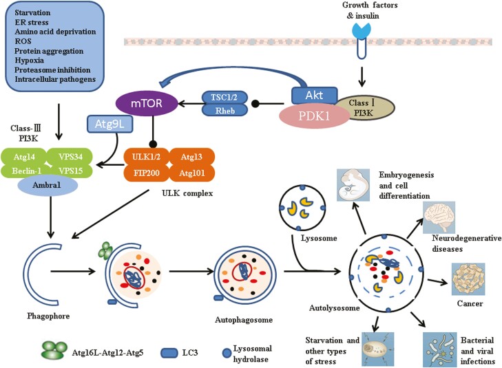
Multiple sclerosis (MS), a white matter demyelinating disease of the central nervous system (CNS), is characterized by neuroinflammatory and neurodegenerative. Experimental autoimmune encephalomyelitis (EAE) is a commonly used animal model for investigating pathogenic mechanisms of MS, representing the destruction of the blood-brain barrier (BBB), the activation of T cells, and the infiltration of myeloid cells. An increasing number of studies have documented that autophagy plays a critical role in the pathogenesis of both MS and EAE. Autophagy maintains CNS homeostasis by degrading the damaged organelles and abnormal proteins. Furthermore, autophagy is involved in inflammatory responses by regulating the activation of immune cells and the secretion of inflammatory factors. However, the specific mechanisms of autophagy involved in MS and EAE are not completely understood. In this review, we will summarize the complex mechanisms of autophagy in MS and EAE, providing potential therapeutic approaches for the management of MS.

Keywords: autophagy; experimental autoimmune encephalomyelitis; immune cells; inflammatory factors; multiple sclerosis.


Figures

Graphical Abstract
Graphical Abstract
Figure 1.
Figure 1.
Major signaling pathways involved in autophagy. Various factors such as growth factors deficiency, hypoxia, reactive oxygen species (ROS), protein aggregation, damaged organelles induce autophagy. The intracellular phagophore extend and envelop the organelles and degradation products to form autophagosomes. The autophagosome outer membrane binds to the lysosomal membrane and the autophagosome intima. The encapsulated substances are degraded by lysosomal enzymes. Class I PI3K/Akt/mTOR, ULK1/Atg13/FIP200 and Class III PI3K/Vps34/Beclin-1 complex are involved in the regulation of autophagy. The LC3 and Atgl6L1-Atg5-Atgl2 complexes are two ubiquitin-like protein systems and mainly responsible for the extension and closure of the phagophore. Autophagy participates in various pathophysiological processes including embryogenesis and cells differentiation, neurodegenerative diseases, cancer, bacterial and viral infections and Starvation, and other types of stress.
Figure 2.
Figure 2.
Autophagy in the pathogenesis of multiple sclerosis. (A) In MS patients and EAE mice, autophagy is required for DCs phagocytized and presented antigens on MHC class I and II molecules to activate T cells. (B) Autophagy is involved in the regulation of the survival, proliferation, and differentiation of lymphocytes. (C) Inflammatory factors interact with autophagy. Inflammatory factors regulate autophagy to clear pathogens and foreign bodies in the CNS, while autophagy regulates the inflammatory response of the CNS and prevents abnormal inflammatory expansion. (D) Autophagy clears depolarized mitochondria and prevents ROS excessive production in microglia and astrocytes. (E) Autophagy attenuates the damage of astrocytes in the pathogenesis of MS and EAE. (G) Autophagy is involved in microglia-mediated biology, ranging from cellular differentiation, polarization, phagocytosis, and cytokines production. (F) Autophagy may regulate the remyelination and the regrowth of axons in the CNS. (H) Autophagy influences the survival, differentiation, and myelination ability of OLs.

Similar articles

Cited by

References

    1. Chu F, Shi M, Zheng C, Shen D, Zhu J, Zheng X, et al. .. The roles of macrophages and microglia in multiple sclerosis and experimental autoimmune encephalomyelitis. J Neuroimmunol 2018, 318, 1–7. doi:10.1016/j.jneuroim.2018.02.015. - DOI - PubMed
    1. Dendrou CA, Fugger L, Friese MA.. Immunopathology of multiple sclerosis. Nat Rev Immunol 2015, 15, 545–58. doi:10.1038/nri3871. - DOI - PubMed
    1. Lassmann H. Pathogenic mechanisms associated with different clinical courses of multiple sclerosis. Front Immunol 2018, 9, 3116. doi:10.3389/fimmu.2018.03116. - DOI - PMC - PubMed
    1. Dobson R, Giovannoni G.. Multiple sclerosis—a review. Eur J Neurol 2019, 26, 27–40. doi:10.1111/ene.13819. - DOI - PubMed
    1. Lahiri V, Hawkins WD, Klionsky DJ.. Watch what you (self-) eat: autophagic mechanisms that modulate metabolism. Cell Metab 2019, 29, 803–26. doi:10.1016/j.cmet.2019.03.003. - DOI - PMC - PubMed