Review
. 2022 Nov;30(11):1056-1071.
doi: 10.1016/j.tim.2022.05.004. Epub 2022 May 28.

Nuclear antiviral innate responses at the intersection of DNA sensing and DNA repair

Affiliations
Review

Nuclear antiviral innate responses at the intersection of DNA sensing and DNA repair

Joshua L Justice et al. Trends Microbiol. 2022 Nov.

Abstract

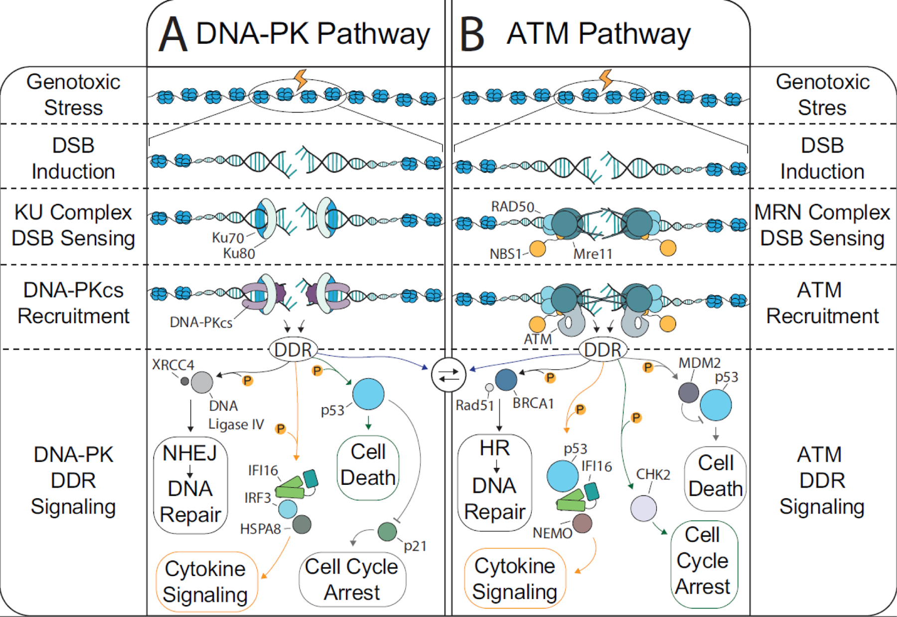
The coevolution of vertebrate and mammalian hosts with DNA viruses has driven the ability of host cells to distinguish viral from cellular DNA in the nucleus to induce intrinsic immune responses. Concomitant viral mechanisms have arisen to inhibit DNA sensing. At this virus-host interface, emerging evidence links cytokine responses and cellular homeostasis pathways, particularly the DNA damage response (DDR). Nuclear DNA sensors, such as the interferon (IFN)-γ inducible protein 16 (IFI16), functionally intersect with the DDR regulators ataxia telangiectasia mutated (ATM) and DNA-dependent protein kinase (DNA-PK). Here, we discuss accumulating knowledge for the DDR-innate immunity signaling axis. Through the lens of this infection-driven signaling axis, we present host and viral molecular strategies acquired to regulate autoinflammation and antiviral responses.

Keywords: DNA damage response; DNA virus infection; antiviral response; interferon; nuclear DNA sensing.


Conflict of interest statement

Declaration of interests No interests are declared.

Figures

Figure 1:
Figure 1:. The DNA damage response and DSB repair.
DSBs arise from endogenous (metabolism, replication fork stalling, transcription, etc.) and exogenous (irradiation, chemicals that inhibit replication proteins, drugs that directly target DNA, etc.) sources. DSBs are primarily repaired by one of four mechanisms: NHEJ, HDR, alternative- EJ, or single strand annealing. The predominant forms of DSB repair, NHEJ and HDR, are induced following a cascade of protein interactions. First, the DSB is detected by the KU (KU70 and KU80) and MRN (Mre11, Rad50, Nbs1) complexes. Afterward, the cell must decide which repair pathway to favor. A) Most of the DSB repair in the body occurs in quiescent cells where NHEJ is the preferred pathway. The KU complex recruits the catalytic subunit of the DNA-PK holoenzyme, which triggers the kinase activity of the DNA-PK complex. The DNA-PK derived DDR can trigger DNA repair, cell death, cell cycle arrest, and interferon signaling; however, the protein players involved in these processes are distinct from that involved in HDR. B) When the cell has multiple copies of DNA available, such as in S Phase, the MRN complex will trigger ATM kinase activity and induce the DDR. There are several cellular mechanisms in place to bias DNA repair either toward HDR or toward NHEJ. For example, when 53BP1 is recruited by RNF8 and RNF168 to a DSB to block end resection and thus block strand invasion, thereby promoting NHEJ. Nevertheless, there is also substantial crosstalk between ATM and DNA-PK during the DDR, particularly when end-trimming is required for NHEJ.
Figure 2:
Figure 2:. DNA sensing throughout the cell.
A) The detection of self-derived DNA and of extranuclear viral DNA can be mediated by any of several protein players: cGAS, Aim2, KU70/80, Mre11/Rad50, DNA-PK, TLR9, and others. After detecting foreign or abnormal DNA, these sensors can trigger inflammatory cytokine expression. There is mounting evidence of crosstalk between the DDR and the DNA sensors throughout the cell. Both ATM and DNA-PK can interact with IFI16 to trigger Type-I IFN responses. There is also evidence that the DDR can antagonize other nucleic acid sensors such as AIM2, cGAS, and hnRNPA2/B1 and vice-versa. B) Outside of the nucleus, the predominate triggers of DNA sensing are from endogenous sources (mitochondrial DNA or damaged nuclear DNA), bacterial DNA, DNA viruses that replicate in the cytosol (e.g., poxvirus), or from replication products encountered during the entry or egress of nuclear-replicating DNA viruses. Several cytosolic DNA sensors (cGAS, KU70/80, etc.) also have a role in nuclear DNA sensing. For simplicity, other DNA sensors that reside outside of the nucleus have not been depicted (e.g. RNAPIII, DDX41, and cytosolic IFI16). C) Several DNA sensors exist within the nucleus, being able to distinguish foreign from host DNA. The nuclear DNA sensors include IFI16, IFIX, and hnRNPA2/B1. Moreover, endogenous triggers of stress, like DNA damage, can activate ATM or DNA-PK to induce cytokine expression. There is crosstalk between the DDR and DNA sensors that can amplify or restrict innate immune signaling. For example, hnRNPA2/B1 and DNA-PK may negatively regulate each other.
Figure 3:
Figure 3:. STING- and IFI16-dependent intrinsic signaling pathways.
A) cGAS is the synthase for cyclic GMP-AMP, which serves as a messenger to activate STING (reviewed in [107]). Upon its activation, STING localizes from the cytosolic face of ER tubules to the Golgi. Thereafter, STING recruits TBK1 to phosphorylate IRF3, a transcription factor. B) Phosphorylated IRF3 dimerizes and translocates to the nucleus to trigger Type-I interferon responses via interaction with CBP/p300. C) STING activates NF-κB to further diversify the cytokine repertoire. D) Multiple routes can activate STING. Bypassing the requirement for STING translocation, recent evidence points to its activation by K63-linked ubiquitination at K150 [108]. Although not depicted, lysosomal targeting of STING, such as via interaction with Neimann-Pick type C1 (NPC1), can regulate STING responses [109]. E) IFI16 is a member of the PYHIN family of proteins that bind to DNA and then self-oligomerize via their HIN-200 and PYRIN domains, respectively. Quiescently, IFI16 resides predominantly in the nucleolus and nucleoplasm. Upon viral genome deposition, IFI16 rapidly localizes to the nuclear periphery (reviewed in [67]). IFI16 undergoes oligomeric assembly on the viral DNA. F) At the viral genome, IFI16 and DNA-PK participate in a feedback loop [5]. IFI16 recruits and/or activates DNA-PK, which in turn phosphorylates IFI16 to promote interferon expression. G) In the context of DNA damage, ATM can trigger IFI16 nuclear export with TRAF6 and p53 to induce STING ubiquitination and subsequent NF-κB activation [6].
Figure 4:
Figure 4:. Viral strategies to subvert the DDR and nuclear DNA sensors.
Nuclear replicating DNA viruses have acquired many mechanisms to overcome the link between DDR and intrinsic immunity. A) Papillomaviruses replication requires ATM pathway components as well as the KU-complex. The papillomavirus oncogenes, E6 and E7, inhibit DNA-PK and IFI16. B) Polyomaviruses replication also utilizes ATM pathway components to amplify the viral DNA. Simultaneously, ATM activation inhibits DNA-PK localization to the viral DNA and the agnoprotein induces KU-complex nuclear export. It is not known if polyomaviruses usurp IFI16. C) Adenoviruses broadly inhibit the DDR via expression of the E1A, E4 and E1B proteins. Adenoviruses also suppress IFI16 expression through an unknown mechanism. Interestingly, the KU-complex, but not DNA-PK, localizes to the Adenoviruses replication complex. D). The α-herpesvirus, HSV-1, extensively reorganizes the DDR. ATM and the MRN complex support HSV-1 replication, but the DNA-PK pathway components are suppressed. The viral E3-ubiquitin ligase, ICP0, promotes both IFI16 and DNA-PK proteolytic degradation. E) Relatively less is understood about how β-herpesviruses, such as HCMV, interact with the DDR. DNA-PK kinase activity is suppressed during HCMV infection. There is conflicting evidence regarding the role and status of ATM during infection. It is possible that the DDR is relevant during HCMV infection given the potent and immediate inhibition of IFI16 by the HCMV tegument protein, pUL83. E) γ-herpesviruses, like EBV and KSHV, suppress DNA-PK kinase activity, while ATM increases viral replication. Interestingly, DNA-PK can inhibit ATM activation during KSHV infection. KSHV lytic infection suppresses IFI16 via an unknown mechanism.

Similar articles

Cited by

References

    1. Majzoub K, et al. (2019) The Innate Antiviral Response in Animals: An Evolutionary Perspective from Flagellates to Humans. Viruses 11 - PMC - PubMed
    1. Gallucci S and Maffei ME (2017) DNA Sensing across the Tree of Life. Trends Immunol 38, 719–732 - PubMed
    1. Härtlova A, et al. (2015) DNA damage primes the type I interferon system via the cytosolic DNA sensor STING to promote anti-microbial innate immunity. Immunity 42, 332–343 - PubMed
    1. Reinert LS, et al. (2016) Sensing of HSV-1 by the cGAS-STING pathway in microglia orchestrates antiviral defence in the CNS. Nat Commun 7, 13348. - PMC - PubMed
    1. Justice JL, et al. (2021) Systematic profiling of protein complex dynamics reveals DNA-PK phosphorylation of IFI16 en route to herpesvirus immunity. Sci Adv 7 - PMC - PubMed

Publication types