. 2022 May 31;13(1):3016.
doi: 10.1038/s41467-022-30787-6.

BRD4 promotes resection and homology-directed repair of DNA double-strand breaks

Affiliations

BRD4 promotes resection and homology-directed repair of DNA double-strand breaks

John K Barrows et al. Nat Commun. .

Abstract

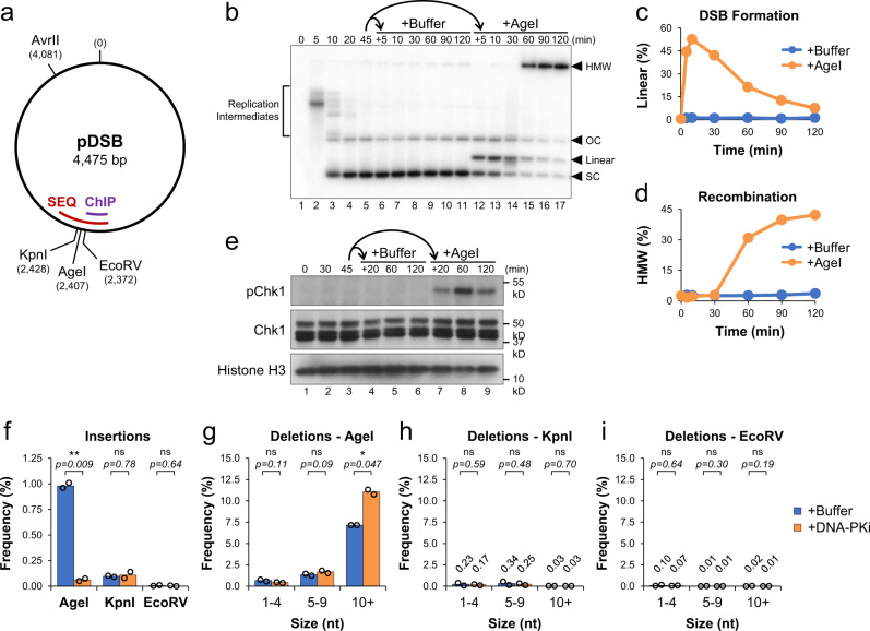
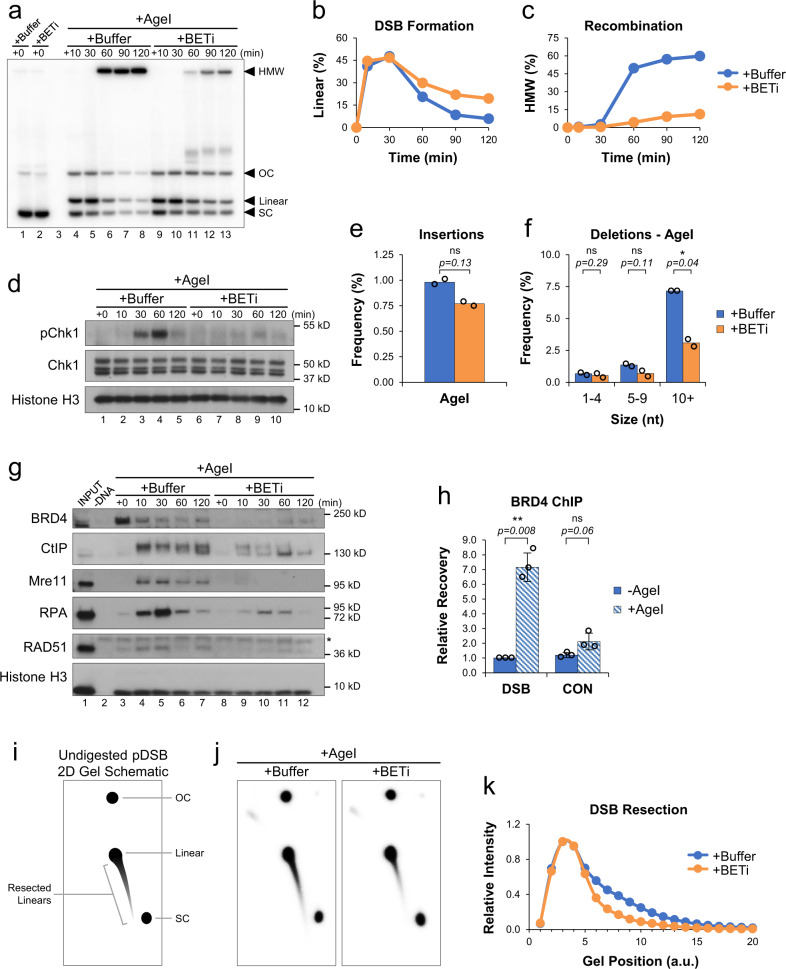
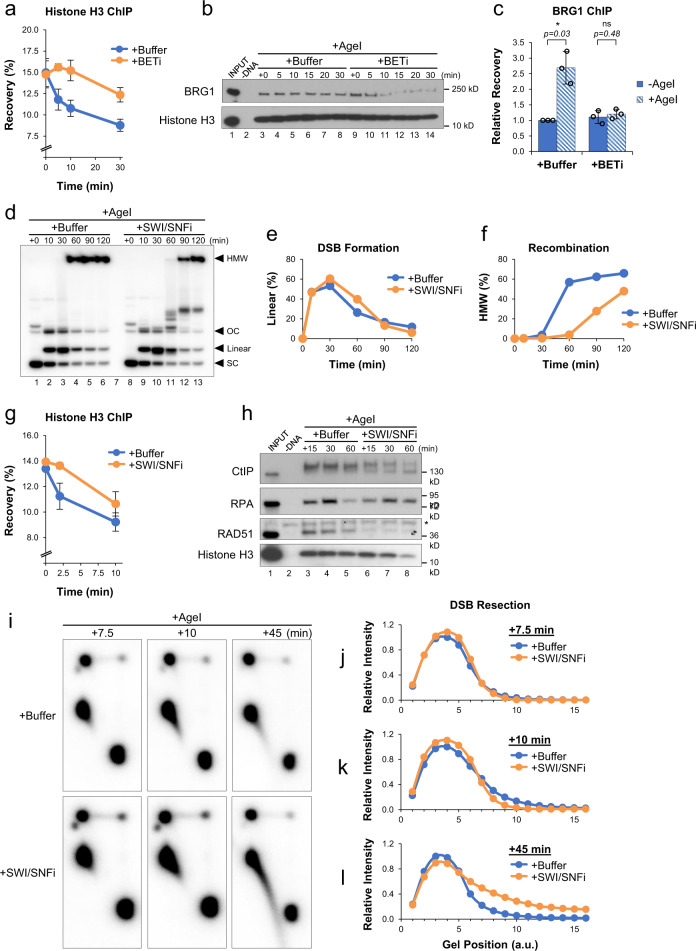
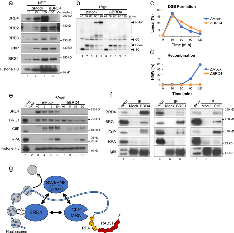
Double-strand breaks (DSBs) are one of the most toxic forms of DNA damage and represent a major source of genomic instability. Members of the bromodomain and extra-terminal (BET) protein family are characterized as epigenetic readers that regulate gene expression. However, evidence suggests that BET proteins also play a more direct role in DNA repair. Here, we establish a cell-free system using Xenopus egg extracts to elucidate the gene expression-independent functions of BET proteins in DSB repair. We identify the BET protein BRD4 as a critical regulator of homologous recombination and describe its role in stimulating DNA processing through interactions with the SWI/SNF chromatin remodeling complex and resection machinery. These results establish BRD4 as a multifunctional regulator of chromatin binding that links transcriptional activity and homology-directed repair.


Conflict of interest statement

The authors declare no competing interests.

Figures

Fig. 1
Fig. 1. Competition between DSB repair pathways in Xenopus egg extracts.
a Schematic of pDSB showing the relative position of restriction enzyme sites and DSB regions amplified for ChIP and amplicon sequencing (SEQ). b pDSB was replicated in the presence of α32P[dATP] for 45 min. The reaction was then split and supplemented with buffer or AgeI. Samples were withdrawn, resolved by 1D gel electrophoresis, and visualized by autoradiography (n = 4 independent experiments). Labels indicate the position of: replication intermediates, supercoiled (SC), linear, and open circular (OC) plasmids, and high molecular weight (HMW) molecules. c, d Quantitation of linear (c) and HMW (d) molecules from (b). e Protein samples from (b) were withdrawn and analyzed by Western blot (n = 2 independent experiments). fi pDSB was replicated in extract supplemented with buffer or NU7441 (DNA-PKi) for 45 min. Reactions were then supplemented with AgeI, KpnI or EcoRV. Samples were withdrawn 30 min after enzyme addition and analyzed by amplicon sequencing (n = 2 independent experiments). Results are graphed to show the frequency of insertion products (f), and the frequency of different deletion products for AgeI, KpnI and EcoRV reactions (gi). Data values are labeled for low frequency deletions in KpnI and EcoRV reactions. Student two-tailed t-test: not significant (ns), p-value < 0.05 (*), p-value < 0.01 (**).
Fig. 2
Fig. 2. BET proteins promote DNA end resection.
a pDSB was replicated with α32P[dATP] in reactions containing buffer or JQ1 (BETi). After 45 min, reactions were supplemented with AgeI and samples were withdrawn for 1D gel electrophoresis (n = 4 independent experiments). b, c Quantitation of linear (b) and HMW (c) molecules from (a). d Protein samples from (a) were withdrawn and analyzed by Western blot (n = 2 independent experiments). e, f pDSB was replicated in extract containing buffer or JQ1 (BETi). After 45 min, reactions were supplemented with AgeI. Samples were withdrawn 30 min after enzyme addition and analyzed by amplicon sequencing (n = 2 independent experiments). Results are graphed to show the frequency of insertion products (e), and the frequency of different deletion products (f). g pDSB was replicated in extract supplemented with buffer or JQ1 (BETi). After 45 min, AgeI was added and DNA-bound proteins were isolated by plasmid pull-down. Samples were analyzed by Western blot with the indicated antibodies (n = 2 independent experiments). Non-specific band (*). h pDSB and a control plasmid lacking AgeI sites were replicated in extract. After 45 min, AgeI was added and samples were withdrawn 5 min later for analysis by BRD4 ChIP (n = 3 independent experiments). i Schematic of undigested 2D gel intermediates. The relative position of open circular (OC), supercoiled (SC), and linear plasmids is indicated. An example of resected linear molecules is also shown. j pDSB was replicated with α32P[dATP] in reactions containing buffer or BETi. After 45 min, AgeI was added and samples were withdrawn 30 min later for 2D gel electrophoresis (n = 2 independent experiments). k Quantitation of linear and resected molecules in (j). Arbitrary units (a.u.). Error bars represent ± one standard deviation from the mean. Student’s two-tailed t test: not significant (ns), p-value < 0.05 (*), p-value < 0.01 (**).
Fig. 3
Fig. 3. BET-mediated regulation of SWI/SNF.
a pDSB was replicated in extract supplemented with buffer or JQ1 (BETi). After 45 min, AgeI was added and samples were withdrawn for analysis by histone H3 ChIP (n = 2 independent experiments). b pDSB was replicated in extract supplemented with buffer or BETi. After 45 min, AgeI was added and DNA-bound proteins were isolated by plasmid pull-down. Samples were analyzed by Western blot with the indicated antibodies (n = 2 independent experiments). c pDSB was replicated in extract supplemented with buffer or JQ1 (BETi). After 45 min, AgeI was added and samples were withdrawn 5 min later for analysis by BRG1 ChIP (n = 3 independent experiments). d pDSB was replicated with α32P[dATP] in extract containing buffer or the BRG1/BRM ATP Inhibitor-1 (SWI/SNFi). After 45 min, reactions were supplemented with AgeI and samples were withdrawn for 1D gel electrophoresis (n = 2 independent experiments). e, f Quantitation of linear (e) and HMW (f) molecules from (d). g pDSB was replicated in extract supplemented with buffer or SWI/SNFi. After 45 min, AgeI was added and samples were withdrawn for analysis by histone H3 ChIP (n = 2 independent experiments). h pDSB was replicated in extract containing buffer or SWI/SNFi. After 45 min, reactions were supplemented with AgeI and DNA-bound proteins were isolated by plasmid pull-down. Samples were analyzed by Western blot with the indicated antibodies (n = 2 independent experiments). Non-specific band (*). i pDSB was replicated with α32P[dATP] in extract containing buffer or SWI/SNFi. After 45 min, reactions were supplemented with AgeI and samples were withdrawn for 2D gel electrophoresis (n = 3 independent experiments). jl Quantitation of linear and resected molecules in (i) at 7.5 (j), 10 (k), and 45 (l) minutes after AgeI addition. Arbitrary units (a.u.). Error bars represent ± one standard deviation from the mean. Student’s two-tailed t test: not significant (ns), p-value < 0.05 (*).
Fig. 4
Fig. 4. BRD4 promotes resection and homology-directed repair.
a Mock-depleted (ΔMock) or BRD4-depleted (ΔBRD4) NPE was analyzed by Western blot using the indicated antibodies (n = 2 independent experiments). b pDSB was replicated with α32P[dATP] in mock- or BRD4-depleted extracts. After 60 min, reactions were supplemented with AgeI and samples were isolated at the indicated time points for 1D gel electrophoresis (n = 2 independent experiments). c, d Quantitation of linear (c) and HMW (d) molecules from (b). e pDSB was replicated in mock- or BRD4-depleted extract. After 60 min, reactions were supplemented with AgeI and DNA-bound proteins were isolated by plasmid pull-down. Samples were analyzed by Western blot with the indicated antibodies (n = 2 independent experiments). f A mixture of HSS and NPE was immunoprecipitated with beads conjugated to mock, BRD4, BRG1, or CtIP antibodies. Bead-bound proteins were analyzed by Western blot with the indicated antibodies (n = 3 independent experiments). g Schematic showing interactions between BRD4-BRG1-CtIP and their role in coordinating DSB repair through nucleosome eviction, DNA end resection, and homologous recombination. Acetylation (Ac); Mre11-Rad50-Nbs1 (MRN).

References

    1. Curtin NJ. DNA repair dysregulation from cancer driver to therapeutic target. Nat. Rev. Cancer. 2012;12:801–817. doi: 10.1038/nrc3399. - DOI - PubMed
    1. Chatterjee N, Walker GC. Mechanisms of DNA damage, repair, and mutagenesis. Environ. Mol. Mutagen. 2017;58:235–263. doi: 10.1002/em.22087. - DOI - PMC - PubMed
    1. Brandsma I, Gent DC. Pathway choice in DNA double strand break repair: observations of a balancing act. Genome Integr. 2012;3:9. doi: 10.1186/2041-9414-3-9. - DOI - PMC - PubMed
    1. Sung P, Klein H. Mechanism of homologous recombination: mediators and helicases take on regulatory functions. Nat. Rev. Mol. Cell Biol. 2006;7:739–750. doi: 10.1038/nrm2008. - DOI - PubMed
    1. Chang HHY, Pannunzio NR, Adachi N, Lieber MR. Non-homologous DNA end joining and alternative pathways to double-strand break repair. Nat. Rev. Mol. Cell Biol. 2017;18:495–506. doi: 10.1038/nrm.2017.48. - DOI - PMC - PubMed

Publication types

MeSH terms