. 2022 May 31;12(1):9039.
doi: 10.1038/s41598-022-12919-6.

Epigenetic regulation of BAF60A determines efficiency of miniature swine iPSC generation

Affiliations

Epigenetic regulation of BAF60A determines efficiency of miniature swine iPSC generation

Hongli Jiao et al. Sci Rep. .

Erratum in

Abstract

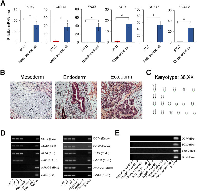
Miniature pigs are an ideal animal model for translational research to evaluate stem cell therapies and regenerative applications. While the derivation of induced pluripotent stem cells (iPSCs) from miniature pigs has been demonstrated, there is still a lack of a reliable method to generate and maintain miniature pig iPSCs. In this study, we derived iPSCs from fibroblasts of Wisconsin miniature swine (WMS), Yucatan miniature swine (YMS), and Göttingen minipigs (GM) using our culture medium. By comparing cells of the different pig breeds, we found that YMS fibroblasts were more efficiently reprogrammed into iPSCs, forming colonies with well-defined borders, than WMS and GM fibroblasts. We also demonstrated that YMS iPSC lines with a normal pig karyotype gave rise to cells of the three germ layers in vitro and in vivo. Mesenchymal stromal cells expressing phenotypic characteristics were derived from established iPSC lines as an example of potential applications. In addition, we found that the expression level of the switch/sucrose nonfermentable component BAF60A regulated by STAT3 signaling determined the efficiency of pig iPSC generation. The findings of this study provide insight into the underlying mechanism controlling the reprogramming efficiency of miniature pig cells to develop a viable strategy to enhance the generation of iPSCs for biomedical research.


Conflict of interest statement

The authors declare no competing interests.

Figures

Figure 1
Figure 1
Generation of iPSCs from fibroblasts of three breeds of miniature pigs. (A) Schematic diagram illustrating the timeline and stepwise procedures to establish iPSC lines. (B) Phase-contrast images showing flat, elongated cells with typical fibroblast morphology. (C) Phase-contrast images showing pig iPSC colonies with well-defined borders at day 21 post transfection. (D) ALP staining of iPSC colonies and corresponding reprogramming efficiency presented by the number of ALP-positive colonies per million cells. ALP-positive colonies in each 100-mm dish were counted. Scale bar = 200 µm. *p < 0.05; n = 3.
Figure 2
Figure 2
Expression of pluripotency markers in pig iPSCs and parental fibroblasts. (A) Immunofluorescence staining detecting the expression of the pluripotency markers NANOG, OCT4, and SOX2 in iPSC lines generated from WMS, YMS, and GM fibroblasts. DAPI stains nuclei (blue). (B) Relative mRNA levels of the pluripotency markers NANOG, OCT4, SOX2, and LIN28 determined by quantitative RT–PCR. Scale bar = 100 µm. *p < 0.05; n = 3.
Figure 3
Figure 3
Derivation of 3 germ layer cells and karyotype of pig iPSCs. (A) Quantitative RT–PCR detecting relative mRNA levels of the germ layer-associated markers TBXT and CXCR4 (mesoderm), PAX6 and NES (ectoderm), and SOX17 and FOXA2 (endoderm) in cells derived from iPSCs after 7 days of germ layer-specific induction. (B) Hematoxylin and eosin (H&E) staining of teratomas derived from a representative pig iPSC line. Tissues of 3 germ layers, including cartilage (mesoderm), gut-like epithelium (endoderm), and neuronal rosette (ectoderm), were present in a teratoma. (C) Chromosome analysis of a representative iPSC line revealing a normal diploid pig cell with a 38, XX karyotype. (D) Expression of exogenous and endogenous pluripotency markers in 3 fibroblast lines (p5) and 3 iPSC lines (p15). (E) Expression of exogenous Yamanaka factors in mesodermal, ectodermal, and endodermal cells derived from iPSCs. Original gels are presented in Supplementary Fig. S4. Scale bar = 200 µm. *p < 0.05; n = 3.
Figure 4
Figure 4
Derivation and characterization of iPSC-MSCs. (A) Phase-contrast images of cells in iPSC culture induced for 21 days of MSC differentiation. (B) Expression of surface markers on iPSC-MSCs. Pink histograms represent surface markers of interest. Blue histograms represent isotype controls. (C) Alizarin red S staining, quantification of calcium deposition, and relative mRNA levels of the bone-associated markers CBFA1, ALP, and OC in cells differentiated from iPSC-MSCs after 21 days of osteogenesis. (D) Alcian blue staining, quantification of GAG production, and relative mRNA levels of the cartilage-associated markers SOX9, COL2, and ACAN in cells differentiated from iPSC-MSCs after 21 days of chondrogenesis. (E) Oil red O staining, quantification of lipid droplets, and relative mRNA levels of the fat-associated markers LPL and PPARG2 in cells differentiated from iPSC-MSCs after 21 days of adipogenesis. Scale bar = 200 µm. *p < 0.05; n = 3.
Figure 5
Figure 5
Changes in telomere and DNA methylation of fibroblasts undergoing cellular reprogramming and MSC differentiation. (A) Telomerase activity of pig fibroblasts before and after cellular reprogramming and iPSCs after differentiation into MSCs. (B) Expression levels of TERT in cells at different stages. (C) Relative telomere length of cells in response to cellular reprogramming and MSC differentiation induction. T/S ratio: the ratio of the copy number of telomere repeats to that of a single control gene. (D) Quantification of global DNA methylation determined by the level of 5mC in different cells. (E) Quantification of global DNA hydroxymethylation determined by the level of 5hmC in different cells. *p < 0.05; n = 3.
Figure 6
Figure 6
Expression levels of SWI/SNF complexes and STAT3 activity in cells of 3 miniature pig breeds. (A) Transcript levels of selected epigenetic enzymes in fibroblasts and iPSCs of different breeds of miniature pigs. (B) Images of western blots and quantification of protein bands detecting BAF60A, pSTAT3, and STAT3 expression in cells. GAPDH was used as a loading control. (C) Images and quantification of western blots of proteins extracted from YMS iPSCs treated with the STAT3 inhibitor CPT for 2 h. GAPDH was used as a loading control. (D) Images and quantification of western blots of proteins extracted from YMS iPSCs treated with BAF60A siRNA or scrambled control. GAPDH was used as a loading control. (E) Interaction of BAF60A and OCT4 in YMS iPSC lines analyzed by co-IP. Original western blots are presented in Supplementary Fig. S5. *p < 0.05; n = 3.
Figure 7
Figure 7
Effects of BAF60A knockdown and overexpression on the reprogramming efficiency of YMS fibroblasts. (A) Transcript levels of BAF60A in scrambled control and BAF60A-knockdown iPSCs (left). Macrographs of iPSC colonies formed in Petri dishes detected by ALP staining 21 days after transfection for BAF60A knockdown (middle). Quantification of ALP-positive colonies normalized to total transfected cells (right). (B) Colony morphology of control and BAF60A-overexpressing iPSCs (top). Transcript levels of BAF60A in empty vector control and BAF60A-overexpressing iPSCs (bottom left). Macrographs of iPSC colonies detected by ALP staining 21 days after transfection for BAF60A overexpression (bottom middle). Quantification of ALP-positive colonies normalized to total transfected cells (bottom right). Scale bar = 200 µm. *p < 0.05; n = 3.

References

    1. Barone AAL, et al. Genetically modified porcine split-thickness skin grafts as an alternative to allograft for provision of temporary wound coverage: Preliminary characterization. Burns. 2015;41(3):565–574. - PubMed
    1. Summerfield A, Meurens F, Ricklin ME. The immunology of the porcine skin and its value as a model for human skin. Mol. Immunol. 2015;66(1):14–21. - PubMed
    1. Branski LK, et al. A porcine model of full-thickness burn, excision and skin autografting. Burns. 2008;34(8):1119–1127. - PMC - PubMed
    1. Lai L, et al. Production of α-1,3-galactosyltransferase knockout pigs by nuclear transfer cloning. Science. 2002;295(5557):1089–1092. - PubMed
    1. Perleberg C, Kind A, Schnieke A. Genetically engineered pigs as models for human disease. Dis. Model. Mech. 2008;11(1):dmm030783. - PMC - PubMed

Publication types