. 2022 May 31;12(1):77.
doi: 10.1186/s13578-022-00802-w.

The two-faced role of ATF2 on cisplatin response in gastric cancer depends on p53 context

Affiliations

The two-faced role of ATF2 on cisplatin response in gastric cancer depends on p53 context

Lingxue Xu et al. Cell Biosci. .

Abstract

Background: Activating transcription factor-2 (ATF2) is a member of the basic leucine zipper family of DNA-binding proteins, which exhibits both oncogenic and tumor suppression activity in different tumors. However, the molecular mechanism of its dual function in cancer chemotherapy especially in gastric cancer has still not been elucidated.

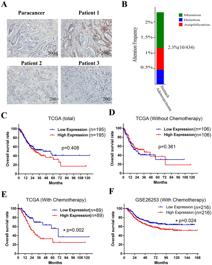
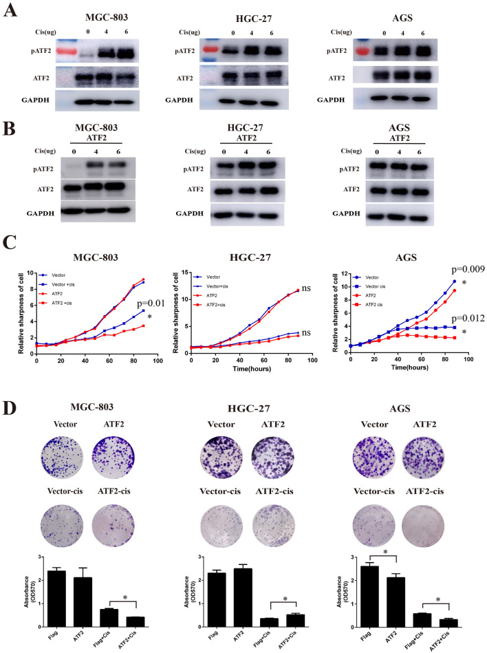
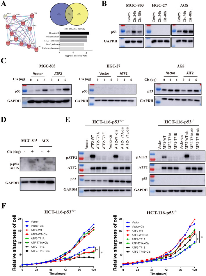
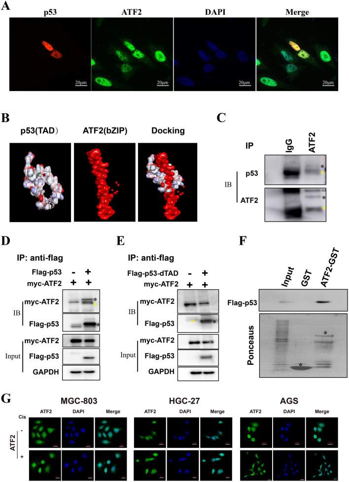
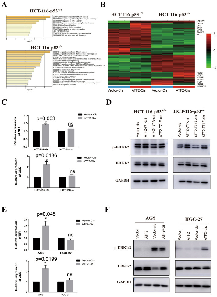
Methods: The protein expression and location of ATF2 in gastric cancer tissues was detected with immunohistochemistry assay, and the clinical significance was analyzed using TCGA and GEO database. The activation and impact of ATF2 in cisplatin treated cells were evaluated with western blot, incucyte live cell analysis, clone formation and tumor xenografts assays. Interaction between ATF2 and p53 was confirmed with immunoprecipitation and GST-pull down. Potential molecular mechanism of ATF2 in different p53 status cells was analyzed with RNA sequencing and real-time quantitative PCR.

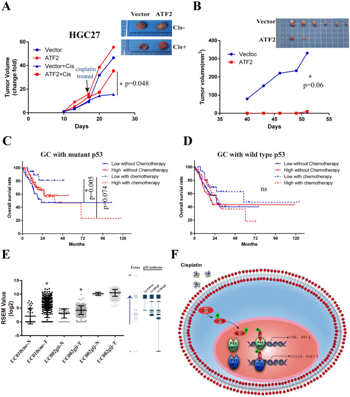
Results: ATF2 mainly located in the nucleus of cancer cells, higher ATF2 level was associated with poor five-year survival of gastric patients, especially in those undergone chemotherapy treatment. Cisplatin treatment significantly activated ATF2 in p53 mutant cells. ATF2 could interact with the trans-activation domain of p53 and enhance cisplatin sensitivity in p53 wild type cell lines, while promoted cell survival in mutant p53 cancer cells by affecting ERK1/2 pathway.

Conclusions: This study confirmed the effect of ATF2 on cisplatin sensitivity was associated with the functional status of p53 in gastric cancer cells. Integrated analysis of ATF2 expression and P53 status could be used to evaluate the chemotherapy sensitivity and prognosis of gastric cancer patients.

Keywords: ATF2; Cisplatin; ERK1/2; Gastric cancer; Prognosis; p53.


Conflict of interest statement

All authors declare that no conflict of interests.

Figures

Fig. 1
Fig. 1
High expression of ATF2 in GC is associated with poor prognosis and chemotherapy response. A Immunohistochemistry staining was performed to evaluate the expression and location of ATF2 in human gastric cancer tissues. The original magnification is 200 ×. B Gene status of ATF2 including mutations (green), amplification (red), and deletions (blue) in GC tissue (n = 434) were analyzed using cBioportal software. C Overall survival of GC patients with ATF2-low or -high expression using TCGA database. D Overall survival of ATF2 low or -high expression group in GC patients without chemotherapy. E Overall survival of ATF2-low or ATF2-high expression group in GC patients with chemotherapy. F Overall survival of ATF2-low or ATF2-high expression group in GC patients with chemotherapy using GSE26253 dataset. Kaplan-Meier and Log-rank analysis is used, p value < 0.05 is considered statistically significant
Fig. 2
Fig. 2
ATF2 is involved in cisplatin induced cell death. A Protein expression of ATF2 and phosphorylated-ATF2-Thr71 (p-ATF2) in different GC cell lines by Western Blotting after treatment with various doses of cisplatin. B Protein expression of ATF2 and p-ATF2 (phospho-Thr71) after treatment with different doses cisplatin in ATF2-overexpressing GC cell lines. C Cell proliferation and cytotoxicity of cisplatin (2 µg/ml) treated ATF2-overexpressing GC cell lines. D Cell colony formation ability of cisplatin (2 µg/ml) treated ATF2-overexpressing GC cell lines. Data are presented as mean ± SEM
Fig. 3
Fig. 3
The genetic status of p53 affects the function of ATF2. A STRING database analysis was performed to explore potential interactions between ATF2 and p53, then the overlapped genes was predicted with Venny software, and the KEGG pathway enrichment of sharing genes were further analyzed with STRING, top 5 pathways were shown. B Protein expression of p53 in GC cells after cisplatin treatment for 24 or 48 h was analyzed by Western Blot assay. C Protein expression of p53 in GC cell lines treated with different doses of cisplatin was analyzed by Western Blot. D The phosphorylation of p53 in cisplatin treating MGC-803 and AGS were detected with Western Blot. E HCT116 p53+/+ or HCT-116 p53−/− cells that over-expressing different ATF2 subtypes (WT, T71A, T71E) were treated with cisplatin, then p53, ATF2 and p-ATF2 (phospho-Thr71) level were detected by Western Blot. ATF2 wild type (ATF2-WT), inactivated mutant type that mutated Thr to Ala (ATF2-T71A), activated mutant type that mutated Thr to Glu (ATF2-T71E). F Cell proliferation and cytotoxicity of cisplatin treated ATF2 subtype colorectal cancer cell lines with incucyte live cell analysis system. Cisplatin was used at a final concentration of 2 µg/ml
Fig. 4
Fig. 4
ATF2 interacts with the p53 TAD domain. A The co-localization of ATF2 (green) and p53 (red) GC cells was analyzed by confocal imaging. DAPI stains nuclear DNA (blue). The photomicrographs were taken at the original magnification 400 ×. B Molecular docking with the crystal structures of ATF2 and p53 were performed by Discovery Studio software. C The interaction between endogenous ATF2 and p53 was assessed with immunoprecipitation (IP) using ATF2 antibody. D Flag-p53 and Myc-ATF2 plasmids were co-transfected into cancer cells, then protein interaction was confirmed with immunoprecipitation (IP) assay. E Flag-p53-dTAD and Myc-ATF2 plasmids were co-transfected and an IP assay was performed. F The interaction between ATF2 and flag-p53 in vitro was elucidated using a GST-pull down assay. G The sub-cellular localization of ATF2 (green) in cisplatin treated GC cell lines was determined using immunofluorescence (400×). The positive band was marked with black star (*), and the heavy chain of antibody in IP was marked with yellow star (*)
Fig. 5
Fig. 5
Regulation of ERK1/2 pathway by ATF2 is associated with p53. A Differential expressing genes between control and ATF2 overexpressing group in HCT-116 p53+/+ or HCT-116 p53−/− cell were identified with R studio, then gene oncology (GO) enrichment of elevated genes (foldchange > = 2) was analyzed with Metascape, and top GO biological processes were displayed. B Cluster analysis was performed using R studio and genes related with MAPK pathway were marked in the heatmap. C Relative expression of CSK and NF1 in cisplatin treated HCT-116 cells were measured with Real-time quantitative PCR assay. D Protein expression of total ERK1/2 and p-ERK1/2 (Thr202/Tyr204, activated) were detected with Western blot assay. E Real-time quantitative PCR assay was performed to evaluate the expression of CSK and NF1 in cisplatin treated GC cells. F Protein expression of total ERK1/2 and p-ERK1/2 (Thr202/Tyr204, activated) in GC cells were detected with Western blot assay
Fig. 6
Fig. 6
ATF2 was a chemotherapy resistance indicator for GC with dysfunctional p53. A Xenografts tumor size of ATF2 over-expressing group and control group with or without cisplatin treatment in HGC-27 cells. The animals were treated with intraperitoneal (i.p.) injection of cisplatin (3 mg/kg) or PBS when the average tumor volume reached 1500 mm3. Cisplatin was injected once weekly for two weeks. Data were presented as mean ± SEM (n = 5/group). B Xenografts tumor size of AGS over-expressing group and control group without cisplatin administration in AGS cells. C The overall survival of high and low ATF2 GC patients with or without chemotherapy when p53 was mutated. D The overall survival of high and low ATF2 GC patients with or without chemotherapy when p53 was wild type. E Statistical analysis of different p53 splicing variants in GC tissues using TCGA database. The right figure represents the number of exons contained in the p53 splicing variants, data provided by TSVdb database. UC010cne isoform lacked the 1st to 8th exons of p53; UC002-gii isoform lacked the1st to 4th exons of p53; UC002-gij isoform retains the full length of p53. N: normal adjacent noncancerous tissues. T tumor tissues. Paired-Samples t test is used, and *p < 0.05, **p < 0.01. F The model of the biological functions played by ATF2 in p53 mutated or wild type GC cells

Similar articles

References

    1. Bray F, Ferlay J, Soerjomataram I, Siegel RL, Torre LA, Jemal A. Global cancer statistics 2018: GLOBOCAN estimates of incidence and mortality worldwide for 36 cancers in 185 countries. CA Cancer J Clin. 2018;68:394–424. doi: 10.3322/caac.21492. - DOI - PubMed
    1. Chen W, Zheng R, Baade PD, Zhang S, Zeng H, Bray F, Jemal A, Yu XQ, He J. Cancer statistics in China, 2015. CA Cancer J Clin. 2016;66:115–32. - PubMed
    1. Kang HM, Kim GH, Jeon HK, Kim DH, Jeon TY, Park DY, et al. Circulating tumor cells detected by lab-on-a-disc: Role in early diagnosis of gastric cancer. PLoS ONE. 2017;12:e0180251. - PMC - PubMed
    1. Lau E, Ronai ZA. ATF2 - at the crossroad of nuclear and cytosolic functions. J Cell Sci. 2012;125:2815–24. - PMC - PubMed
    1. Lopez-Bergami P, Lau E, Ronai Z. Emerging roles of ATF2 and the dynamic AP1 network in cancer. Nat Rev Cancer. 2010;10:65–76. - PMC - PubMed