. 2022 May 31;15(1):185.
doi: 10.1186/s13071-022-05295-7.

Depletion of Toxoplasma adenine nucleotide translocator leads to defects in mitochondrial morphology

Affiliations

Depletion of Toxoplasma adenine nucleotide translocator leads to defects in mitochondrial morphology

Yihan Wu et al. Parasit Vectors. .

Abstract

Background: Adenine nucleotide translocase (ANT) is a protein that catalyzes the exchange of ADP/ATP across the inner mitochondrial membrane. Beyond this, ANT is closely associated with cell death pathways and mitochondrial dysfunction. It is a potential therapeutic target for many diseases. The function of the ANT in Toxoplasma gondii is poorly understood.

Methods: The CRISPR/CAS9 gene editing tool was used to identify and study the function of the ANT protein in T. gondii. We constructed T. gondii ANT transgenic parasite lines, including endogenous tag strain, knockout strain and gene complement strain, to clarify the function and location of TgANT. Mitochondrial morphology was observed by immunofluorescence and transmission electron microscopy.

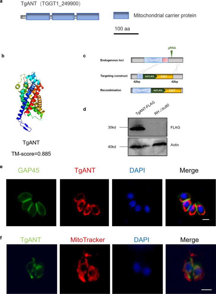
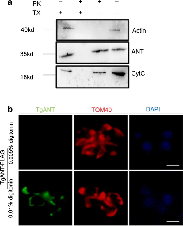
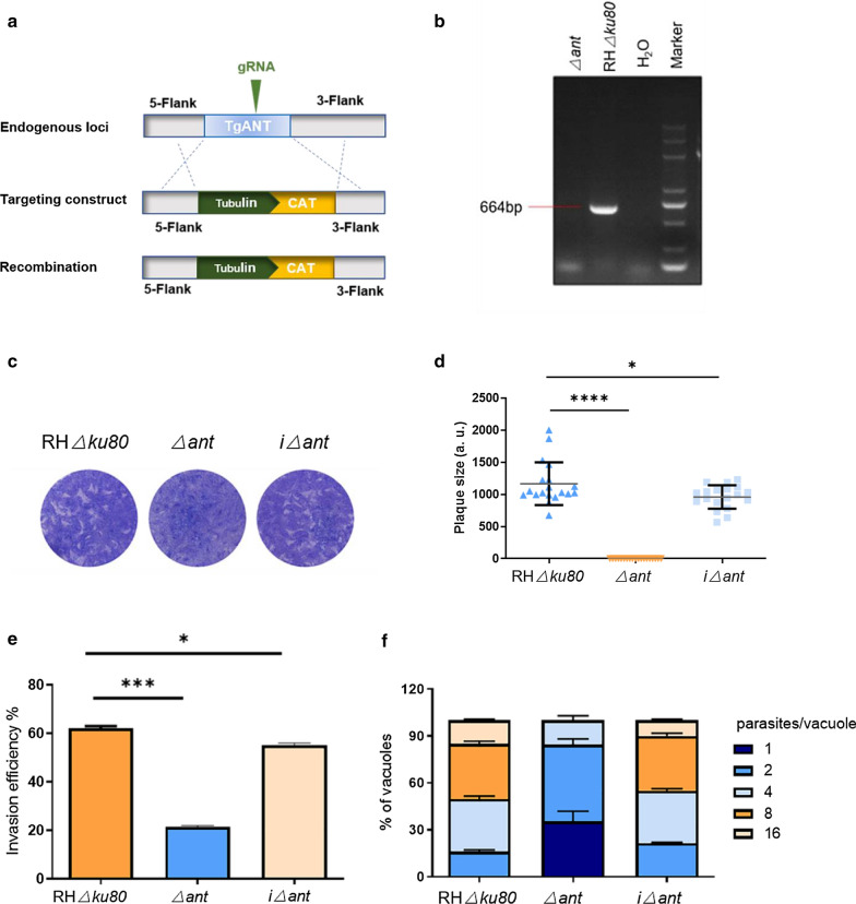
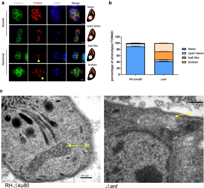
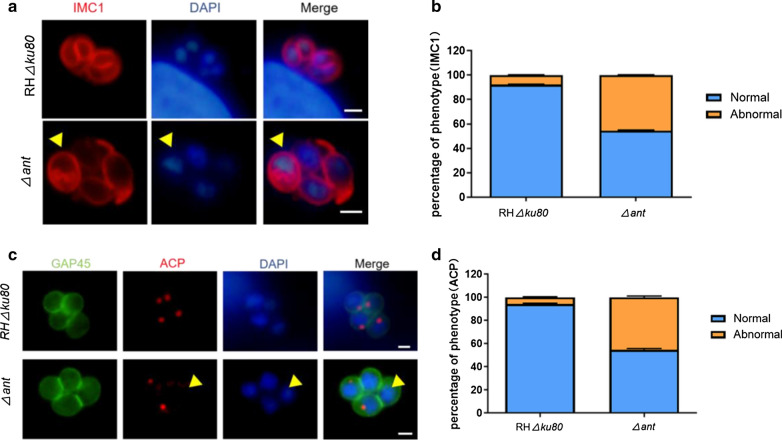
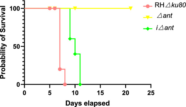
Results: Toxoplasma gondii was found to encode an ANT protein, which was designated TgANT. TgANT localized to the inner mitochondrial membrane. The proliferation of the Δant strain was significantly reduced. More important, depletion of TgANT resulted in significant changes in the morphology and ultrastructure of mitochondria, abnormal apicoplast division and abnormal cytoskeletal daughter budding. In addition, the pathogenicity of the Δant strain to mice was significantly reduced.

Conclusions: Altogether, we identified and characterized the ANT protein of T. gondii. Depletion of TgANT inhibited parasite growth and impaired apicoplast and mitochondrial biogenesis, as well as abnormal parasite division, suggesting TgANT is important for parasite growth.

Keywords: Adenine nucleotide translocase; Mitochondria; Toxoplasma gondii.


Conflict of interest statement

The authors declare that the research was conducted in the absence of any commercial or financial relationships that could be construed as a potential conflict of interest.

Figures

Fig. 1
Fig. 1
Identification and localization of TgANT protein. a Domain architecture of TgANT in Toxoplasma gondii. The domains were predicted by databases of protein families (Pfam). b Three-dimensional (3D) structures of TgANT predicted by Ab Initio Modeling using trRosetta. The quality of trRosetta modeling results is mainly based on its TM-score, which is between 0 and 1. The closer to 1, the higher the accuracy of the modeling and the greater the credibility. c Strategy for the construction of the strain with the FLAG endogenous epitope tag. d Western blot confirmed the expression of FLAG-tagged TgANT in parasites. Actin was used as a control. e IFA assays indicated that TgANT showedthe typical lasso-shaped mitochondria-like localization in intracellular tachyzoites. Parasites were labeled with rabbit anti-GAP45 (green), mouse anti-FLAG (red) and DAPI (blue). Scale bar, 2 μm. f IFA results showed the colocalization of anti-TgANT signal (green) and MitoTracker (red). MitoTracker was used as a mitochondrial marker. Scale bar: 2 μm. Abbreviations: CAT, chloramphenicol; DAPI,4′,6-Diamidino-2-phenylindole; IFA, immunofluorescence assay; TgANT, adenine nucleotide translocase in T. gondii
Fig. 2
Fig. 2
TgANT localizes to the IMM. a The PK protection assay was used to determine the accessibility of TgANT to 0.1 mg/ml PK in the presence or absence of the detergent 0.5% TX. After permeabilization by TX, PK could easily pass through organelle membranes to the stroma and digest stroma proteins nonspecifically. Actin was used as a cytoplasmic protein marker, and PK did not need the assistance of TX permeabilization solution for its nonspecific degradation process. CytC is a marker of mitochondrial intermembrane space, and its degradation by PK requires the synergistic effect of the TX permeabilizer. b The TgANT-FLAG-TOM40-HA strain is permeabilized with 0.005% or 0.01% digitonin. TOM40 was used as the markers of the mitochondrial outer membrane. Using 0.005% digitonin only allowed the detection of TOM40, while using 0.01% digitonin, both TOM40 and TgANT were detected, suggesting that TgANT is associated with the IMM. Parasites were labeled with mouse anti-FLAG (green), rabbit anti-HA (red) and DAPI (blue). Scale bar: 2 μm. Abbreviations: CytC, cytochrome c; IMM, inner mitochondrial membrane; PK, proteinase K; TX, Triton-X-100
Fig. 3
Fig. 3
Lack of TgANT decreased the growth of parasites. a Schematic of CRISPR/Cas9 strategy used for gene deletion. b PCR analysis confirmed knockout of the ant gene in the gene deletion mutant (Δant). c Plaque assays were used to compare the overall growth ability of the RHΔku80, Δant and iΔant strains. Each well was infected with 150 parasites, and plaques were stained after 7 days. d Plaque areas were measured by counting pixel points in Photoshop C6S software (Adobe Inc.). The data are compiled from three independent experiments. The plaque assay showed that Δant strains had distinct growth defects. Asterisks indicate significant differences at *P < 0.05 and ****P < 0.0001. e Parasite invasion into HFFs was examined. The invasion rate of the Δant strain was 51%, which was significantly different from that of the RHΔku80 strain (62%). Data are presented as the mean ± standard deviation of the results from three assays. Asterisks indicate significant differences at*P < 0.05 and ***P < 0.001. f Comparison of intracellular replication ability of Δant, iΔant, and RHΔku80 strains. In the Δant strain, the proportions of PVs containing 1, 2 and 4 parasites was 35, 49 and 51%, respectively, and it was almost impossible to observe PVs containing > 4 parasites. The proportions of PVs containing 1, 2, 4, 8 and 16 parasites in the RHΔku80 strain was 0, 1%, 34, 35 and 15%, respectively. HFFs, Human foreskin fibroblasts; PVs, parasitophorous vacuoles; Δ, gene knockout; iΔ, gene complement
Fig. 4
Fig. 4
Depletion of TgANT leads to disruption of mitochondrial morphology. a HA endogenous tags were added to the C terminal of the mitochondrial outer membrane protein TOM40 of the Δant and RHΔku80 strains. Mitochondrial outer membrane morphology was visualized by IFA. Two different mitochondrial phenotypes were identified in Δant, which we named ball-like and broken (yellow arrowhead). GAP45 (green) was used as a marker of the parasite membrane; TOM40 (red) was used as a marker of the parasite mitochondrial outer membrane; DAPI (blue) was used to stain nuclei. Mitochondrial morphology of intracellular parasites was scored as indicated. Scale bar: 2 μm. b Randomly selected vacuoles (n = 100) from two independent experiments were quantified. No significant changes were seen in the RHΔku80 strain, but the mitochondrion of the ant gene knockout parasites showed severe morphological defects, classified as “broken” (prevalence: appox. 27%) and “ball-like” (prevalence: approx. 26%). c Representative transmission electron microscopy images of Δant and RHΔku80 parasites. The deletion of ant resulted in a loss of density and a reduced number of mitochondrial cristae. The mitochondria of RHΔku80 parasites had a regular morphology with a clear cristae structure. M, Mitochondrion. Scale bars: 200 nm
Fig. 5
Fig. 5
Loss of TgANT results in asynchronous replication. a IMC1 (red), the marker of different stages of cell division; DAPI (blue) was used to stain nuclei. Arrowheads indicate parasites with abnormal cytoskeletal daughter budding. Scale bar: 2 μm. b Rate of asynchronous PVs in Δant (approx. 45%) and RHΔku80 (approx. 7%). Randomly selected vacuoles (n = 100) from two independent experiments were quantified. c Apicoplast of Δant and RHΔku80 strains was determined by immunofluorescence. ACP was used to mark the apicoplast. Arrows indicate parasites with abnormal apicoplast division. Scale bar: 2 μm. d Loss rate of the apicoplast in Δant (appox. 44%) and RHΔku80 (approx. 7%). Randomly selected vacuoles (n = 100) from two independent experiments were quantified. Abbreviations: ACP, Acyl carrier protein; IMC1, Inner membrane complex protein 1
Fig. 6
Fig. 6
TgANT knockout parasites show significantly reduced pathogenicity to mice. Five BALB/c mice were infected with 100 RHΔku80, Δant or iΔant tachyzoites, and their survival was assessed over 21 days

Similar articles

Cited by

References

    1. Lourido S. Toxoplasma gondii . Trends Parasitol. 2019;35:944–5. doi: 10.1016/j.pt.2019.07.001. - DOI - PubMed
    1. Melo EJ, Attias M, De Souza W. The single mitochondrion of tachyzoites of Toxoplasma gondii. J Struct Biol. 2000;130:27–33. doi: 10.1006/jsbi.2000.4228. - DOI - PubMed
    1. de Souza W, Attias M, Rodrigues JCF. Particularities of mitochondrial structure in parasitic protists (apicomplexa and kinetoplastida) Int J Biochem Cell Biol. 2009;41:2069–80. doi: 10.1016/j.biocel.2009.04.007. - DOI - PubMed
    1. Sheiner L, Vaidya AB, McFadden GI. The metabolic roles of the endosymbiotic organelles of Toxoplasma and Plasmodium spp. Curr Opin Microbiol. 2013;16:452–8. doi: 10.1016/j.mib.2013.07.003. - DOI - PMC - PubMed
    1. van Dooren GG, Stimmler LM, McFadden GI. Metabolic maps and functions of the Plasmodium mitochondrion. FEMS Microbiol Rev. 2006;30:596–630. doi: 10.1111/j.1574-6976.2006.00027.x. - DOI - PubMed

Substances

LinkOut - more resources