. 2022 May 31;22(1):204.
doi: 10.1186/s12935-022-02620-z.

Mechanisms of miR-3189-3p-mediated inhibition of c-MYC translation in triple negative breast cancer

Affiliations

Mechanisms of miR-3189-3p-mediated inhibition of c-MYC translation in triple negative breast cancer

Cecilia Vittori et al. Cancer Cell Int. .

Abstract

Background: Triple negative breast cancer (TNBC) is an aggressive subtype of breast cancer characterized by the lack of estrogen receptor, progesterone receptor, and HER2. Our lab previously characterized miR-3189-3p as a microRNA with potent anti-cancer activity against glioblastoma. Here, we hypothesized a similar activity in TNBC cells. As miR-3189-3p is predicted to target a variety of RNA binding proteins, we further hypothesized an inhibitory effect of this miRNA on protein synthesis.

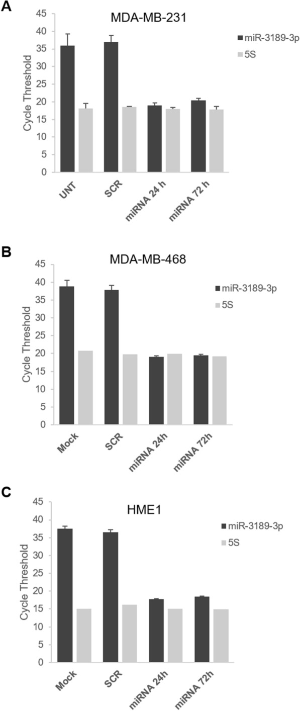
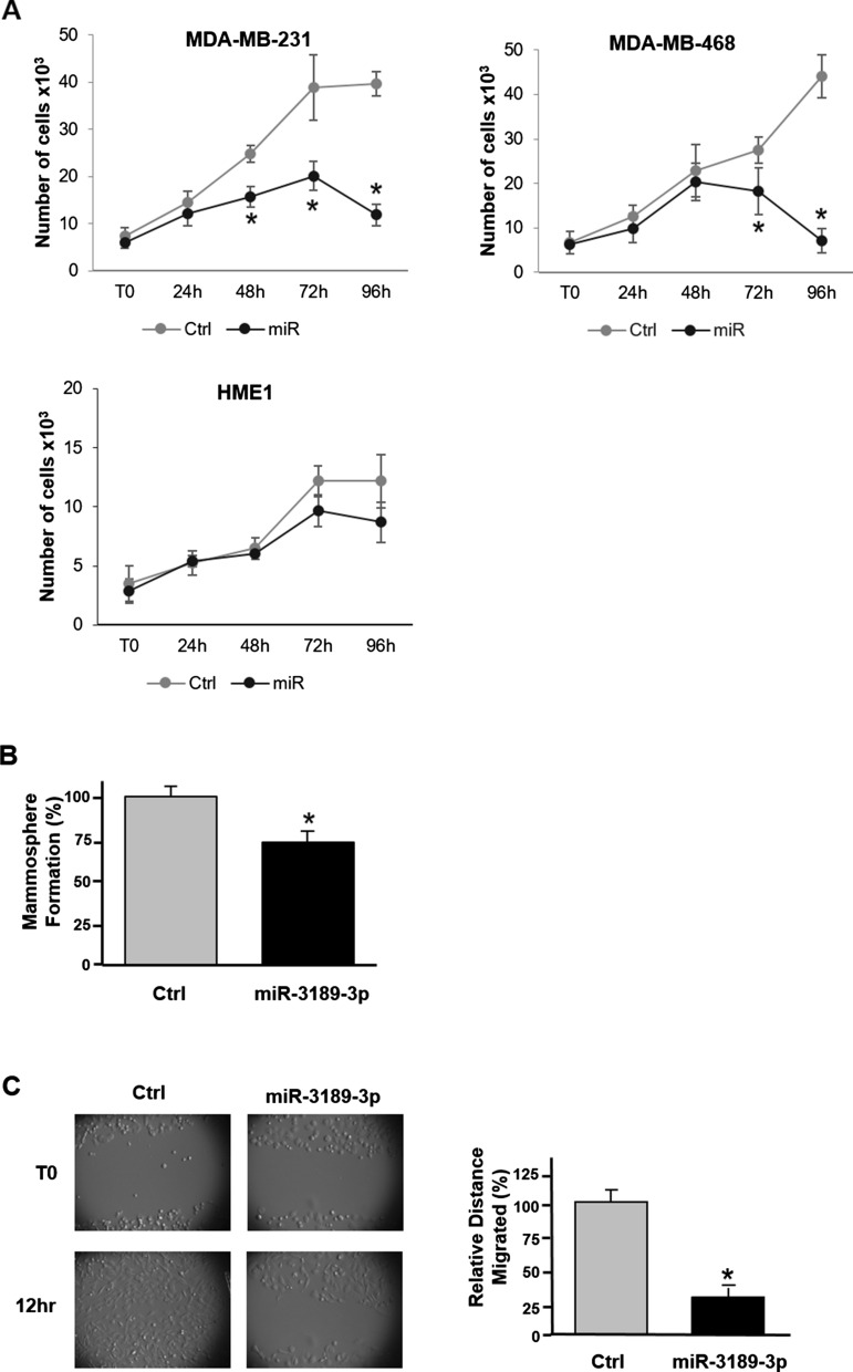
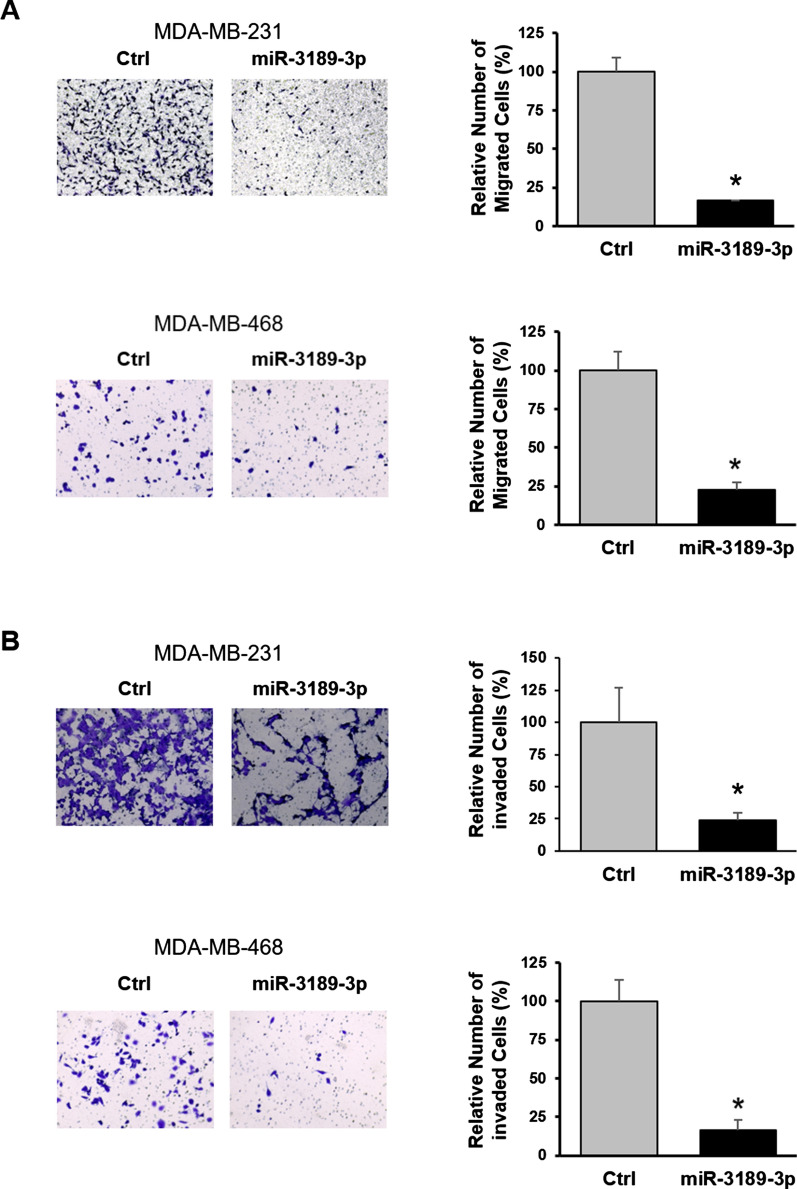
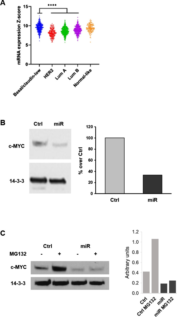
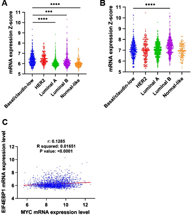
Methods: MDA-MB-231 and MDA-MB-468 cells were used to investigate the effect of miR-3189-3p on cell proliferation, migration, and invasion. TGCA database was used to analyze the expression of miR-3189-3p, c-MYC, 4EPB1, and eIF4E in breast cancer. Western blotting and RT-qPCR assays were used to assess the expression of selected proteins and RNAs after transfections.

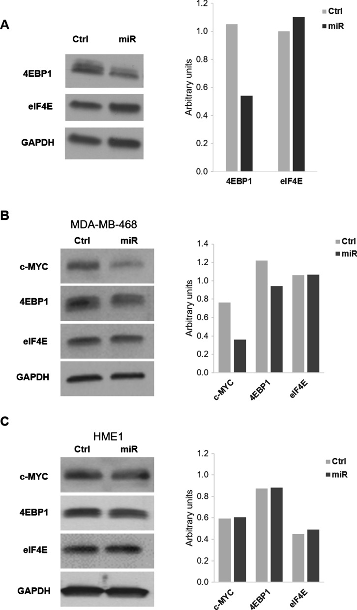
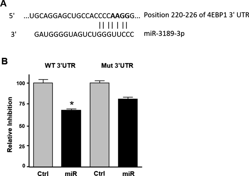
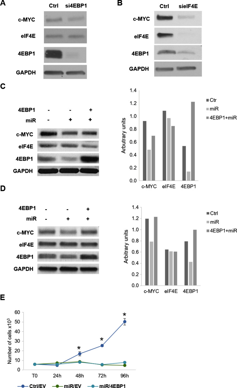
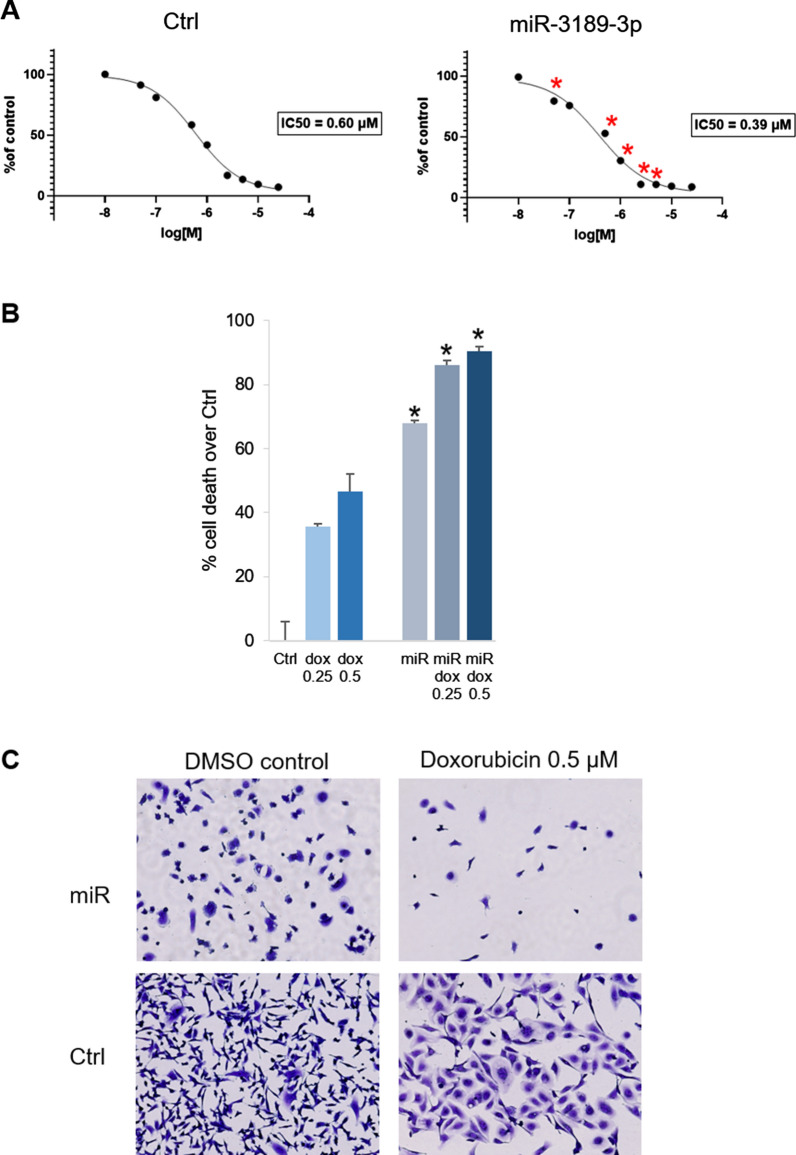
Results: Although c-MYC is not a predicted gene target for miR-3189-3p, we discovered that c-MYC protein is downregulated in miRNA-treated TNBC cells. We found that the downregulation of c-MYC by miR-3189-3p occurs in both normal growth conditions and in the absence of serum. The mechanism involved the direct inhibition of eIF4EBP1 by miR-3189-3p. Additionally, we found that miR-3189-3p could negatively affect cap-independent translation mediated by internal ribosome entry sites (IRES) or by m6A. Finally, miR-3189-3p sensitized TNBC cells to doxorubicin.

Conclusion: Overall, results indicated that miR-3189-3p exerts its anti-tumor activity through targeting translational regulatory proteins leading to an impairment in c-MYC translation, and possibly other oncogenic factors, suggesting that miR-3189-3p, alone or in combination, could be a valuable therapeutic approach against a malignancy with few treatment options.

Keywords: 4EBP1; Breast cancer; Translation; c-MYC; miR-3189-3p.


Conflict of interest statement

The authors declare no conflicts of interest.

Figures

Fig. 1
Fig. 1
Relative expression of miR-3189-3p in TNBC after transfection with the miRNA mimic. Bar graphs showing cycle threshold values of mature miR-3189-3p expression in MBA-MD-231 (A), MDA-MB-468 (B), and HME1 (C) cells at 24 and 72 h post-transfection. For reference, expression of the small RNA 5S is also indicated
Fig. 2
Fig. 2
MiR-3189-3p impairs proliferation and migration of TNBC cells. A Plot indicating proliferation of MDA-MB-231, MDA-MB-468, and HME1 cells at the indicated time points. B Cell proliferation assay for mammospheres obtained from MDA-MB-231 cells, performed 72 h post-transfection with the miR-3189-3p or the scramble control. Results are expressed as percent of the number of mammospheres obtained from cells transfected with miR-3189-3p versus the number of mammospheres from control cells. C Representative images of a scratch assay 72 h post-transfection with miR-3189-3p or scramble control. Migration of cells toward the cell-free area was monitored by time-lapse imaging using an Olympus VivaView system. Original magnification 10×. The bar graph represents quantification of the migration obtained from three experiments, each in duplicate. In all graph, the asterisks indicate statistical significance
Fig. 3
Fig. 3
MiR-3189-3p impairs migration/invasion of TNBC cells. A Representative images of a transwell migration assay performed using MDA-MB-231 (upper panel) and MDA-MB-468 (lower panel) 72 h post-transfection with the miR-3189-3p or scramble control (Ctrl). Migrated cells were stained with crystal violet and pictures were acquired with an optical microscope. Original magnification 20×. Quantification of the migrated cells was obtained from three different random image fields per insert and averaged from at least three biological replicates. B Representative images of a transwell invasion assay performed using MDA-MB-231 (upper panel) and MDA-MB-468 (lower panel) and analyzed 72 h post-transfection with the miR-3189-3p or scramble control (Ctrl). Transwells were stained and pictures were acquired with an optical microscope. Original magnification 20×. The bar graph indicates the average number of cells migrated through the Matrigel calculated from three different random image fields per insert and averaged from at least three biological replicates. * Indicates statistical significance
Fig. 4
Fig. 4
miR-3189-3p mediates c-MYC downregulation at translational level. A Expression of c-MYC mRNA in different subtypes of breast cancer (METABRIC). * Indicates statistical significance. B Western blot for c-MYC protein from MDA-MB-231 cells transfected with miR-3189-3p (miR) or scramble control (Ctrl) for 72 h. Densitometric analysis of the bands relative to c-MYC was done using Image J. C Western blots performed on MDA-MB-231 cells that were transfected with scramble control (Ctrl) or miR-3189-3p (miR) in the absence or presence of the proteasome inhibitor MG132. 14–3-3 was used as loading control
Fig. 5
Fig. 5
Overexpression of miR-3189-3p downregulates c-MYC and 4EBP1 protein levels in TNBC cells but not in normal epithelial cells. A Western blot to detect 4EBP1 and eIF4E proteins in MDA-MB-231 cells transfected with miR-3189-3p (miR) or scramble control (Ctrl). B, C Representative Western blots showing expression of c-MYC, 4EBP1, and eIF4E in MDA-MB-468 (b) and HME1 normal epithelial cells. GAPDH was used as loading control
Fig. 6
Fig. 6
MiR-3189-3p directly targets 4EBP1 3′UTR. A Alignment of 4EBP1 3′UTR sequence with miR-3189-3p. In bold are indicated the mutated bases. B Luciferase assay of MDA-MB-231 cells co-transfected with miR-3189-3p (miR) or control (Ctrl) and either 4EBP1 3′UTR (WT 3′UTR) or the mutated 4EBP1 3′UTR (Mut 3′UTR). The scramble sequence of the miRNA was used as control. * Indicates statistical significance
Fig. 7
Fig. 7
METABRIC data set analysis shows increased expression of 4EBP1, but not eIF4E, in TNBC compared to normal-like controls. Expression of 4EBP1 (A) and eIF4E (B) mRNAs in different subtypes of breast cancer (METABRIC). * indicates statistical significance. C Correlation between 4EBP1 and c-MYC mRNA expression in breast cancer tumors (METABRIC). * Indicates statistical significance. The association was measured using the Pearson correlation coefficient (R) and the P-value
Fig. 8
Fig. 8
Role of 4EBP1 in the miR-3189-3p-mediated c-MYC downregulation. Western blots showing levels of the indicated proteins after silencing of 4EBP1 (A) or eIF4E (B). Cells transfected with the scramble RNA were used as control (Ctrl). Western blot showing c-MYC, eIF4E and 4EBP1 proteins in MDA-MB-231 cells transfected with miR-3189-3p or scramble control in the presence of serum (C) or in serum-free conditions (D). Where indicated, cells were co-transfected with 4EBP1 expression vector. GAPDH was used as a loading control for every blot. E Scatter plot showing proliferation of MDA-MB-231 cells transfected with miR-3189-3p (miR) and either pcDNA3 EV (EV) or 4EBP1. Cells transfected with scramble RNA and EV served as control. * Indicates statistical significance between Ctrl/EV and either miR/EV or miR/4EBP1
Fig. 9
Fig. 9
miR-3189-3p impairs IRES-dependent translation and m6A-dependent translation. Western blots to detect IRES-dependent translation of ECFP after co-transfection of MDA-MB-231 cells with pYIC/EMCV-IRES (A) or pYIC/c-MYC-IRES (B) and miR-3189-3p (miR) or scramble control (Ctrl). C Western blot to detect IRES-dependent translation of ECFP after co-transfection of MDA-MB-231 cells with pYIC/EMCV-IRES and miR-3189-3p or scramble control. Where indicated, cells were co-transfected also with 4EBP1 expression vector. EMCV- or MYC-IRES were detected using ECFP antibody. D Western blot of MDA-MB-231 cells co-transfected with miR-3189-3p (miR) or scramble control (Ctrl) together with a plasmid encoding a circular RNA containing the protein E7 under m6A translation (E7) or the empty vector (EV) for 24 and 48 h. GAPDH was used as a loading control for every blot
Fig. 10
Fig. 10
miR-3189-3p sensitizes MDA-MB-231 cells to doxorubicin. A Graphs indicating IC50 of doxorubicin in control-transfected (Ctrl) and miR-3189-3p-transfected (miR) cells. B Bar graph showing the percentage of cell death at the indicated concentrations of doxorubicin (dox) in control- or miR-transfected cells. C Representative images of Ctrl and miR-transfected cells after treatment with 0.5 µM doxorubicin or DMSO control. Asterisks in A and B indicate statistical significance between miR and control at the indicated concentrations of doxorubicin

References

    1. Harbeck N, Penault-Llorca F, Cortes J, Gnant M, Houssami N, Poortmans P, Ruddy K, Tsang J, Cardoso F. Breast cancer. Nat Rev Dis Primers. 2019;5(1):66. doi: 10.1038/s41572-019-0111-2. - DOI - PubMed
    1. Jia LY, Shanmugam MK, Sethi G, Bishayee A. Potential role of targeted therapies in the treatment of triple-negative breast cancer. Anticancer Drugs. 2016;27(3):147–155. doi: 10.1097/CAD.0000000000000328. - DOI - PubMed
    1. Kawaji H, Kubo M, Yamashita N, Yamamoto H, Kai M, Kajihara A, Yamada M, Kurata K, Kaneshiro K, Harada Y. Comprehensive molecular profiling broadens treatment options for breast cancer patients. Cancer Med. 2020;10(2):529–539. doi: 10.1002/cam4.3619. - DOI - PMC - PubMed
    1. Schmid P, Adams S, Rugo HS, Schneeweiss A, Barrios CH, Iwata H, Diéras V, Hegg R, Im S-A, Shaw Wright G. Atezolizumab and nab-paclitaxel in advanced triple-negative breast cancer. N Engl J Med. 2018;379(22):2108–2121. doi: 10.1056/NEJMoa1809615. - DOI - PubMed
    1. Castellvi J, Garcia A, Rojo F, Ruiz-Marcellan C, Gil A, Baselga J, Ramon y Cajal S. Phosphorylated 4E binding protein 1: a hallmark of cell signaling that correlates with survival in ovarian cancer. Cancer. 2006;107(8):1801–1811. doi: 10.1002/cncr.22195. - DOI - PubMed