. 2022 Jul 1;128(1):28-39.
doi: 10.1152/jn.00102.2022. Epub 2022 Jun 1.

Cardiovascular deconditioning increases GABA signaling in the nucleus tractus solitarii

Affiliations

Cardiovascular deconditioning increases GABA signaling in the nucleus tractus solitarii

Ludmila Lima-Silveira et al. J Neurophysiol. .

Abstract

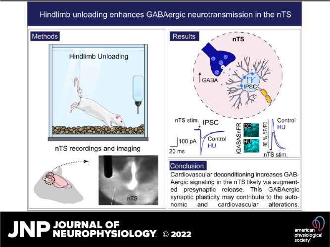
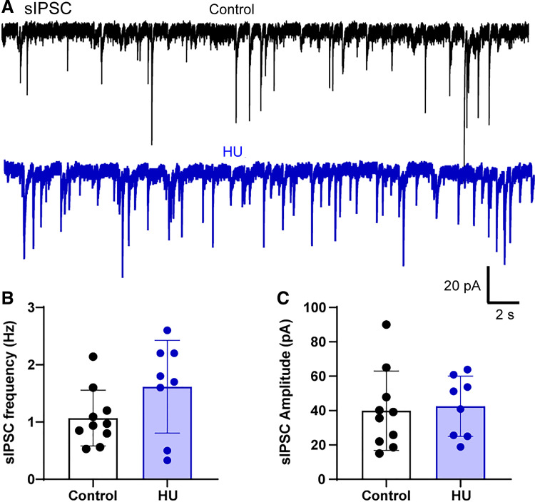
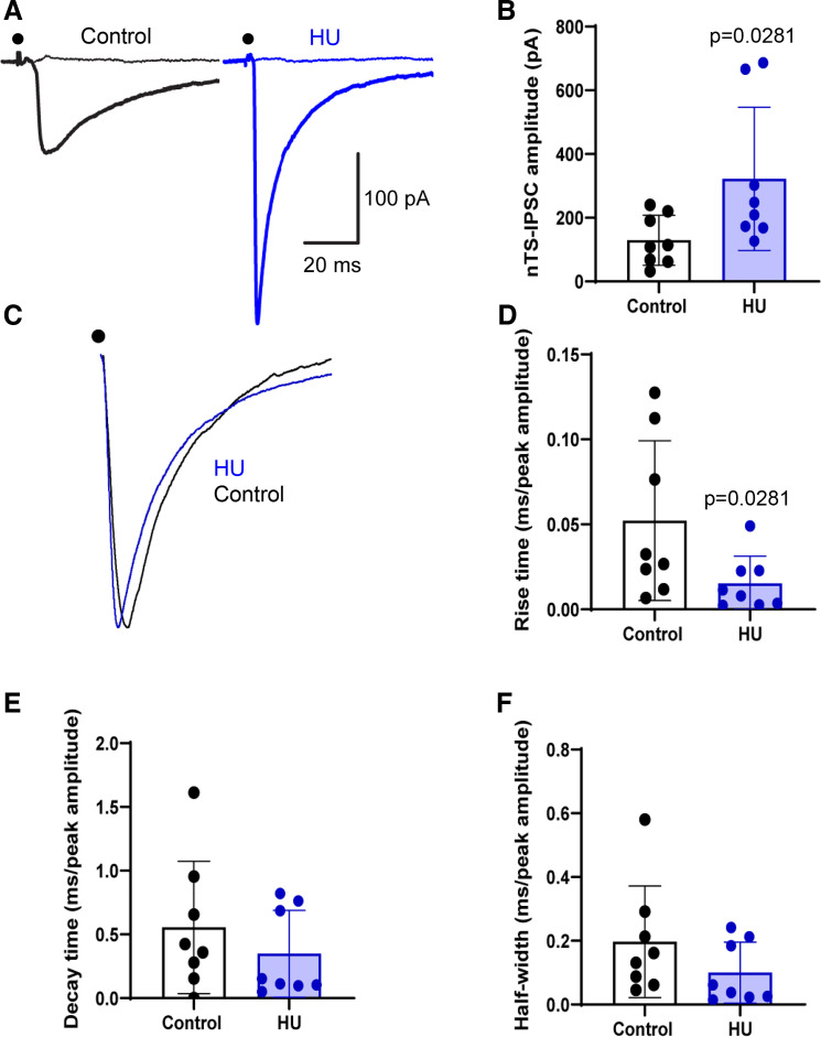
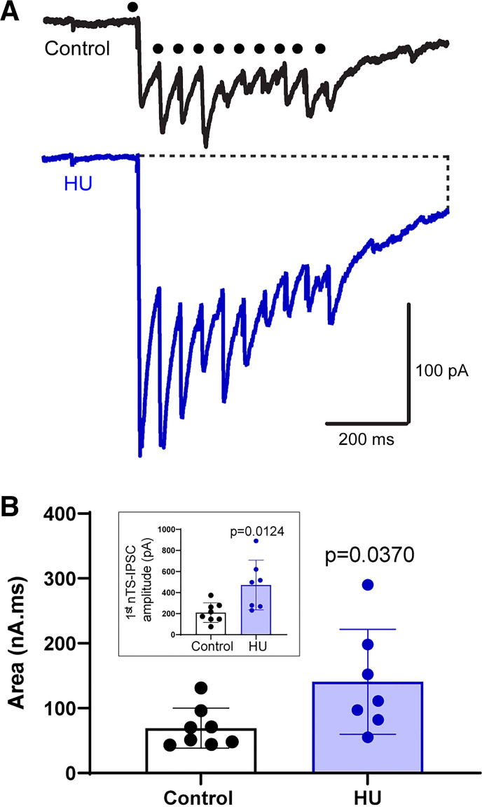
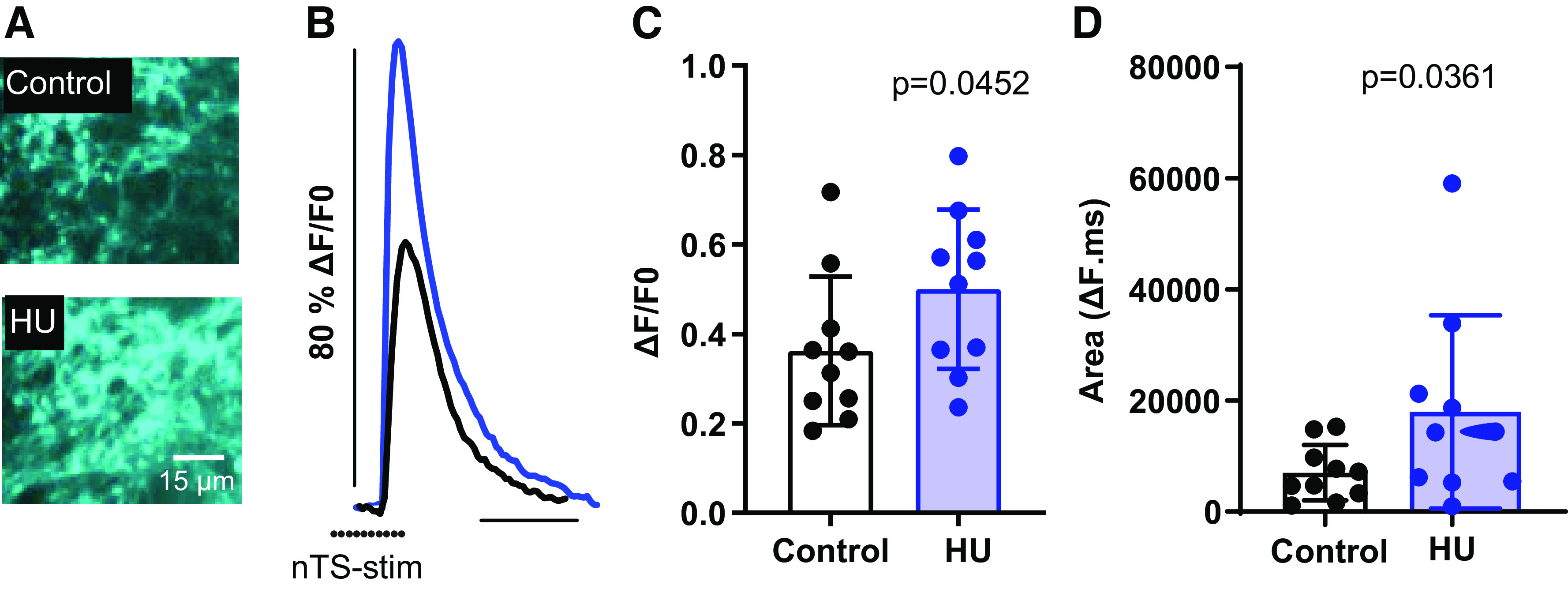
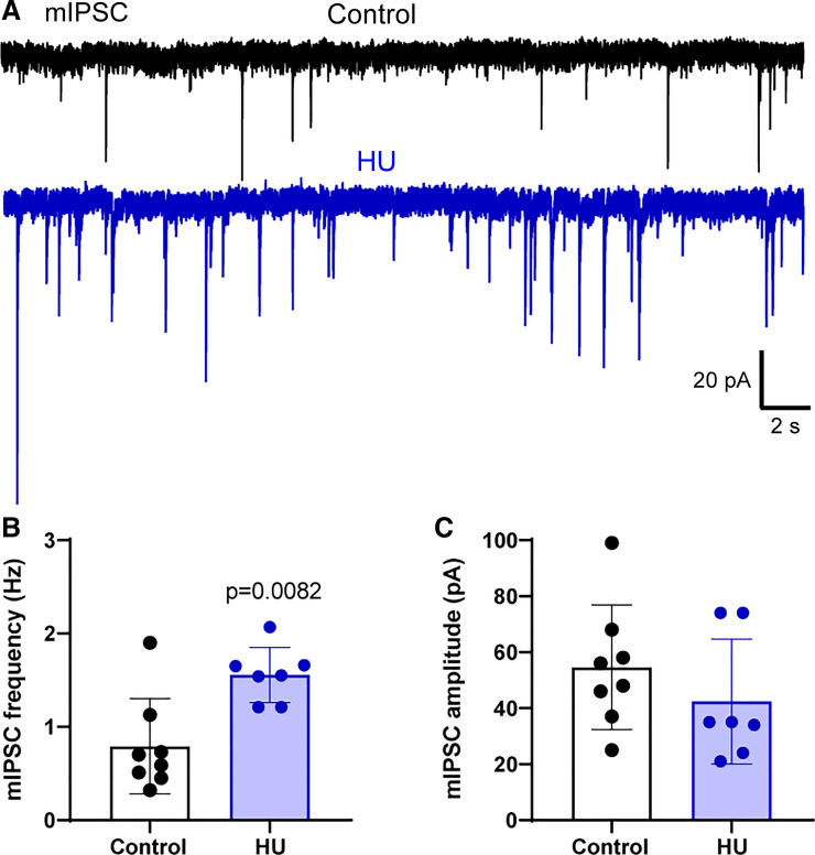
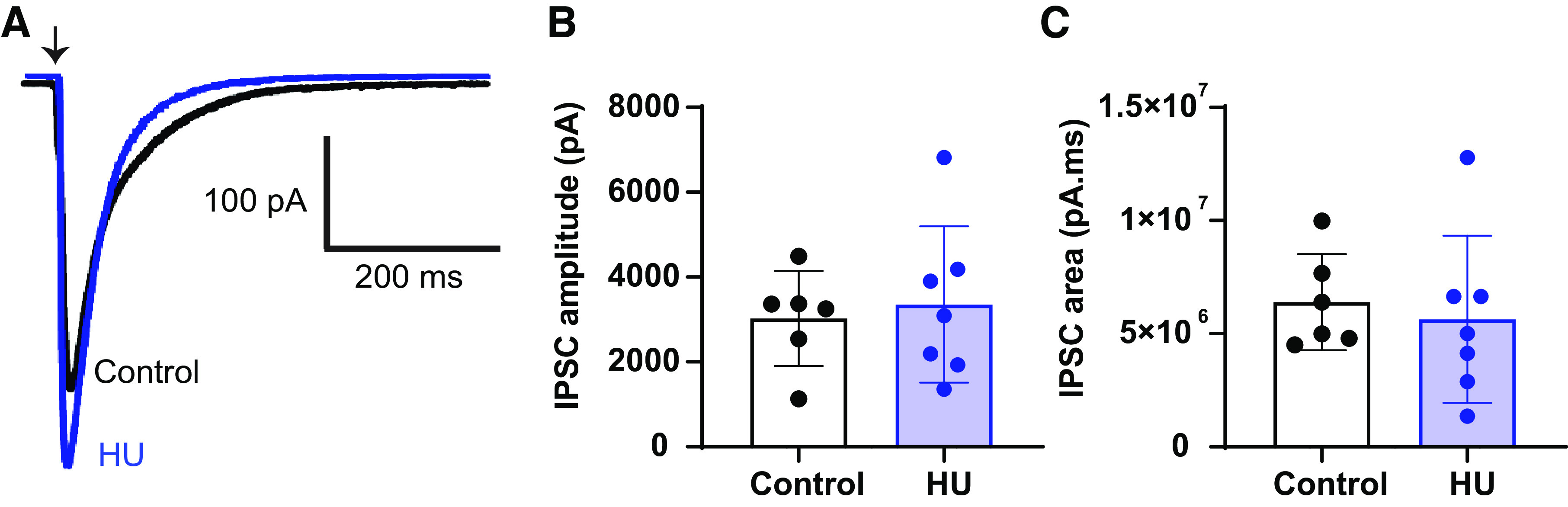
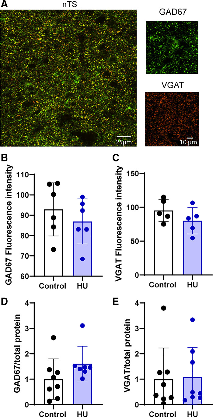
The nucleus tractus solitarii (nTS) is the major integrative brainstem region for autonomic modulation and processing of cardiovascular reflexes. GABA and glutamate are the main inhibitory and excitatory neurotransmitters, respectively, within this nucleus. Alterations in the GABA-glutamate regulation in the nTS are related to numerous cardiovascular comorbidities. Bedridden individuals and people exposed to microgravity exhibit dysautonomia and cardiovascular deconditioning that are mimicked in the hindlimb unloading (HU) rat model. We have previously shown in the nTS that HU increases glutamatergic neurotransmission yet decreases neuronal excitability. In this study, we investigated the effects of HU on nTS GABAergic neurotransmission. We hypothesized that HU potentiates GABA signaling via increased GABAergic release and postsynaptic GABA receptor expression. Following HU or control postural exposure, GABAergic neurotransmission was assessed using whole cell patch clamp whereas the magnitude of GABA release was evaluated via an intensity-based GABA sensing fluorescence reporter (iGABASnFR). In response to GABA interneuron stimulation, the evoked inhibitory postsynaptic current (nTS-IPSC) amplitude and area, as well as iGABASnFR fluorescence, were greater in HU than in control. HU also elevated the frequency but not the amplitude of spontaneous miniature IPSCs. Picoapplication of GABA produced similar postsynaptic current responses in nTS neurons of HU and control. Moreover, HU did not alter GABAA receptor α1 subunit expression, indicating minimal alterations in postsynaptic membrane receptor expression. These results indicate that HU increases GABAergic signaling in the nTS likely via augmented release of GABA from presynaptic terminals. Altogether, our data indicate GABA plasticity contributes to the autonomic and cardiovascular alterations following cardiovascular deconditioning (CVD).NEW & NOTEWORTHY Gravity influences distribution of blood volume and autonomic function. Microgravity and prolonged bed rest induce cardiovascular deconditioning (CVD). We used hindlimb unloading (HU), a rat analog for bed rest, to investigate CVD-induced neuroplasticity in the brainstem. Our data demonstrate that HU increases GABA modulation of nucleus tractus solitarii (nTS) neurons via presynaptic plasticity. Given the importance of nTS in integrating cardiovascular reflexes, this study provides new evidence on the central mechanisms behind CVD following HU.

Keywords: IPSC; autonomic nervous system; hindlimb unloading; synaptic transmission.


Conflict of interest statement

No conflicts of interest, financial or otherwise, are declared by the authors.

Figures

None
Graphical abstract
Figure 1.
Figure 1.
Hindlimb unloading (HU) does not affect spontaneous inhibitory postsynaptic currents (sIPSCs) in nucleus tractus solitarii (nTS). A: example traces of sIPSC recordings in nTS neurons from a control and HU rat. Average data showing frequency (B) and amplitude (C) of sIPSCs. HU tended to increase sIPSC frequency, but it was not statistically significant (P = 0.0921, control: n = 10, N = 10; HU: n = 8, N = 8, unpaired t test).
Figure 2.
Figure 2.
Hindlimb unloading (HU) enhances evoked GABAergic neurotransmission in the nucleus tractus solitarii (nTS). A: representative data showing the increased nTS-evoked inhibitory postsynaptic currents (IPSCs) amplitude in HU compared with control. Dot indicates the point of stimulus. Flat lines indicate the absence of currents after GABAR blockade with gabazine and bicuculline. B: mean data revealing the significant increase in nTS-IPSC amplitude in HU group (control: n = 8, N = 8; HU: n = 8, N = 8). C and D: re-scaled current traces showing that HU reduced the rise time of nTS-IPSCs but had no effect on their decay time (E) and half-width (F). Rise time, decay time, and half-width were normalized to the nTS-IPSC amplitude. B and D, Mann–Whitney test.
Figure 3.
Figure 3.
Hindlimb unloading (HU) increases overall nucleus tractus solitarii-evoked inhibitory postsynaptic currents (nTS-IPSC) signaling. A: example traces of nTS-IPSCs produced by consecutive stimulation of nTS neuropil at higher frequency (20 Hz). Note the enhanced inward current in HU compared to control (control: n = 8, N = 10; HU: n = 7, N = 7). Dot indicates time of nTS stimulation. B: the area of IPSCs produced by nTS stimulation at 20 Hz was greater in HU vs. control. Inset: the amplitude of the initial nTS-IPSCs was greater in HU compared with control. B, Mann–Whitney test’ inset, unpaired t test.
Figure 4.
Figure 4.
Hindlimb unloading (HU) enhances presynaptic release of GABA in the nucleus tractus solitarii (nTS). A: example of intensity-based GABA sensing fluorescence reporter (iGABASnFR) after nTS stimulation. B: iGABASnFR fluorescence response was greater in HU than control. C: quantitative data of the peak iGABASnFR fluorescence reported as change relative to pre-stimulation baseline (ΔF/F0). D: average fluorescence area showing the increased response in HU compared with control [control: 10 averaged regions of interests (ROIs), N = 3; HU: 10 averaged ROIs, N = 4]. Increased iGABASnFR fluorescence indicates greater presynaptic release of GABA. Dots indicate the point of stimulus. C and D, unpaired t test.
Figure 5.
Figure 5.
Hindlimb unloading (HU) enhances the action potential-independent release of GABA. A: representative recordings of miniature inhibitory postsynaptic currents (mIPSCs) in the presence of tetrodotoxin (TTX, 1 µM) in nTS neurons from a control and HU rat. B: mean data showing the increased frequency of mIPSCs in HU compared with control. C: quantitative data demonstrating that the amplitude of mIPSCs was comparable between the groups (control; n = 9, N = 4; HU: n = 7, N = 4). B, Mann–Whitney test.
Figure 6.
Figure 6.
Hindlimb unloading (HU) has no effect on postsynaptic responses to GABA in nucleus tractus solitarii (nTS) neurons. A: puffer application of GABA (100 µM, 15 ms) produces similar postsynaptic responses in nTS neurons of HU and control (control: n = 6, N = 4; HU: n = 7, N = 4). B and C: average data demonstrating the amplitude and area of inward current generated by puffer application of GABA in HU and control. Arrow represents the point of puffer application of GABA. IPSC, inhibitory postsynaptic currents.
Figure 7.
Figure 7.
Hindlimb unloading (HU) does not change the expression of glutamic acid decarboxylase 67 (GAD67) and vesicular GABA transporter (VGAT). A: immunofluorescence image of GAD67 (green) and VGAT (orange) in the nucleus tractus solitarii (nTS) region of a representative rat from control group. B and C: mean data showing comparable values of fluorescent intensity of GAD67 (control: N = 6, HU: N = 6) and VGAT (control: N = 5, HU: N = 5) in HU and control. Immunoblot quantification of GAD67/total protein ratio (D, control: N = 8, HU: N = 7) and VGAT/total protein ratio (E, control: N = 8, HU: N = 8) confirmed that expression of these proteins was not altered by HU.

Similar articles

Cited by

References

    1. Andresen MC, Kunze DL. Nucleus tractus solitarius—gateway to neural circulatory control. Annu Rev Physiol 56: 93–116, 1994. doi:10.1146/annurev.ph.56.030194.000521. - DOI - PubMed
    1. Andresen MC, Doyle MW, Bailey TW, Jin YH. Differentiation of autonomic reflex control begins with cellular mechanisms at the first synapse within the nucleus tractus solitarius. Braz J Med Biol Res 37: 549–558, 2004. doi:10.1590/s0100-879x2004000400012. - DOI - PubMed
    1. Mifflin SW, Felder RB. Synaptic mechanisms regulating cardiovascular afferent inputs to solitary tract nucleus. Am J Physiol Heart Circ Physiol 259: H653–H661, 1990. doi:10.1152/ajpheart.1990.259.3.H653. - DOI - PubMed
    1. Talman WT, Perrone MH, Reis DJ. Evidence for l-glutamate as the neurotransmitter of baroreceptor afferent nerve fibers. Science 209: 813–815, 1980. doi:10.1126/science.6105709. - DOI - PubMed
    1. Andresen MC, Yang MY. Non-NMDA receptors mediate sensory afferent synaptic transmission in medial nucleus tractus solitarius. Am J Physiol Heart Circ Physiol 259: H1307–H1311, 1990. doi:10.1152/ajpheart.1990.259.4.H1307. - DOI - PubMed

Publication types

LinkOut - more resources