. 2022;14(3):527-551.
doi: 10.1016/j.jcmgh.2022.05.007. Epub 2022 May 26.

HIRA Supports Hepatitis B Virus Minichromosome Establishment and Transcriptional Activity in Infected Hepatocytes

Affiliations

HIRA Supports Hepatitis B Virus Minichromosome Establishment and Transcriptional Activity in Infected Hepatocytes

Maëlle Locatelli et al. Cell Mol Gastroenterol Hepatol. 2022.

Abstract

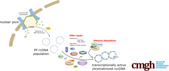
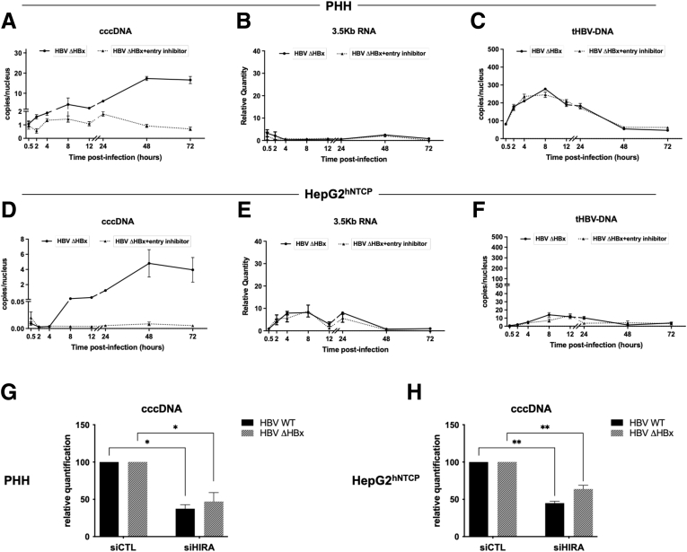
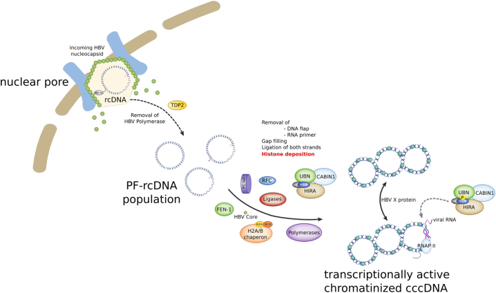
Background & aims: Upon hepatitis B virus (HBV) infection, partially double-stranded viral DNA converts into a covalently closed circular chromatinized episomal structure (cccDNA). This form represents the long-lived genomic reservoir responsible for viral persistence in the infected liver. Although the involvement of host cell DNA damage response in cccDNA formation has been established, this work investigated the yet-to-be-identified histone dynamics on cccDNA during early phases of infection in human hepatocytes.

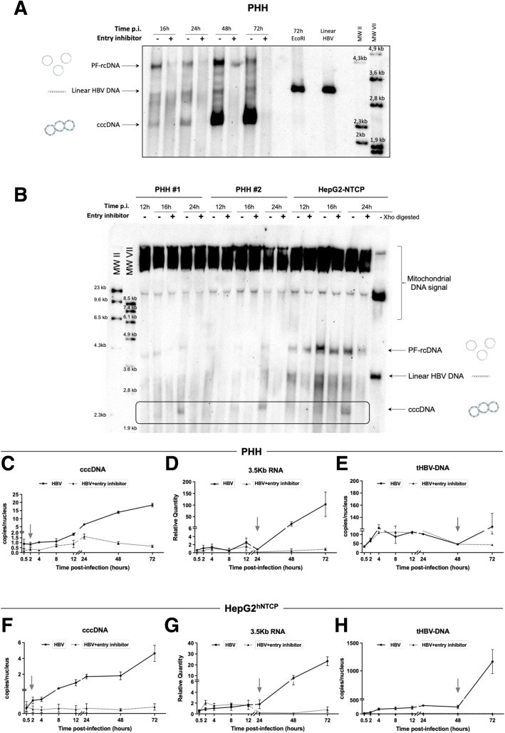
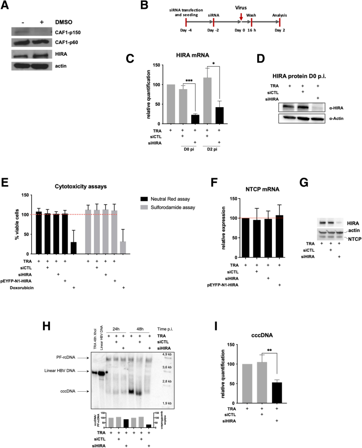
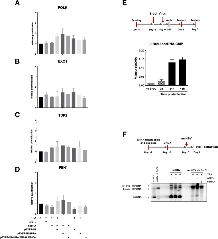
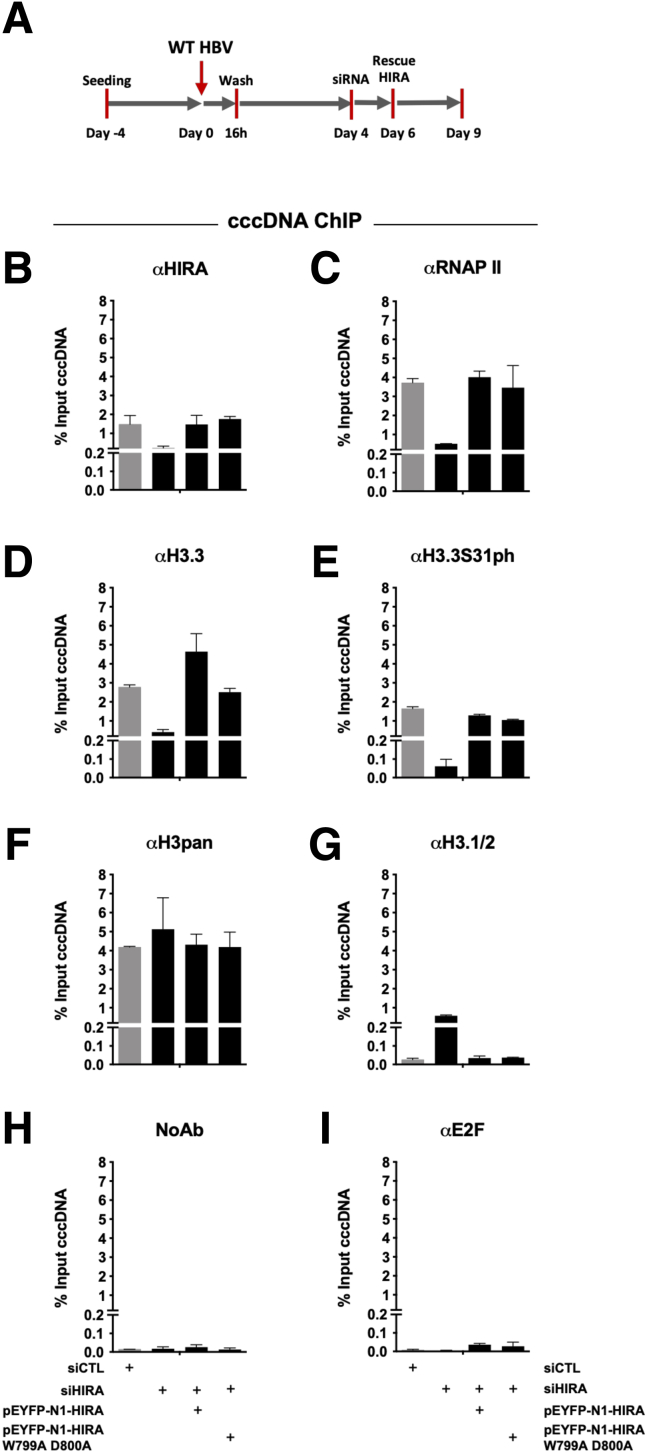
Methods: Detailed studies of host chromatin-associated factors were performed in cell culture models of natural infection (ie, Na+-taurocholate cotransporting polypeptide (NTCP)-overexpressing HepG2 cells, HepG2hNTCP) and primary human hepatocytes infected with HBV, by cccDNA-specific chromatin immunoprecipitation and loss-of-function experiments during early kinetics of viral minichromosome establishment and onset of viral transcription.

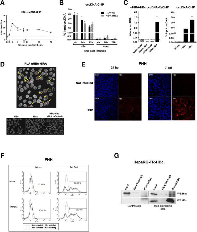
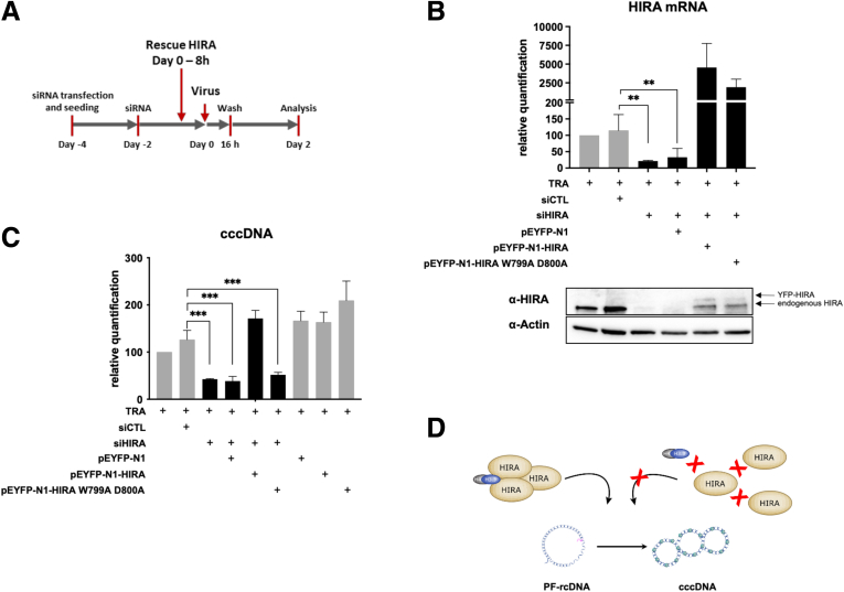
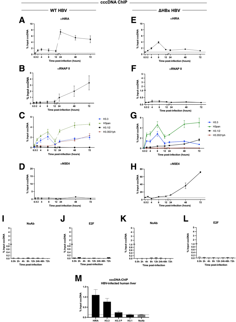
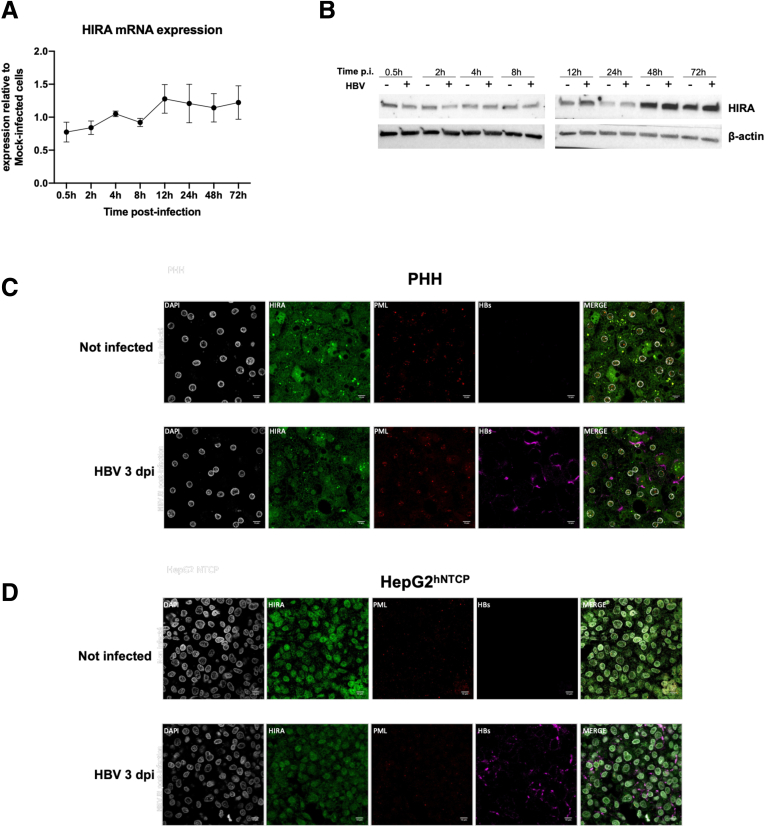
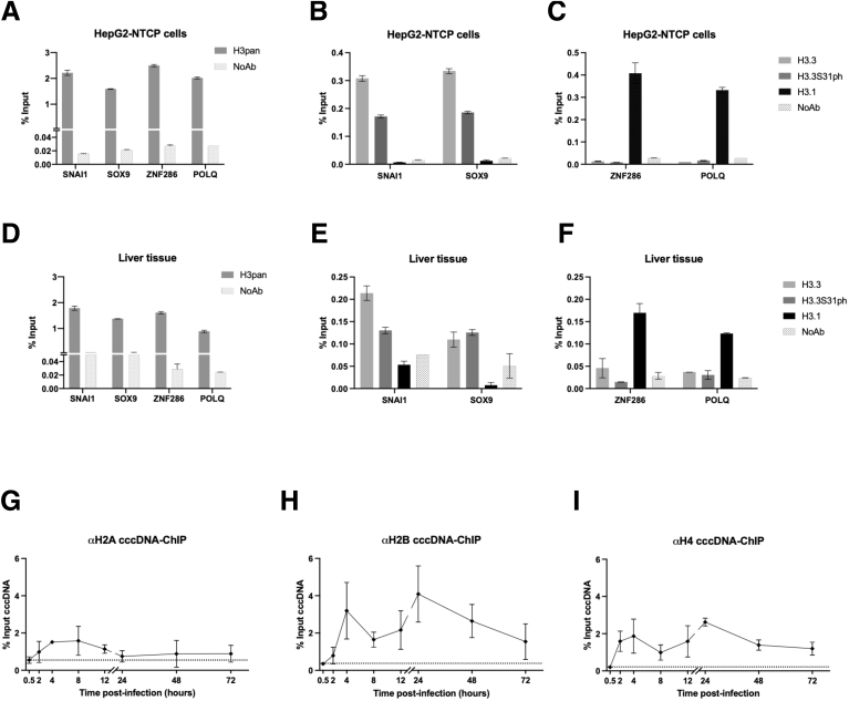
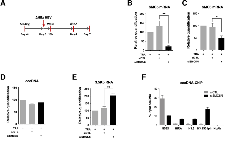
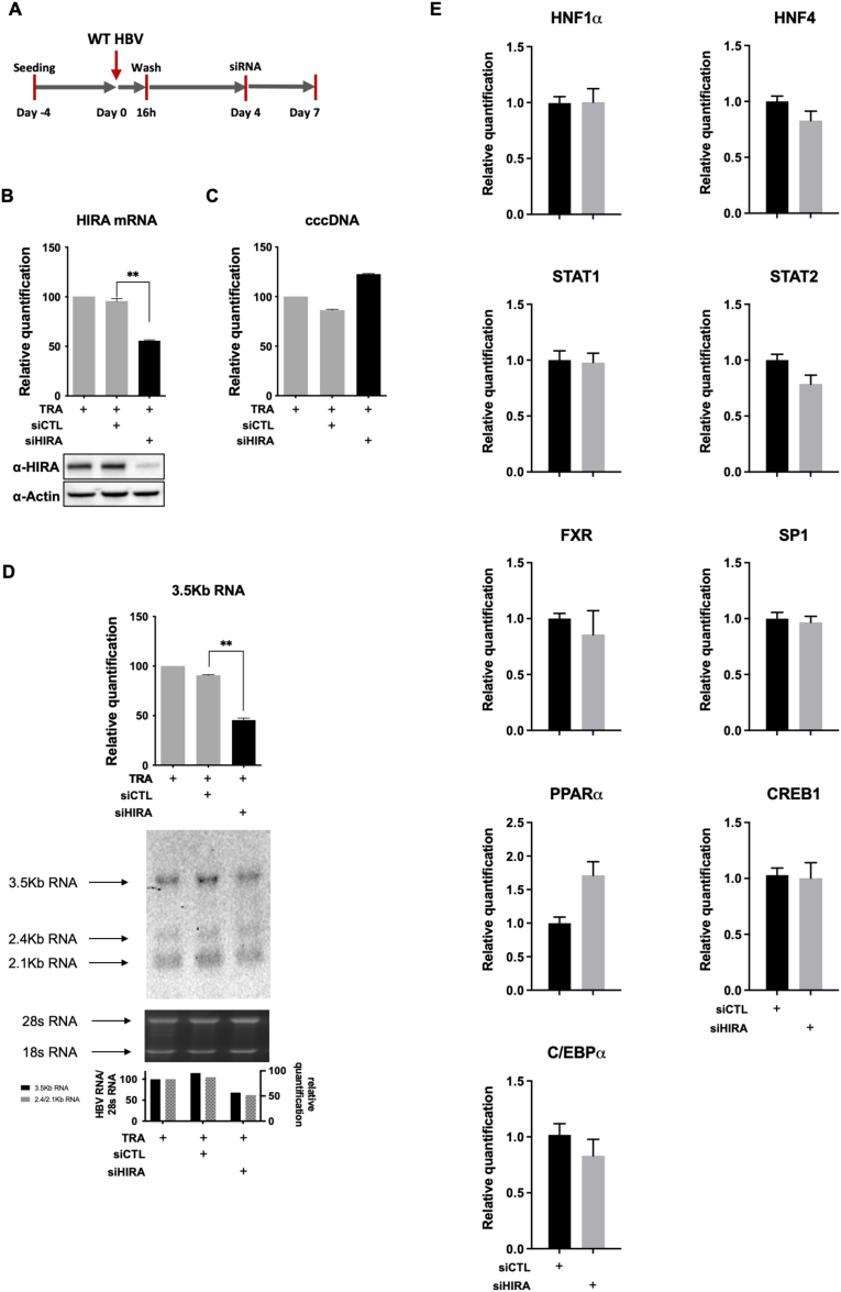
Results: Our results show that cccDNA formation requires the deposition of the histone variant H3.3 via the histone regulator A (HIRA)-dependent pathway. This occurs simultaneously with repair of the cccDNA precursor and independently from de novo viral protein expression. Moreover, H3.3 in its S31 phosphorylated form appears to be the preferential H3 variant found on transcriptionally active cccDNA in infected cultured cells and human livers. HIRA depletion after cccDNA pool establishment showed that HIRA recruitment is required for viral transcription and RNA production.

Conclusions: Altogether, we show a crucial role for HIRA in the interplay between HBV genome and host cellular machinery to ensure the formation and active transcription of the viral minichromosome in infected hepatocytes.

Keywords: H3.3; HBV; HIRA; cccDNA; chromatin.


Figures

None
Graphical abstract
Figure 1
Figure 1
HBV minichromosome establishment occurs very rapidly after infection. (A and B) Southern blot analysis of cccDNA appearance kinetic. PHHs or HepG2hNTCP cells were infected with HBV in the presence or not of 100 nmol/L preS1-mimicking peptide for up to 16 hours and then harvested at the indicated times points. Mitochondrial DNA was used as an internal loading control. The specificity of the cccDNA band is shown by linearization after digestion with EcoRI or XhoI restriction enzymes. (C–H) qPCR quantification of viral cccDNA, 3.5-kb RNA, and total HBV DNA (tHBV-DNA) in (C–E) PHHs and (F–H) HepG2hNTCP-infected cells. Cells were inoculated with HBV for up to 16 hours and then harvested at the indicated time points. cccDNA and tHBV-DNA quantification were normalized over β-globin quantity, while the relative 3.5-kb RNA amount was normalized over the housekeeping gene GUSb expression. Graphs represent the means ± SEM of at least 3 independent experiments. MW, molecular weight; p.i., post infection.
Figure 2
Figure 2
Full cccDNA supercoiling in de novo infected cells requires HIRA protein expression. (A) Detection by Western blot analysis of CAF-1 (subunits p150 and p60) and HIRA before and after 72 hours of 2.5% DMSO addition to HepG2hNTCP cells. (B) HepG2hNTCP cells were transfected twice with siRNA anti-HIRA or a nontargeting siRNA (siCTL) and then inoculated for 16 hours with HBV. Cells were harvested for analysis 2 dpi. (C and D) HIRA messenger RNA (mRNA) and protein expression after siRNA transfection was determined by (B) real-time qPCR and (C) Western blot, respectively. β-actin served as Western blot loading control. HIRA mRNA was normalized over housekeeping GUSb gene levels and expressed as relative to the control condition treated only with the transfection reagent (TRA). (E–G) HepG2hNTCP cells were transfected with siRNA against HIRA according to the timelines shown in Figure 6A and inoculated for 16 hours with HBV at 250 viral genome equivalents/cell. The cells were harvested for analysis 2 dpi. (E) Neutral red and sulforhodamide cytotoxicity assay analysis. Doxorubicin treatment served as positive control for cell mortality. (F) mRNA and (G) protein levels of hNTCP receptor. β-actin served as Western blot loading control. (H and I) cccDNA amount at 2 dpi was measured by (H) Southern blot and (I) qPCR. cccDNA quantification was normalized over β-globin quantity and expressed as relative to TRA control. The 2-tailed P value was calculated for a risk threshold of .05 using the 2/K sample permutation test with Monte Carlo resampling approximation. ∗P < .05, ∗∗P < .01, and ∗∗∗P < .001. p.i., post infection.
Figure 3
Figure 3
Full cccDNA supercoiling in de novo infected cells occurs concomitantly to HBV genome repair. (A–D) Messenger RNA levels of (A) POLK, (B) EXO1, (C) TDP2, and (D) FEN1 were quantified by real-time qPCR assay and expressed as a percentage of TRA-treated cells after normalization over GUSb housekeeping gene expression. Data represent the means ± SEM of at least 3 independent experiments. (E) ChIP analysis of BrdU-containing cccDNA molecules. HepG2hNTCP cells were treated with 20 μmol/L for 24 hours before HBV infection and cultured for the indicated time points. cccDNA-ChIP qPCR using no antibody or anti-E2F antibody served as ChIP technical negative controls (Figure 8A and B), and the signal at 0.5 hpi was considered a specific qPCR background for cccDNA quantification (Figure 1F). Data are expressed as a percentage of enrichment with respect to initial input chromatin. (F) mcHBV constructs digested or not with Nb.BsrDI were transfected into HepG2hNTCP cells 48 hours after transfection with siHIRA or siCTL. Cells were harvested 24 hours after mcHBV transfection and the cccDNA amount was measured by Southern blot. Graphs represent the means ± SEM of at least 3 independent experiments. EXO1, Exonuclease 1; FEN-1, flap structure-specific endonuclease 1; OC (rc)-HBV DNA, open circular (relaxed circular) HBV DNA; POLK, DNA polymerase kappa; TDP2, tyrosyl-DNA phosphodiesterase-2.
Figure 4
Figure 4
HBV protein neosynthesis is not required for HIRA-dependent cccDNA formation. (A–F) qPCR quantification of viral (A and D) cccDNA, (B and E) 3.5-kb RNA, and (C and F) total HBV DNA (tHBV-DNA) in (A–C) PHH and (B–F) HepG2hNTCP cells infected with ΔHBx-HBV in the presence or not of 100 nmol/L pre-S1 mimicking peptide for up to 16 hours and then harvested at the indicated times points. cccDNA and tHBV-DNA quantification was normalized over β-globin quantity, while the relative 3.5-kb RNA amount was normalized over the housekeeping gene GUSb expression. (G) PHH and (H) HepG2hNTCP cells were transfected with siRNA against HIRA according to the experimental timeline shown in Figure 2B and inoculated for 16 hours with either WT or ΔHBx HBV. Cells were harvested for analysis 2 dpi. cccDNA amount was measured by qPCR, normalized over β-globin quantity, and expressed as a percentage of siCTL-treated cells. Graphs represent the means ± SEM of at least 3 independent experiments. The 2-tailed P value was calculated for a risk threshold of .05 using the 2/K sample permutation test with Monte Carlo resampling approximation. ∗P < .05 and ∗∗P < .01.
Figure 5
Figure 5
Incoming HBV core protein associates to cccDNA and HIRA early after infection. (A and B) HepG2hNTCP cells were infected with either WT or ΔHBx HBV for up to 16 hours and then extensively washed and cultured for the indicated time points before ChIP analysis using an antibody against HBc. The levels of HBc on cccDNA were analyzed through the infection kinetics by ChIP-qPCR and expressed as the percentage of input chromatin. cccDNA-ChIP qPCR using no antibody (NoAb) or anti-E2F antibody served as technical negative controls (Figure 8A–D), and the signal at 0.5 hpi was considered a specific qPCR background for cccDNA quantification (Figure 1F). (C) the simultaneous presence of HIRA and HBc on the same cccDNA molecule was assessed by sequential ChIP-qPCR 24 hpi using an antibody against HIRA first and then an antibody directed against anti-HBc for immunoprecipitation. NoAb-NoAb, NoAb-HBc, and HIRA-NoAb combinations of sequential immunoprecipitation served as negative controls, and IP with single HBc and HIRA served as positive controls. Graphs represent the means ± SEM of at least 3 independent experiments. (D) Proximity between HBc and HIRA was assessed by PLA in HBV-infected PHHs at 24 hpi. The PLA signal is indicated by arrows. Uninfected and infected PHHs stained with only HBc or HIRA antibodies were used as negative controls (lower panels). (E) Immunofluorescence and (F) flow cytometry analysis of HBc-positive PHHs at 24 hpi and 7 dpi. Immunofluorescence was performed with antibody against HBc (red) and nuclei are stained by 4′,6-diamidino-2-phenylindole (DAPI) signal (blue). (G) Western blot analysis of HIRA-HBc immunoprecipitation in HepaRG cells inducible for HBc expression. Immunoprecipitation was performed with an anti-HBc antibody, using HepaRG-TR-HBc noninduced cells as a control (left panel). Western blot with anti-HIRA antibody showed a specific band in the immunoprecipitated fraction in the presence of HBc (right panel).
Figure 6
Figure 6
HIRA trimerization is required for HBV minichromosome establishment. (A) HepG2hNTCP cells were transfected twice with siHIRA or siCTL and then transfected with plasmids encoding for either WT HIRA (pEYFP-N1-HIRA) or for a trimerization-incompetent HIRA mutant (pEYFP-N1-HIRA W799A D800A) before inoculation with HBV. The cells were harvested for analysis 2 dpi. (B) HIRA messenger RNA (mRNA) and protein expression after siRNA transfection and transcomplementation was determined by real-time qPCR and Western blot. β-actin served as Western blot loading control. HIRA mRNA was normalized over housekeeping GUSb gene expression and expressed as relative to the control treated only with the TRA. (C) cccDNA levels at 2 dpi were measured by qPCR. The cccDNA amount was normalized over β-globin quantity and then expressed as relative to the control treated only with TRA. (D) Schematic representation indicating that HIRA trimerization, required for H3.3 deposition, is necessary for full PF-rcDNA to cccDNA conversion in living infected hepatocytes. Graphs represent the means ± SEM of at least 3 independent experiments. The 2-tailed P value was calculated for a risk threshold of .05 using the 2/K sample permutation test with Monte Carlo resampling approximation. ∗∗P < .01, and ∗∗∗P < .001.
Figure 7
Figure 7
Dynamics of HIRA and H3.3 recruitment and phosphorylation of H3.3S31 during de novo cccDNA formation and transcription of established cccDNA pool. HepG2hNTCP cells were infected with (A–D) WT or (E–H) ΔHBx-HBV for up to 16 hours and then extensively washed and cultured for the indicated time points before ChIP-qPCR analysis using antibodies against (A and E) histone chaperone HIRA, (B and F) RNAP II, (C and G) total H3 (H3pan) and histone variants H3.3 and H3.1/2 and H3.3S31ph, and (D and H) SMC5/6 complex subunit NSE4. (I–L) cccDNA-ChIP qPCR using no antibody (NoAb) or anti-E2F antibody served as technical negative controls. The signal at 0.5 hpi was considered a specific qPCR background for cccDNA quantification (Figure 1F). (M) Snap-frozen liver samples from 3 chronically infected male patients were subjected to cccDNA-ChIP with antibodies against HIRA, H3.3, H3.3S31ph, and H3.1/2. NoAb immunoprecipitation served as ChIP negative control. Data are expressed as a percentage of enrichment with respect to initial input chromatin and represent the means ± SEM of at least 3 independent experiments.
Figure 8
Figure 8
HIRA expression levels and cellular localization are not affected by HBV infection. HepG2hNTCP cells or PHHs were cultured in 2.5% DMSO containing medium for 72 hours, HBV infected at 250 viral genome equivalents/cell for up to 16 hours and then extensively washed and cultured for the indicated time points. HIRA messenger RNA (mRNA) and protein expression were analyzed throughout the infection kinetic by (A) real-time qPCR, (B) Western blot, and (C and D) immunofluorescence. HIRA mRNA levels were normalized over the housekeeping gene GUSb. β-actin signal served as loading control for Western blot analysis. Graphs represent the means ± SEM of at least 3 independent experiments. Immunofluorescence was performed with antibodies against HIRA (green), promyelocytic leukemia protein (PML, red), and HBV S protein (violet). Nuclei were stained by 4′,6-diamidino-2-phenylindole (DAPI) signal in grey.
Figure 9
Figure 9
Recruitment of the nucleosome core histones onto cccDNA. ChIP-qPCR analysis of genomic target sites shown to be preferentially bound by either H3.3 (SNAI1 and SOX9 promoters) or H3.1/2 (ZNF286 promoter and POLQ gene body) by using antibodies recognizing all H3 variants (H3pan), H3.3, H3.3S31ph, and H3.1/2 in (A–C) HBV-infected HepG2hNTCP cells at 72 hpi and (D–F) human liver tissue derived from 3 chronically HBV-infected patients. No antibody (NoAb) immuniprecipitation served as ChIP technical negative control. (G–I) HepG2hNTCP cells were infected at 250 viral genome equivalents/cell for up to 16 hours and then extensively washed and cultured for the indicated time points before ChIP-qPCR analysis using antibodies against histones H2A (G), H2B (H) and H4 (I). Data are expressed as a percentage of enrichment with respect to initial input chromatin and represent the means ± SEM of at least 3 independent experiments.
Figure 10
Figure 10
ΔHBx-HBV transcription recovery is associated to enhanced HIRA and H3.3 recruitment to cccDNA. (A) Experimental timeline for SMC5/6 knock-down after HBV infection. HepG2hNTCP cells were infected for 16 hours and then extensively washed and transfected with siSMC5 and siSMC6 or siCTL at 4 dpi. Cells were harvested for analysis at 7 dpi. Messenger RNA (mRNA) expression of (B) SMC5 and (C) SMC6 after siRNA transfection was determined by real-time qPCR. (D) cccDNA and (E) 3.5-kb RNA amount was measured by qPCR at 7 dpi. cccDNA quantification was normalized over β-globin quantity, while relative 3.5-kb RNA amount was normalized over the housekeeping gene GUSb expression. (F) cccDNA-ChIP analysis using anti-NSE4, HIRA, H3.3, and H3.3S31ph antibodies at 9 dpi in CTL vs SMC5/6-depleted conditions. NoAb served as ChIP technical negative control. Graphs represent the means ± SEM of at least 3 independent experiments. The 2-tailed P value was calculated for a risk threshold of .05 using the 2/K sample permutation test with Monte Carlo resampling approximation. ∗P < .05 and ∗∗P < .01.
Figure 11
Figure 11
HIRA is required for the maintenance of cccDNA transcriptional activity. (A) Experimental timeline for HIRA knock-down after HBV infection. HepG2hNTCP cells were infected for 16 hours and then extensively washed and transfected with siHIRA or siCTL at 4 dpi. Cells were harvested for analysis at 7 dpi. (B) HIRA messenger RNA (mRNA) and protein expression after siRNA transfection was determined by real-time qPCR and Western blot. β-actin served as Western blot loading control. (C) cccDNA and (D) 3.5-kb RNA amount was measured by qPCR and Northern blot at 7 dpi. cccDNA quantification was normalized over β-globin quantity, while relative 3.5-kb RNA amount was normalized over the housekeeping gene GUSb expression. (E) HepG2hNTCP cells were transfected with siRNA against HIRA according to the timeline shown in Figure 2B and inoculated for 16 hours with HBV at 250 viral genome equivalents/cell. The cells were harvested for analysis at 7 dpi. mRNA levels of hepatocyte nuclear factor-1α (HNF1α), hepatocyte nuclear factor-4α (HNF4α), signal transducer and activator of transcription 1 and 2 (STAT1, STAT2), nuclear receptor farnesoid X (FXR), specificity protein 1 (SP1), peroxisome proliferator activated receptor α (PPARα), CAMP responsive element binding protein 1 (CREB1), and CCAAT/enhancer-binding protein α (C/EBPα) were quantified by real-time qPCR assay and expressed as a percentage of TRA-treated cells after normalization over GUSb housekeeping gene expression. Data represent the means ± SEM of at least 3 independent experiments. Graphs represent the means ± SEM of at least 3 independent experiments. The 2-tailed P value was calculated for a risk threshold of .05 using the 2/K sample permutation test with Monte Carlo resampling approximation. ∗∗P < .01.
Figure 12
Figure 12
HIRA is required for recruitment of H3.3, RNAP II, and H3.3S31ph to established cccDNA. (A–I) cccDNA-ChIP analysis at 9 dpi in CTL vs HIRA-depleted and transcomplemented conditions (WT or W799A D800A mutant constructs). cccDNA ChIP was performed with (B) antibodies against HIRA, (C) cellular RNAP II, (D) H3.3, and its (E) phosphorylated form H3.3S31ph, (F) H3pan, and (G) H3.1/2. (H and I) No antibody (NoAb) or anti-E2F antibody served as ChIP technical negative controls. Graphs represent the means ± SEM of at least 3 independent experiments.
Figure 13
Figure 13
Schematic representation of HIRA involvement in cccDNA formation and transcriptional activity in infected hepatocytes. Once entered in the cell, the HBV nucleocapsid is shuttled to the nuclear pore, where the viral genome, the naked, partially double-stranded rcDNA covalently attached to the HBV polymerase, is released in the nucleoplasm together with the HBV core protein (HBc). Host DNA repair cellular machinery components, comprising tyrosyl-DNA phosphodiesterase-2 (TDP2), flap structure-specific endonuclease 1 (FEN-1), replication factor C (RFC), proliferating cell nuclear antigen (PCNA), translesion DNA polymerases, topoisomerases, and components of the ATR-CHK1 pathway are heavily involved in the biological reactions leading to viral polymerase and RNA primer eviction from rcDNA, as well as in viral DNA strand completion and ligation. The HIRA complex, through de novo H3.3 deposition, first ensures the building of HBV genome chromatin structure, allowing the establishment of the cccDNA pool, and then contributes to cccDNA active transcription, which is associated to H3.3 phosphorylation on S31. CABIN1, Calcineurin Binding Protein 1; UBN, Ubinuclein 1.

Comment in

References

    1. Seeger C., Mason W.S. Molecular biology of hepatitis B virus infection. Virology. 2015;479–480C:672–686. - PMC - PubMed
    1. Lieberman P.M. Chromatin regulation of virus infection. Trends Microbiol. 2006;14:132–140. - PubMed
    1. Xia Y., Guo H. Hepatitis B virus cccDNA: formation, regulation and therapeutic potential. Antiviral Res. 2020;180 - PMC - PubMed
    1. Fanning G.C., Zoulim F., Hou J., Bertoletti A. Therapeutic strategies for hepatitis B virus infection: towards a cure. Nat Rev Drug Discov. 2019;18:827–844. - PubMed
    1. Saper G., Kler S., Asor R., Oppenheim A., Raviv U., Harries D. Effect of capsid confinement on the chromatin organization of the SV40 minichromosome. Nucleic Acids Res. 2013;41:1569–1580. - PMC - PubMed

Publication types