. 2022 Jul;298(7):102073.
doi: 10.1016/j.jbc.2022.102073. Epub 2022 May 26.

High-resolution structures of the SAMHD1 dGTPase homolog from Leeuwenhoekiella blandensis reveal a novel mechanism of allosteric activation by dATP

Affiliations

High-resolution structures of the SAMHD1 dGTPase homolog from Leeuwenhoekiella blandensis reveal a novel mechanism of allosteric activation by dATP

Bradley P Klemm et al. J Biol Chem. 2022 Jul.

Abstract

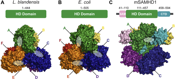
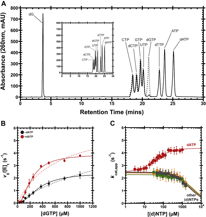
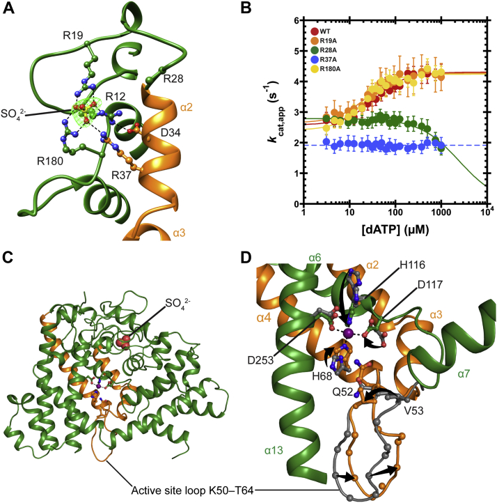
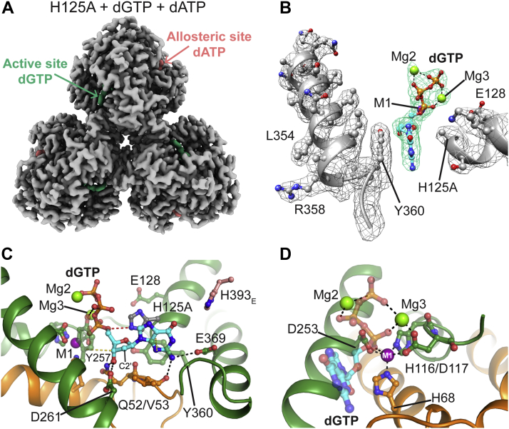
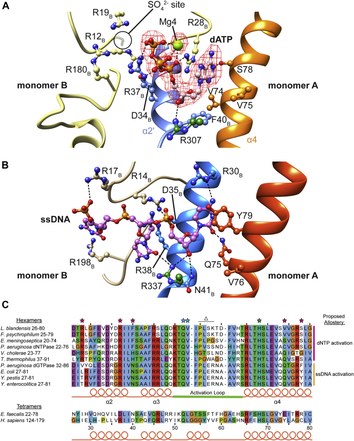
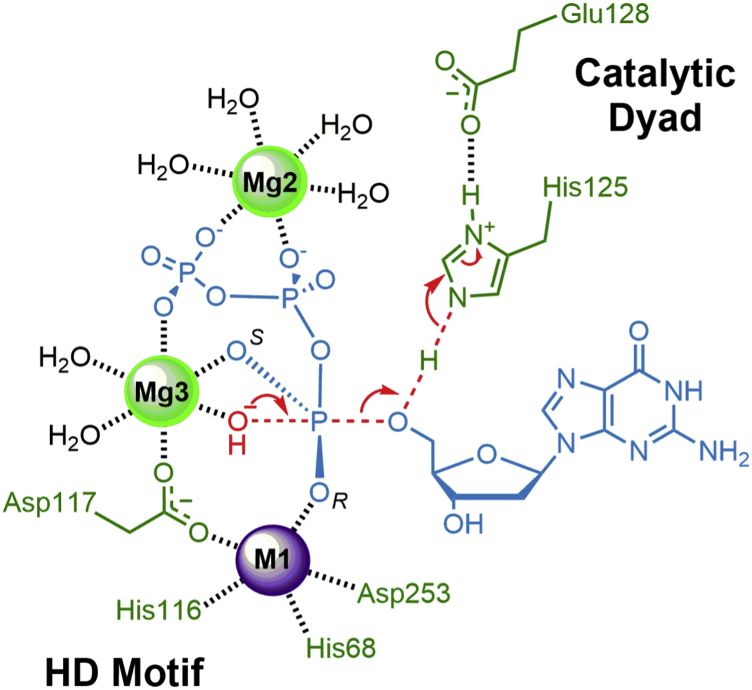
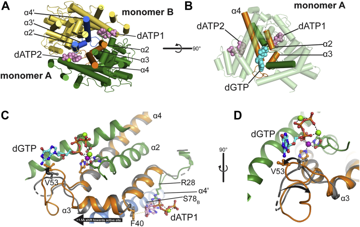
Deoxynucleoside triphosphate (dNTP) triphosphohydrolases (dNTPases) are important enzymes that may perform multiple functions in the cell, including regulating the dNTP pools and contributing to innate immunity against viruses. Among the homologs that are best studied are human sterile alpha motif and HD domain-containing protein 1 (SAMHD1), a tetrameric dNTPase, and the hexameric Escherichia coli dGTPase; however, it is unclear whether these are representative of all dNTPases given their wide distribution throughout life. Here, we investigated a hexameric homolog from the marine bacterium Leeuwenhoekiella blandensis, revealing that it is a dGTPase that is subject to allosteric activation by dATP, specifically. Allosteric regulation mediated solely by dATP represents a novel regulatory feature among dNTPases that may facilitate maintenance of cellular dNTP pools in L. blandensis. We present high-resolution X-ray crystallographic structures (1.80-2.26 Å) in catalytically important conformations as well as cryo-EM structures (2.1-2.7 Å) of the enzyme bound to dGTP and dATP ligands. The structures, the highest resolution cryo-EM structures of any SAMHD1-like dNTPase to date, reveal an intact metal-binding site with the dGTP substrate coordinated to three metal ions. These structural and biochemical data yield insights into the catalytic mechanism and support a conserved catalytic mechanism for the tetrameric and hexameric dNTPase homologs. We conclude that the allosteric activation by dATP appears to rely on structural connectivity between the allosteric and active sites, as opposed to the changes in oligomeric state upon ligand binding used by SAMHD1.

Keywords: allosteric regulation; cryo-EM; crystallography; enzyme mechanism; enzyme structure; nucleoside/nucleotide metabolism; structure–function.


Conflict of interest statement

Conflict of interest The authors declare that they have no conflicts of interest with the contents of this article.

Figures

Figure 1
Figure 1
Crystal structures of SAMHD1 homologs from three species. Generated in PyMOL. A, Leeuwenhoekiella blandensis homolog (PDB ID: 3BG2). B, Escherichia coli dGTPase (PDB ID: 4XDS). C, mouse SAMHD1 isoform 1 (PDB ID: 6BRG). A domain architecture diagram is shown for each crystal structure, with monomer A colored accordingly. All other monomers are labeled and colored for each homolog. The visible active-site cavities for each homolog are indicated by dashed ovals. We define the HD domain as the entire protein for E. coli dGTPase (505 amino acids in total) and L. blandensis dGTPase homolog (444 amino acids in total). For mouse SAMHD1, we define the HD domain boundaries by structural conservation with the hexameric homologs, whereas the start of the SAM domain and end of the CTD are defined here by the residues that are ordered in the crystal structure shown. The domain numbering is listed above the diagram (626 amino acids in total). CTD, C-terminal domain; HD, histidine–aspartate; PDB, Protein Data Bank; SAM, sterile alpha motif; SAMHD1, sterile alpha motif and HD domain–containing protein 1.
Figure 2
Figure 2
Leeuwenhoekiella blandensis dGTPase activity and allosteric activation.A, 1-h hydrolysis of a mixture containing all eight standard dNTPs and NTPs by the dNTPase homolog from L. blandensis. The eight (d)NTPs were reacted together (1 mM each), with the substrates and products separated by HPLC (solid line). A control reaction at time 0 is shown as a dashed line and separately in the inset, for clarity. B, v0/[E]tversus [dGTP] plots with or without 500 μM dATP. Data are presented as the mean and SEM of three independent experiments. The solid lines are the fit of Equation 1 to the data with nH = 2, whereas the dashed lines are the fit for nH = 1. For dGTP-only, kcat = 2.6 ± 0.1 s−1, KM = 390 ± 30 μM, and nH = 1.7 ± 0.1. With 500 μM dATP, kcat = 3.8 ± 0.1 s−1, KM = 224 ± 9 μM, and nH = 2.2 ± 0.1. C, dGTPase activity at 1 mM dGTP, titrating the remaining seven standard (d)NTPs. We refer to this substrate concentration as an apparent kcat (kcat,app) because it is above the threshold required to bring the turnover within 20% of the kcat, thereby approximating conditions with saturating substrate. Data are plotted as the mean and SEM of four independent experiments. Activation was observed for dATP (red circles), with K1/2 = 36 ± 3 μM and nH = 1.3 ± 0.1. Inhibitory effects are observed for dTTP (orange circles), dCTP (green circles), ATP (red squares), UTP (orange squares), CTP (green squares), and GTP (gray squares). The IC50s are estimated to be >2 mM. dNTP, deoxynucleoside triphosphate; dNTPase, dNTP triphosphohydrolase.
Figure 3
Figure 3
Crystal structures of Leeuwenhoekiella blandensis dGTPase.A, close-up of a sulfate-binding site showing the position of nearby arginine residues. Sulfate FoFc omit map contoured to 6σ (green mesh). Alpha helices α2 and α3 are important for our proposed allosteric activation model (discussed later), so they are colored orange and labeled for reference. Generated in Chimera. B, dATP titrations at 1 mM dGTP. Data are plotted as the mean and SEM of four independent experiments. Relative to the WT (red; data are replotted from Fig. 2C for reference), R19A (orange) and R180A (purple) have minimal effect on activation by dATP (K1/2 = 36 ± 3 μM, 23 ± 3 μM, and 21 ± 2 μM, respectively). R28A (green) and R37A (blue) eliminate activation by dATP, with dATP seemingly inhibiting R28A at higher concentrations and R37A activity entirely independent of dATP in the concentration range we tested. C, overview of monomer A. The orange helices (α2, α3, and α4) are colored as such for reference. Generated in PyMOL. D, the active site of R37A dGTPase. We use the R37A crystals for this analysis because loop residues 57 to 60 are disordered in the WT crystals, whereas we have a complete picture of the active-site loop in the R37A structures. Crystals were soaked with Mn2+ prior to freezing (solid, orange, and green) or frozen without soaking Mn2+ (gray). Structures were aligned by the HD motif in PyMOL (RMSD = 0.347 over the four α-carbon atoms). C-alpha atoms on the loop are shown as spheres. Arrows are highlighting residue displacement between the structures. Generated in PyMOL. HD, histidine–aspartate.
Figure 4
Figure 4
Cryo-EM structures of dGTPase with dGTP bound in the active site.A, H125A dGTPase (gray) bound to both dGTP substrate (teal) and dATP activator (pink) (th 0.11). The postprocessed map was generated using DeepEMhancer, whereas the image was generated in ChimeraX. B, electron density for alpha helices in the active site (gray) and the dGTP substrate (teal) in monomer A in the H125A + dGTP structure (th 0.1). The postprocessed map was generated using DeepEMhancer, whereas the image was generated in Chimera. C, monomer A from the H125A + dGTP structure highlighting the guanine- and 2′-deoxyribose-binding residues. The point of view is from the left side of B. dGTPase residues are colored green/orange as for previous figures, dGTP is cyan, and each is colored by heteroatom; M1 is purple, whereas Mg2 and Mg3 are green. Hydrogen bonds are black dashed lines, whereas van der Waals/stacking interactions with Y257 and Y360 are yellow dashed lines. H125 (gray) was added back in silico using the Mutagenesis Wizard in PyMOL with backbone-dependent rotamers imposed. The H125 nitrogen NE2 would be 3.1 Å from the 5′-oxygen atom and in line to protonate it during catalysis (red dashed line). D, monomer A from the H125A + dGTP structure highlighting the positions of the three metal ions. The point of view is approximately the position of the helix containing H125 and E128 in C. Black dashed lines are metal-ligand interactions.
Figure 5
Figure 5
Leeuwenhoekiella blandensis dGTPase allosteric site compared with Escherichia coli dGTPase. Generated in Chimera. A, dATP-binding residues from the H125A L. blandensis dGTPase + dGTP + dATP structure (th 0.11). Monomer A residues are colored green/orange, monomer B is colored blue/yellow, dATP is pink, M3 is green, and all are colored by heteroatom. There is additional density (at left) found in the same position as the sulfate ion from our crystal structures. The postprocessed map was generated using DeepEMhancer. B, the equivalent ssDNA-binding site in the E. coli dGTPase (Protein Data Bank [PDB] ID: 4X9E). C, alignment of dGTPase homologs, limited to the sequence containing the residues that we identified as important for allosteric activation in the L. blandensis homolog. The residues that directly interact with dATP are indicated by magenta stars. Residues that bind the dGTP 2′-deoxyribose are indicated by cyan stars. One of the two histidine–aspartate (HD) motif histidine residues is also present in this sequence, indicated by the green star. The residues that we deleted from the active-site loop in the Δ55 to 58 mutation are indicated by the Δ. Proposed allosteric site specificity for the hexameric homologs is labeled at right. The sequences included in the alignment are: L. blandensis dGTPase, EAQ51213; Flavobacterium psychrophilum dGTPase, WP_011962456; Enterococcus meningoseptica dNTPase, WP_078793617; Pseudomonas aeruginosa dNTPase (PA3043), NP_251733; Vibrio cholerae VC1979, Q9KQL9; Thermus thermophilus dGTPase (TT1383), BAC98488; P. aeruginosa dGTPase (PA1124), NP_249815.1; E. coli dGTPase, P15723; Yersinia pestis dGTPase, WP_002209369; Yersinia enterocolitica dGTPase, WP_075339176; Enterococcus faecalis dNTPase, WP_002357999; and Homo sapiens SAMHD1, Q9Y3Z3. SAMHD1, sterile alpha motif and HD domain–containing protein 1.
Figure 6
Figure 6
Transition state of the proposed reaction mechanism for SAMHD1-related dNTPase homologs, with residues numbered for the Leeuwenhoekiella blandensis dGTPase. The α-phosphate pro-RP and pro-SP oxygens are labeled R and S, accordingly. SAMHD1, sterile alpha motif and HD domain–containing protein 1.
Figure 7
Figure 7
Model for dGTPase activation by dATP. dATP binding to the allosteric site is proposed to activate dGTPase by way of the connections between the orange structural elements and the active-site loop. Generated in PyMOL. A, side-on view of the dimer containing monomers A and B. Monomer A is green, with helices α2, α3, and α4 colored orange. Monomer B is yellow, with helices α2', α3′, and α4′ colored blue. The dATP molecules are colored pink; the two M4 atoms are green spheres. B, overview of monomer A (transparent) highlighting the physical linkage between the active site and the two dATP sites (orange) and other important active-site structural elements (green), otherwise colored as in A with the dGTP colored cyan. C, displacement of helix α3 in dATP site 1 is coupled to the active-site loop movement in monomer A. dATP is pink, dGTP is cyan, and both are colored by heteroatom; M1 is shown as a purple sphere, whereas M2 and M3 are green spheres. R28 and F40 binding to dATP1 causes helix α3 to be displaced by >1.5 Å further into the active site (relative to the apo structure). There is a corresponding movement in the active-site loop displacing the V53 side chain (black arrow). The apo EM structure (gray) is aligned by the metal-coordinating residues of the monomer A histidine–aspartate (HD) motif. D, rotated 90° relative to C to highlight the loop movement in the active site (black arrows). V53 is “pushed” 3.6 Å further into the active site by α3 (as measured by the β-carbon), whereas P55 is “pulled” >5.5 Å in the opposite direction (as measured by the α-carbon).

Similar articles

Cited by

References

    1. Coggins S.A., Mahboubi B., Schinazi R.F., Kim B. Mechanistic crosstalk between DNA/RNA polymerase enzyme kinetics and nucleotide substrate availability in cells: implications for polymerase inhibitor discovery. J. Biol. Chem. 2020;295:13432–13443. - PMC - PubMed
    1. Yao N.Y., Schroeder J.W., Yurieva O., Simmons L.A., O’Donnell M.E. Cost of rNTP/dNTP pool imbalance at the replication fork. Proc. Natl. Acad. Sci. U. S. A. 2013;110:12942–12947. - PMC - PubMed
    1. Kumar D., Abdulovic A.L., Viberg J., Nilsson A.K., Kunkel T.A., Chabes A. Mechanisms of mutagenesis in vivo due to imbalanced dNTP pools. Nucl. Acids Res. 2011;39:1360–1371. - PMC - PubMed
    1. Ahluwalia D., Bienstock R.J., Schaaper R.M. Novel mutator mutants of E. coli nrdAB ribonucleotide reductase: insight into allosteric regulation and control of mutation rates. DNA Repair (Amst) 2012;11:480–487. - PMC - PubMed
    1. Ahluwalia D., Schaaper R.M. Hypermutability and error catastrophe due to defects in ribonucleotide reductase. Proc. Natl. Acad. Sci. U. S. A. 2013;110:18596–18601. - PMC - PubMed

Publication types

MeSH terms

Substances

Supplementary concepts