. 2022 Sep 1;1868(9):166447.
doi: 10.1016/j.bbadis.2022.166447. Epub 2022 May 25.

Methamphetamine induces thoracic aortic aneurysm/dissection through C/EBPβ

Affiliations

Methamphetamine induces thoracic aortic aneurysm/dissection through C/EBPβ

Bao-Ying Luo et al. Biochim Biophys Acta Mol Basis Dis. .

Abstract

Aims: Thoracic aortic aneurysm/dissection (TAAD) is a life-threatening disease with diverse clinical manifestations. Although the association between methamphetamine (METH) and TAAD is frequently observed, the causal relationship between METH abuse and aortic aneurysm/dissection has not been established. This study was designed to determine if METH causes aortic aneurysm/dissection and delineate the underlying mechanism.

Methods and results: A new TAAD model was developed by exposing METH to SD rats pre-treated with lysyl oxidase inhibitor β-aminopropionitrile (BAPN). Combination of METH and BAPN caused thoracic aortic aneurysm/dissection in 60% of rats. BAPN+METH significantly increased the expression and activities of both matrix metalloproteinase MMP2 and MMP9, consistent with the severe elastin breakage and dissection. Mechanistically, METH increased CCAAT-enhancer binding protein β (C/EBPβ) expression by enhancing mothers against decapentaplegic homolog 3 (Smad3) and extracellular regulated protein kinase (ERK1/2) signaling. METH also promoted C/EBPβ binding to MMP2 and MMP9 promoters. Blocking C/EBPβ significantly attenuated METH+BAPN-induced TAAD and MMP2/MMP9 expression. Moreover, BAPN+METH promoted aortic medial smooth muscle cell (SMC) apoptosis through C/EBPβ-mediated IGFBP5/p53/PUMA signaling pathways. More importantly, the expression of C/EBPβ, MMP2/MMP9, and apoptosis-promoting proteins was increased in the aorta of human patients with thoracic aortic dissection, suggesting that the mechanisms identified in animal study could be relevant to human disease.

Conclusions: Our study demonstrated that METH exposure has a casual effect on TAAD. C/EBPβ mediates METH-introduced TAAD formation by causing elastin breakage, medial cell loss and degeneration. Therefore, C/EBPβ may be a potential factor for TAAD clinical diagnosis or treatment.

Keywords: Apoptosis; C/EBPβ; MMP2/9; Methamphetamine; Thoracic aortic aneurysm/dissection.


Figures

Figure 1.
Figure 1.
METH exposure induced thoracic aortic aneurysm/dissection (TAAD) in BAPN-preconditioned SD rats. (A) Flowchart of animal drug delivery. (B) METH and/or BAPN treatment promoted the formation of TAAD in rat aorta. Red arrows indicate the TAAD in BAPN+METH-treated aorta. (C-D) Prevalence of TAAD in different groups. White area in BAPN+METH group in D denotes the TAAD with aortic rupture. The probability value was adjusted with the Bonferroni method for pairwise comparisons (p<0.05, n=3). (E) H&E and EVG staining of aorta sections from the rectangle areas shown in B. Dissection and elastin fragmentation were observed in METH-treated rats with BAPN-pretreatment.
Figure 2.
Figure 2.
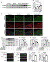
METH with BAPN-pretreatment increased MMP9/MMP2 production and activities in rat aorta tissues. (A) Western blot analyses of MMP9 and MMP2 protein expression in aortic tissues from rats treated with vehicle, BAPN, METH, or BAPN+METH. (B-C) Quantification of MMP9 and MMP2 expression by normalized to β-actin (*p<0.05, **p<0.01, ***p<0.005, n=3). (D) Immunostaining of MMP9 in rat aortic tissues exposed to BAPN and/or METH. (E) MMP9 and MMP2 activities in rat aortic tissues were detected by Gelatin-zymography electrophoresis. (F-G) Quantitative analyses of MMP9 and MMP2 activities by normalizing to pro-MMP9 and -MMP2, respectively (**p<0.01, ***p<0.005, n=3). All data were analyzed by factorial design ANOVA followed by LSD post hoc analyses.
Figure 3.
Figure 3.
C/EBPβ mediated METH-induced MMP9/MMP2 expression in aorta SMCs. (A) C/EBPβ expression in rat aorta with BAPN and/or METH treatment was examined by Western blot. (B) Quantification of C/EBPβ expression shown in A by normalized to β-actin. (*p<0.05, factorial design ANOVA followed by LSD post hoc analyses, n=3). (C) C/EBPβ expression in rat aorta was detected by IF staining. Arrows indicate SMCs with positive fluorescent signaling. (D) Aortic SMCs were transfected with control (Ctrl siRNA) or C/EBPβ siRNA (siC/EBPβ) and then treated with vehicle or 1.0 mM METH as indicated. C/EBPβ, MMP9, and MMP2 protein expression were detected by Western blot. (E-G) Quantification of protein expression shown in D by normalized to β-actin (*p<0.05, **p<0.01, ***p<0.005, ****p<0.001, one-way ANOVA followed by LSD post hoc analyses, n=3). (H-I) ChIP assay detected C/EBPβ binding to MMP9 and MMP2 promoters in a chromatin setting. (J-K) C/EBPβ binding enrichment on MMP9 and MMP2 promoters, respectively (***p<0.005, Student’s t test, n=3).
Figure 4.
Figure 4.
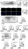
METH regulated C/EBPβ expression through Smad3 and ERK signaling. (A) Smad3 and ERK phosphorylation in aorta of rats treated with BAPN and/or METH were detected by Western blot. (B-C) Quantification of p-Smad3 and p-ERK levels shown in A by normalized to total Smad3 and ERK, respectively (*p<0.05, **p<0.01, ***p<0.005, factorial design ANOVA followed by LSD post hoc analyses, n=3). (D-G) Aortic SMCs were treated with Smad3 inhibitor SIS3 or ERK inhibitor U0126 for 30 min followed by vehicle or METH treatment for 8 hours as indicated. p-SMAD3, p-ERK1/2, and C/EBPβ levels were measured by Western blot. (E-F & H-I) Quantification of p-SMAD3, p-ERK and C/EBPβ levels shown in D or G, respectively (**p<0.01, ***p<0.005, ****p<0.001, one-way ANOVA followed by LSD post hoc analyses, n=3).
Figure 5.
Figure 5.
Silencing C/EBPβ inhibited BAPN+METH-induced TAAD and MMP9/2 production in rat aortas. Rats were treated with METH+BAPN along with adenoviral delivery of AD-shGFP or AD-shC/EBPβ as described in methods. (A) Flowchart of animal drug delivery. (B) Representative thoracic aorta with METH and/or BAPN treated. TAAD is indicated by the arrow in BAPN+METH-treated aorta. (C-D) Prevalence of TAAD in different groups, the probability value was adjusted with the Bonferroni method for pairwise comparisons (*p<0.05, n=3). (E) H&E and EVG staining of aorta sections from the rectangle areas shown in B. Aortic dissection and elastin fragmentation displayed in METH/BAPN-treated aorta were blocked in Ad-shC/EBPβ-treated aorta. (F) Western blot analyses of C/EBPβ, MMP9 and MMP2 protein expression in rat aortas with different treatments. (G-I) Quantification of C/EBPβ, MMP2, and MMP9 levels shown in F by normalized to β-actin (*p<0.05, **p<0.01, ***p<0.005, one-way ANOVA followed by LSD post hoc analyses, n=3).
Figure 6.
Figure 6.
C/EBPβ mediated METH-induced cell apoptosis by regulating the expression of IGFBP5-P53-PUMA cascade. (A) METH and METH+BAPN induced PARP and caspase3 cleavage in rat aortas as detected by Western blot analyses. (B-C) Quantification of cleaved PARP and caspase3 levels shown in A by normalized to β-actin (*p<0.05, **p<0.01, ***p<0.005, ****p<0.001, factorial design ANOVA followed by LSD post hoc analyses, n=3). (D) METH and METH+BAPN induced cells apoptosis in aorta media as shown by the TUNEL staining. (E) Quantification of apoptotic cells relative to the total cell numbers in aorta sections (***p<0.005, ****p<0.001, factorial design ANOVA followed by LSD post hoc analyses, n=3). (F) Aortic SMC were transfected with control (AD-shGFP) or C/EBPβ siRNA (AD-shC/EBPβ) followed by treatment with vehicle or BAPN+METH (1.0 mM) as indicated. PARP and caspase3 cleavage levels and IGFBP5/PUMA/p53 protein expression were detected by Western blot. (G-K) Quantification of IGFBP5/PUMA/p53 as well as cleaved PARP and caspase3 levels shown in F by normalized to β-actin (*p<0.05, **p<0.01, factorial design ANOVA followed by LSD post hoc analyses, n=3). (L-Q) Knockdown of C/EBPβ attenuated the expression of apoptosis-regulating proteins and decreased PARP and caspase3 cleavage. IGFBP5, p53, PUMA, cleaved-PARP, and cleaved-caspase3 levels were detected by Western blot and quantified by normalized to β-actin (**p<0.01, ***p<0.005, ****p<0.001, factorial design ANOVA followed by LSD post hoc analyses, n=3).
Figure 7.
Figure 7.
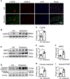
C/EBPβ expression was increased in human aorta with dissection. (A) Immunostaining of C/EBPβ in human thoracic aorta dissection specimens. Arrow indicates C/EBPβ-positive staining. (B, D, G) C/EBPβ, MMP9, MMP2 protein expression and the cleaved PARP and caspase3 levels in human aorta dissection specimens were detected by Western blot. (C, E, F, H, I) Quantification of C/EBPβ, MMP9, MMP2, cleaved PARP and cleaved caspase3 levels shown in B, D, and G by normalizing to P-actin, respectively (**p<0.01, Student’s t test, n=3).

References

    1. Senturk T, Antal A and Gunel T. Potential function of microRNAs in thoracic aortic aneurysm and thoracic aortic dissection pathogenesis. Mol Med Rep. 2019;20:5353–5362. - PubMed
    1. El-Hamamsy I and Yacoub MH. Cellular and molecular mechanisms of thoracic aortic aneurysms. Nat Rev Cardiol. 2009;6:771–86. - PubMed
    1. LeMaire SA and Russell L. Epidemiology of thoracic aortic dissection. Nat Rev Cardiol. 2011;8:103–13. - PubMed
    1. Wu D, Shen YH, Russell L, Coselli JS and LeMaire SA. Molecular mechanisms of thoracic aortic dissection. J Surg Res. 2013;184:907–24. - PMC - PubMed
    1. Liu R, Lo L, Lay AJ, Zhao Y, Ting KK, Robertson EN, Sherrah AG, Jarrah S, Li H, Zhou Z, Hambly BD, Richmond DR, Jeremy RW, Bannon PG, Vadas MA and Gamble JR. ARHGAP18 Protects Against Thoracic Aortic Aneurysm Formation by Mitigating the Synthetic and Proinflammatory Smooth Muscle Cell Phenotype. Circ Res. 2017;121:512–524. - PubMed

Publication types