. 2022 May 28;23(1):136.
doi: 10.1186/s12931-022-02056-z.

Imbalanced prostanoid release mediates cigarette smoke-induced human pulmonary artery cell proliferation

Affiliations

Imbalanced prostanoid release mediates cigarette smoke-induced human pulmonary artery cell proliferation

Abdullah A Alqarni et al. Respir Res. .

Abstract

Background: Pulmonary hypertension is a common and serious complication of chronic obstructive pulmonary disease (COPD). Studies suggest that cigarette smoke can initiate pulmonary vascular remodelling by stimulating cell proliferation; however, the underlying cause, particularly the role of vasoactive prostanoids, is unclear. We hypothesize that cigarette smoke extract (CSE) can induce imbalanced vasoactive prostanoid release by differentially modulating the expression of respective synthase genes in human pulmonary artery smooth muscle cells (PASMCs) and endothelial cells (PAECs), thereby contributing to cell proliferation.

Methods: Aqueous CSE was prepared from 3R4F research-grade cigarettes. Human PASMCs and PAECs were treated with or without CSE. Quantitative real-time RT-PCR and Western blotting were used to analyse the mRNA and protein expression of vasoactive prostanoid syhthases. Prostanoid concentration in the medium was measured using ELISA kits. Cell proliferation was assessed using the cell proliferation reagent WST-1.

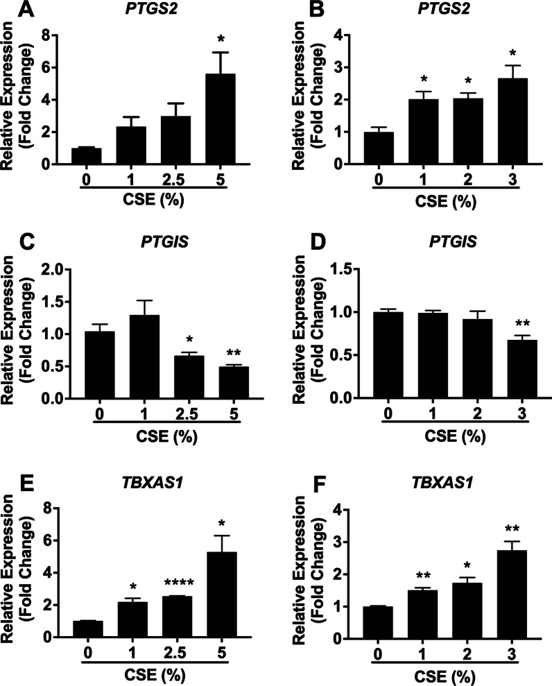
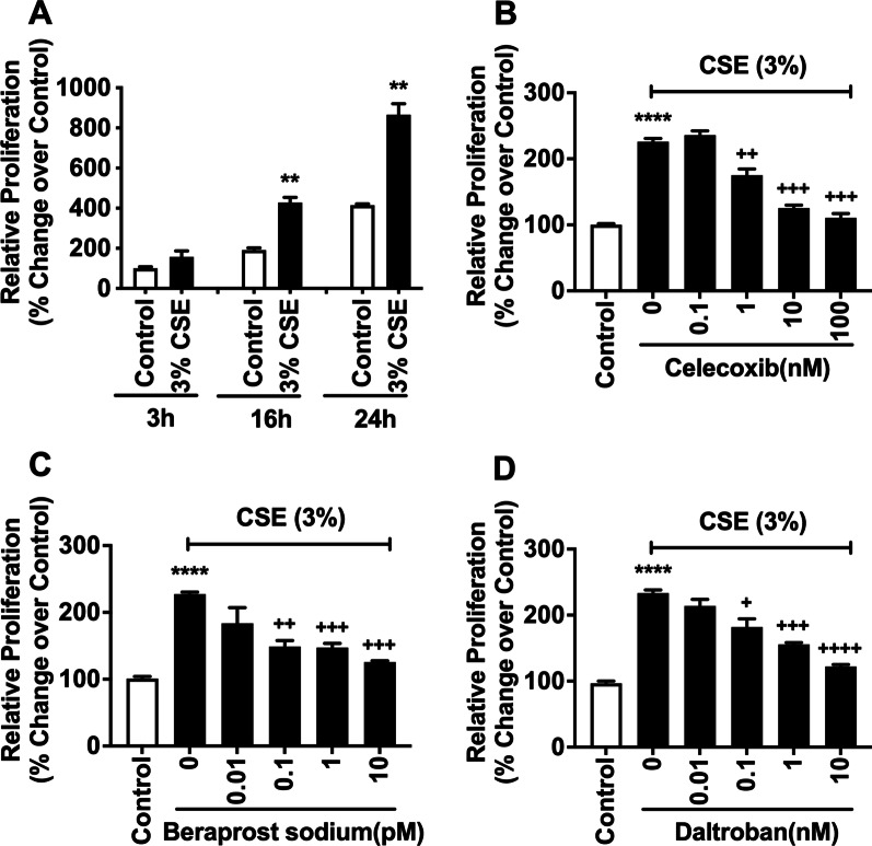
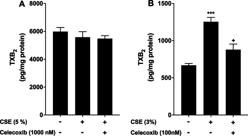
Results: We demonstrated that CSE induced the expression of cyclooxygenase-2 (COX-2), the rate-limiting enzyme in prostanoid synthesis, in both cell types. In PASMCs, CSE reduced the downstream prostaglandin (PG) I synthase (PGIS) mRNA and protein expression and PGI2 production, whereas in PAECs, CSE downregulated PGIS mRNA expression, but PGIS protein was undetectable and CSE had no effect on PGI2 production. CSE increased thromboxane (TX) A synthase (TXAS) mRNA expression and TXA2 production, despite undetectable TXAS protein in both cell types. CSE also reduced microsomal PGE synthase-1 (mPGES-1) protein expression and PGE2 production in PASMCs, but increased PGE2 production despite unchanged mPGES-1 protein expression in PAECs. Furthermore, CSE stimulated proliferation of both cell types, which was significantly inhibited by the selective COX-2 inhibitor celecoxib, the PGI2 analogue beraprost and the TXA2 receptor antagonist daltroban.

Conclusions: These findings provide the first evidence that cigarette smoke can induce imbalanced prostanoid mediator release characterized by the reduced PGI2/TXA2 ratio and contribute to pulmonary vascular remodelling and suggest that TXA2 may represent a novel therapeutic target for pulmonary hypertension in COPD.

Keywords: COPD; Cigarette smoke; Prostanoids; Pulmonary artery cell proliferation; Pulmonary hypertension.


Conflict of interest statement

All authors declare that they have no competing interests.

Figures

Fig. 1
Fig. 1
CSE treatment differentially modulates the mRNA expression of PTGS2, PTGIS, and TBXAS1 in human PASMCs and PAECs. Confluent human PASMCs (A, C, and E) and PAECs (B, D, and F) were treated with different concentrations of CSE for 72 h and 24 h, respectively. Total RNA was isolated, and mRNA levels of PTGS2 (A, B), PTGIS (C, D), TBXAS1 (E, F), and the internal control β2M were determined by real-time RT- PCR. Results are calculated as the ratio of target gene mRNA and β2M mRNA and are expressed as fold change over untreated (0% CSE) cells. Each data point represents mean ± SEM from three independent experiments using PASMCs (A, C, and E) from three different donors and PAECs (B, D, and F) from one donor. *P < 0.05, **P < 0.01, ****P < 0.0001 compared with corresponding untreated cells
Fig. 2
Fig. 2
CSE treatment differentially modulates the protein expression of key enzymes of prostanoid synthesis in human PASMCs and PAECs. Confluent human PASMCs (A, C, and E) and PAECs (B, D, and E) were treated with different concentrations of CSE for 72 h and 24 h, respectively. Total cell lysates were collected, and protein levels of COX-2 (A, B), PGIS (C, D), TXAS (E), and the internal control GAPDH were analyzed by Western blot. Total cell lysates from immortalized human bronchial epithelial (BEAS-2B) cells treated with or without CSE were used as a positive control (E). Irrelavant parts of the Western blotting images were cropped. Optical densitometry analysis of Western blotting bands was then conducted. Results are calculated as the ratio of target protein and GAPDH and are expressed as fold change over untreated (0% CSE) cells. Each data point represents mean ± SEM from three independent experiments using PASMCs (A, C, and E) from three different donors and PAECs (B, D, and E) from one donor. *P < 0.05, **P < 0.01, ***P < 0.001 compared with corresponding untreated cells
Fig. 3
Fig. 3
CSE treatment induces 6-keto PGF1α/TXB2 imbalance and elicits opposing effects on PGE2 production in human PASMCs and PAECs. Confluent human PASMCs (A, C, E, and G) and PAECs (B, D, F, and H) were treated with different concentrations of CSE for 72 h and 24 h, respectively. Medium was collected, and levels of PGE2 (A, B), 6-keto PGF1α (C, D) and TXB2 (E, F) were determined by ELISA and 6-keto PGF1α/TXB2 ratio (G and H) was calculated. Results were normalised with total cell protein and are expressed as pg/mg protein (AF) or 6-keto PGF1α/TXB2 ratio (G, H). Each data point represents mean ± SEM from three independent experiments using PASMCs (A, C, E and G) from three different donors and PAECs (B, D, F, and H) from one donor. *P < 0.05, **P < 0.01, ***P < 0.001 compared with corresponding untreated cells
Fig. 4
Fig. 4
CSE treatment downregulates the protein expression of mPGES-1 in human PASMCs. Confluent human PASMCs were treated with CSE (5%) for 72 h. Total cell lysates were collected, and protein levels of mPGES-1 and the internal control GAPDH were analyzed by Western blot. Irrelavant parts of the Western blotting images were cropped. Optical densitometry analysis of Western blotting bands was then conducted. Results are calculated as the ratio of mPGES-1 and GAPDH and are expressed as fold change over untreated (0% CSE) cells. Each data point represents mean ± SEM from three independent experiments using PASMCs from three different donors. *P < 0.05 compared with untreated cells
Fig. 5
Fig. 5
CSE treatment stimulates proliferation of human PASMCs, and celecoxib, beraprost sodium and daltroban inhibit the proliferation. (A) confluent human PASMCs were treated with or without CSE for up to 72 h, and then WST-1 assay was conducted. (BD) confluent human PASMCs were pre-treated with or without different concentrations of celecoxib (B), beraprost sodium (C) and daltroban (D) for 1 h before being treated with CSE for 24 h. WST-1 was then conducted. Data are expressed as relative proliferation (% change over control). Each data point represents mean ± SEM from three independent experiments using PASMCs from three different donors. *P < 0.05, ***P < 0.001 compared with control; +P < 0.05, ++P < 0.01, +++P < 0.001 compared with CSE alone
Fig. 6
Fig. 6
CSE treatment stimulates proliferation of human PAECs, and celecoxib, beraprost sodium, and daltroban potently inhibit the proliferation. (A) confluent human PAECs were treated with or without CSE for up to 24 h, and then WST-1 assay was conducted. (BD) confluent human PAECs were pre-treated with or without different concentrations of celecoxib (B), beraprost sodium (C), and daltroban (D) for 1 h before being treated with CSE for 24 h. WST-1 was then conducted. Data are expressed as relative proliferation (% change over control). Each data point represents mean ± SEM from three independent experiments using PAECs from one donor **P < 0.01, ****P < 0.0001 compared with control; +P < 0.05, ++P < 0.01, +++P < 0.001, ++++P < 0.0001 compared with CSE alone
Fig. 7
Fig. 7
Selective COX-2 inhibition reduces CSE-induced TXB2 production in human PAECs, but not in PASMCs. Confluent human PASMCs (A) and PAECs (B) were pre-treated with one concentration of celecoxib for 1 h before being treated with CSE for 24 h. Medium was collected, and TXB2 concentration was determined by ELISA. Results were normalised with total cell protein and are expressed as pg/mg protein. Each data point represents mean ± SEM from three independent experiments using PASMCs (A) from three different donors and PAECs (B) from one donor. ***P < 0.001 compared with corresponding untreated cells; +P < 0.05 compared with CSE alone

References

    1. Smith MC, Wrobel JP. Epidemiology and clinical impact of major comorbidities in patients with COPD. Int J Chron Obstruct Pulmon Dis. 2014;9:871–888. doi: 10.2147/COPD.S49621. - DOI - PMC - PubMed
    1. Bazan IS, Fares WH. Pulmonary hypertension: diagnostic and therapeutic challenges. Ther Clin Risk Manag. 2015;11:1221–1233. - PMC - PubMed
    1. Barst RJ. Pulmonary hypertension: past, present and future. Ann Thorac Med. 2008;3:1–4. doi: 10.4103/1817-1737.37832. - DOI - PMC - PubMed
    1. Galie N, Humbert M, Vachiery JL, Gibbs S, Lang I, Torbicki A, et al. 2015 ESC/ERS guidelines for the diagnosis and treatment of pulmonary hypertension: the Joint Task Force for the Diagnosis and Treatment of Pulmonary Hypertension of the European Society of Cardiology (ESC) and the European Respiratory Society (ERS): endorsed by: Association for European Paediatric and Congenital Cardiology (AEPC), International Society for Heart and Lung Transplantation (ISHLT) Eur Heart J. 2016;37:67–119. doi: 10.1093/eurheartj/ehv317. - DOI - PubMed
    1. Ghofrani HA, D'Armini AM, Grimminger F, Hoeper MM, Jansa P, Kim NH, et al. Riociguat for the treatment of chronic thromboembolic pulmonary hypertension. New Eng J Med. 2013;369:319–329. doi: 10.1056/NEJMoa1209657. - DOI - PubMed