. 2022 May 28;12(1):8983.
doi: 10.1038/s41598-022-12995-8.

AXL/CDCP1/SRC axis confers acquired resistance to osimertinib in lung cancer

Affiliations

AXL/CDCP1/SRC axis confers acquired resistance to osimertinib in lung cancer

Yuichi Murakami et al. Sci Rep. .

Abstract

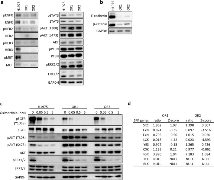
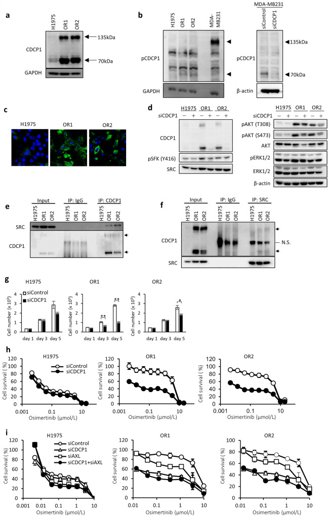
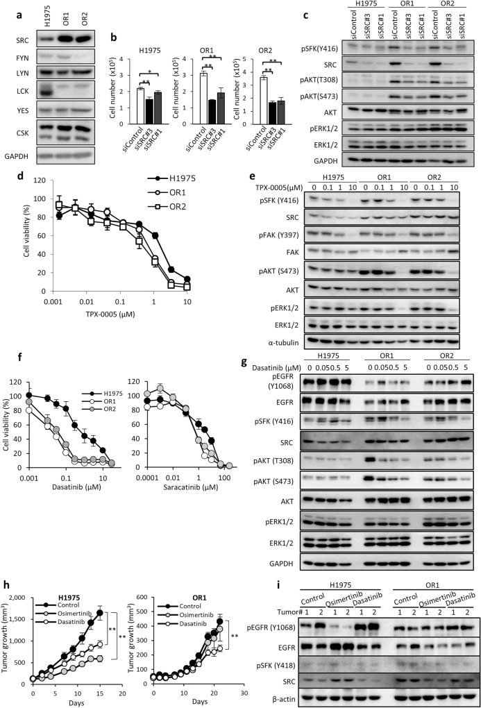
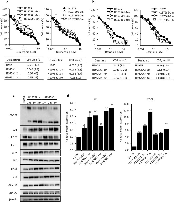
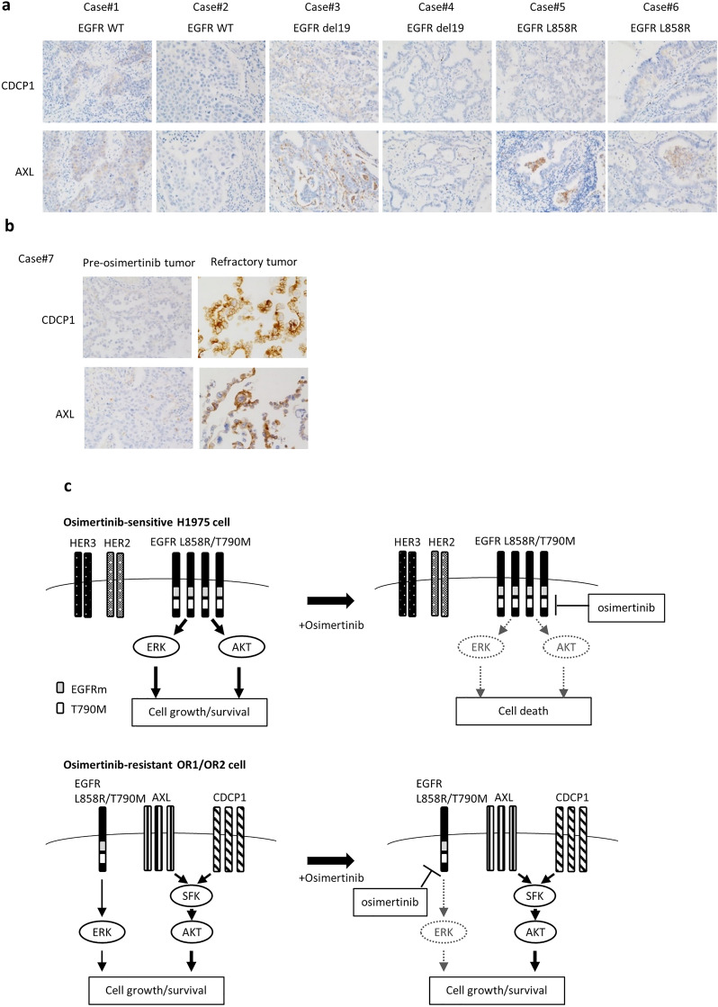
Osimertinib, a third-generation EGFR-TKI, has nowadays been applied to non-small cell lung cancer harboring activated EGFR mutation with or without T790M, but ultimately develop resistance to this drug. Here we report a novel mechanism of acquired resistance to osimertinib and the reversal of which could improve the clinical outcomes. In osimertinib-resistant lung cancer cell lines harboring T790M mutation that we established, expression of multiple EGFR family proteins and MET was markedly reduced, whereas expression of AXL, CDCP1 and SRC was augmented along with activation of AKT. Surprisingly, AXL or CDCP1 expression was induced by osimertinib in a time-dependent manner up to 3 months. Silencing of CDCP1 or AXL restored the sensitivity to osimertinib with reduced activation of SRC and AKT. Furthermore, silencing of both CDCP1 and AXL increased the sensitivity to osimertinib. Either silencing of SRC or dasatinib, a SRC family kinase (SFK) inhibitor, suppressed AKT phosphorylation and cell growth. Increased expression of AXL and CDCP1 was observed in refractory tumor samples from patients with lung cancer treated with osimertinib. Together, this study suggests that AXL/SFK/AKT and CDCP1/SFK/AKT signaling pathways play some roles in acquired osimertinib resistance of non-small cell lung cancer.


Conflict of interest statement

The authors declare no competing interests.

Figures

Figure 1
Figure 1
Osimertinib-resistant cell lines, OR1 and OR2, show reduced EGFR expression with constitutive activation of AKT. (a) Western blot analysis of EGFR and other molecules in lysates of H1975, OR1 and OR2 cells. (b) Western blot analysis of E-cadherin and β-catenin in lysates of H1975, OR1 and OR2 cells. (c) Expression levels of EGFR and other molecules analyzed after treatment with various doses of osimertinib for 6 h. (d) mRNA expression levels of 9 SFK genes by microarray analysis. Relative-fold changes (OR1 or OR2 vs H1975 cells) are presented with expression levels of each gene in H1975 cells normalized to 1.0. NULL, no significant expression.
Figure 2
Figure 2
Enhanced expression of AXL is closely associated with OR1 and OR2 cell growth. (a) Protein expression levels of AXL in H1975, OR1 and OR2 cells by western blot analysis. (b) AXL protein expression levels in three cell lines treated with control or AXL siRNA for 3 days. (c) Effects of AXL silencing on growth of three cell lines. Each value is the mean ± SD of triplicate dishes. *P < 0.05, **P < 0.01, two-tailed Student’s t-test. (d) Effects of AXL silencing on expression of pSFK, pAKT, and pERK in the three cell lines. Cell were treated with an AXL siRNA for 3 days followed by western blot analysis. (e) Combination of osimertinib and AXL siRNA on OR1 and OR2 cell growth. Cells were exposed to 0.4 μM osimertinib and/or AXL siRNA for 3 days. Each value is the mean ± SD of triplicate dishes. *P < 0.05, **P < 0.01. (f) Expression levels of pSFK, pAKT, and pERK were analyzed after treatment with various doses of R428 for 2 days. (g) Sensitivity to osimertinib with or without R428. Cells were exposed for 3 days and subjected to a water-soluble tetrazolium salt (WST) assay. Each value is the mean ± SD of triplicate dishes.
Figure 3
Figure 3
Enhanced expression of CDCP1 in OR1 and OR2 cells is closely associated with cell growth and activation of SFK and AKT. (a) Expression levels of CDCP1 protein in three cell lines by western blot analysis. Arrows indicate two forms (135 kDa and 70 kDa) of CDCP1 protein. (b) Expression levels of phosphorylated CDCP1 protein in three cell lines and effects of CDCP1 silencing on phosphorylation of CDCP1 by western blot analysis. Arrows indicate two forms (135 kDa and 70 kDa) of phosphorylated CDCP1 protein. MDA-MB231 cells were used as positive control. (c) Immunofluorescence analysis of CDCP1 in H1975, OR1, and OR2 cells. (d) Effects of CDCP1 silencing on activation of SFK (pSFK) and AKT (pAKT) by western blot analysis when treated with siRNA for 4 days. (e,f) Co-immunoprecipitation assays to determine interactions of SRC and CDCP1. SRC was detected after immunoprecipitation with anti-CDCP1 antibody (e), and CDCP1 was detected after immunoprecipitation with anti-SRC antibody (f). (g) Effects of CDCP1 silencing on growth of three cell lines. Each value is the mean ± SD of triplicate dishes. *P < 0.05, **P < 0.01, two-tailed Student’s t-test. (h) Effects of combination of osimertinib and CDCP1 silencing on cell growth in three cell lines. Cells were exposed to various doses of osimertinib with or without CDCP1 siRNA for 3 days, followed by WST assays. Each value is the mean ± SD of triplicate dishes. (i) Combination effect of both siRNAs of CDCP1 and AXL on cell growth in the presence of osimertinib. Cells were exposed to various doses of osimertinib for 3 days, followed by WST assays. Each value is the mean ± SD of triplicate dishes.
Figure 4
Figure 4
Osimertinib-resistant cells exhibit enhanced SRC expression, growth dependency on SRC, and collateral sensitivity to dasatinib. (a) Western blot analysis showing 6 SFK genes in three cell lines. (b) SRC knockdown induced cell growth inhibition at differential levels in H1975, OR1, and OR2 cells when treated with siRNAs for 4 days. Each bar shows the mean ± SD of triplicate wells, *P < 0.05, **P < 0.01, two-tailed Student’s t-test. (c) Effect of SRC siRNA on activation of SFK and AKT analyzed after treatment with SRC siRNAs for 3 days. GAPDH served as loading control. (d) Dose response curves of three cell lines to TPX-0005 when treated for 3 days. Each value is the mean ± SD of triplicate dishes. (e) Western blot analysis showing pSFK, pFAK, pAKT, and pERK in H1975, OR1 and OR2 cells when treated with TPX-0005 for 6 h. (f) Dose response curves of H1975, OR1, and OR2 cells to various doses of dasatinib and saracatinib when exposed for 3 days. Each value is the mean ± SD of triplicate dishes. (g) Western blot analysis showing EGFR and other molecules when treated with various doses of dasatinib for 6 h. (h) Animals bearing H1975 or OR1 xenografts were treated with osimertinib and dasatinib. Results are shown as mean tumor volumes and SD at each timepoint. **P < 0.01, two-tailed Student’s t-test. (i) Western blot analysis of H1975 and OR1 tumors after 15 (H1975) and 22 (OR1) days of treatment with osimertinib or dasatinib.
Figure 5
Figure 5
Acquisition of osimertinib resistance and expression of AXL and CDCP1 by H1975 cells exposed for 1, 2, and 3 months to osimertinib. (a,b) Cellular sensitivity to osimertinib (a) and dasatinib (b) of H1975#1, H1975#1-1m, H1975#1-2m and H1975#1-3m, and H1975#2, H1975#2-1m, H1975#2-2m and H1975#2-3m cells. (c) Expression of AXL, CDCP1, EGFR, pEGFR, pSFK, and pAKT detected by western blot analysis. (d) Expression of AXL and CDCP1 mRNA by RT-qPCR analysis. Each value is the mean ± SD of triplicate dishes. *P < 0.05, **P < 0.01 vs H1975 (one-way ANOVA, Dunnett’s test).
Figure 6
Figure 6
Expression of AXL and CDCP1 in lung tumors of patients. (a) IHC analysis of AXL or CDCP1 expression in surgically resected tumors of 6 patients (cases #1–6) before chemotherapy. (b) IHC analysis of AXL and CDCP1 in tumors before EGFR TKI treatment and after recurrence (cases #7). (c) Hypothetical model of how osimertinib resistance is acquired. In osimertinib-sensitive cells, the PI3K/AKT pathway driven by EGFR harboring mutEGFR and T790M is highly susceptible to osimertinib. By contrast, in osimertinib resistant cells, expression of the driver EGFR is reduced, accompanied by SRC activation together with enhanced expression of AXL and CDCP1. Enhanced expression of AXL and CDCP1 activates SFK and AKT signaling pathway, resulting in promotion of cell growth and survival.

References

    1. Mok TS, et al. Gefitinib or carboplatin-paclitaxel in pulmonary adenocarcinoma. N. Engl. J. Med. 2009;361:947–957. doi: 10.1056/NEJMoa0810699. - DOI - PubMed
    1. Rosell R, et al. Erlotinib versus standard chemotherapy as first-line treatment for European patients with advanced EGFR mutation-positive non-small-cell lung cancer (EURTAC): A multicentre, open-label, randomised phase 3 trial. Lancet Oncol. 2012;13:239–246. doi: 10.1016/S1470-2045(11)70393-X. - DOI - PubMed
    1. Sequist LV, et al. Phase III study of afatinib or cisplatin plus pemetrexed in patients with metastatic lung adenocarcinoma with EGFR mutations. J. Clin. Oncol. 2013;31:3327–3334. doi: 10.1200/JCO.2012.44.2806. - DOI - PubMed
    1. Oxnard GR, et al. New strategies in overcoming acquired resistance to epidermal growth factor receptor tyrosinekinase inhibitors in lung cancer. Clin. Cancer Res. 2011;17:5530–5537. doi: 10.1158/1078-0432.CCR-10-2571. - DOI - PMC - PubMed
    1. Yu HA, et al. Analysis of tumor specimens at the time of acquired resistance to EGFR-TKI therapy in 155 patients with EGFR-mutant lung cancers. Clin. Cancer Res. 2013;19:2240–2247. doi: 10.1158/1078-0432.CCR-12-2246. - DOI - PMC - PubMed

Publication types

MeSH terms