Review
. 2022 Jul-Aug;97(4):478-490.
doi: 10.1016/j.abd.2021.09.008. Epub 2022 May 25.

Update on the pathogenesis of vitiligo

Affiliations
Review

Update on the pathogenesis of vitiligo

Helena Zenedin Marchioro et al. An Bras Dermatol. 2022 Jul-Aug.

Abstract

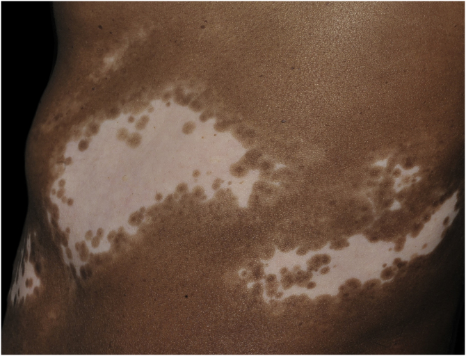
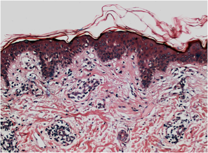
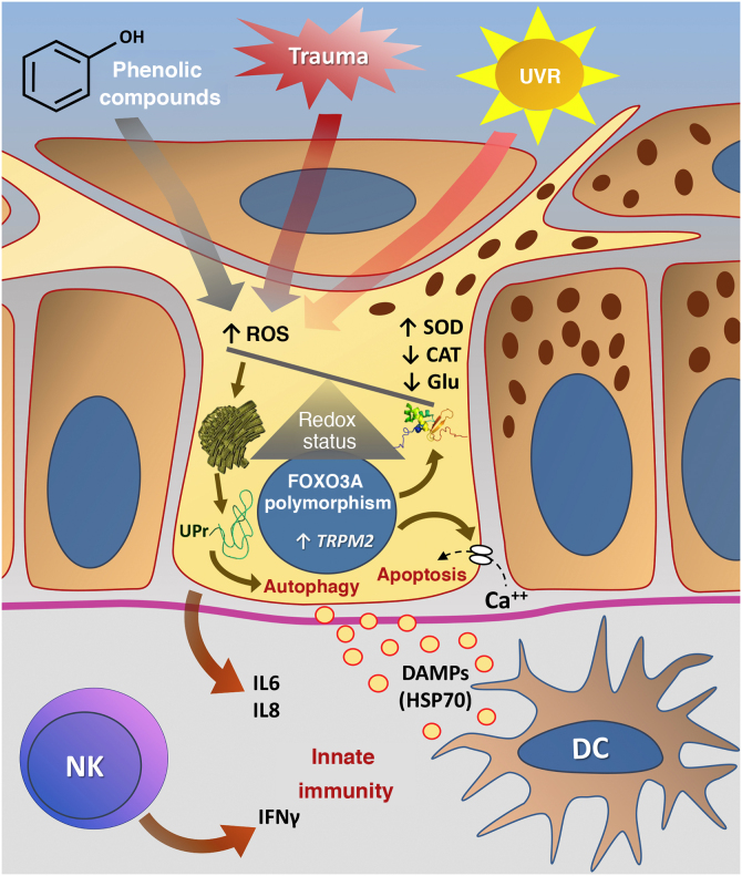
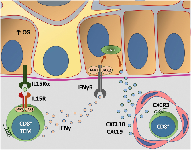
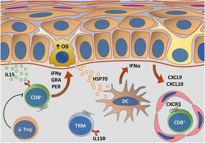
Vitiligo is a complex disease whose pathogenesis results from the interaction of genetic components, metabolic factors linked to cellular oxidative stress, melanocyte adhesion to the epithelium, and immunity (innate and adaptive), which culminate in aggression against melanocytes. In vitiligo, melanocytes are more sensitive to oxidative damage, leading to the increased expression of proinflammatory proteins such as HSP70. The lower expression of epithelial adhesion molecules, such as DDR1 and E-cadherin, facilitates damage to melanocytes and exposure of antigens that favor autoimmunity. Activation of the type 1-IFN pathway perpetuates the direct action of CD8+ cells against melanocytes, facilitated by regulatory T-cell dysfunction. The identification of several genes involved in these processes sets the stage for disease development and maintenance. However, the relationship of vitiligo with environmental factors, psychological stress, comorbidities, and the elements that define individual susceptibility to the disease are a challenge to the integration of theories related to its pathogenesis.

Keywords: Autoimmunity; Oxidative stress; Pigmentation; Vitiligo.


Figures

Figure 1
Figure 1
Segmental vitiligo on the flank of a dark-skinned patient. Achromic macula, with zosteriform distribution; the periphery of the lesion shows a leukomelanoderma band, with several follicular repigmentation points – Source: authors’ file.
Figure 2
Figure 2
Active vitiligo. Histopathology: perivascular lymphocytic infiltrate, with epidermal aggression and basal layer vacuolar degeneration foci (Hematoxylin &eosin, ×40). Picture is a courtesy of Dr. Lismary Mosque.
Figure 3
Figure 3
Representation of Oxidative Stress (OS) and activation of innate immunity in vitiligo. The effects of ultraviolet radiation (UVR), phenolic compounds and trauma (Köbner) increase the production of reactive oxygen species (ROS). In parallel, genetic predisposition (such as mutations in the FOXO3A gene) lead to the inefficiency of antioxidant mechanisms, observed by an increase in the superoxide dismutase (SOD) enzyme, reduction in catalase (CAT) and glutathione (Glu), causing an imbalance in the redox status. OS also causes an accumulation of defective proteins in the endoplasmic reticulum, resulting in a phenomenon called the response to unfolded proteins (UPr), contributing to the process of autophagy leading to the production of proinflammatory interleukins (IL6 and IL8). The increased expression of TRPM2 (transient receptor potential cation channel subfamily M member 2), also induced by OS, promotes an influx of calcium into the melanocyte, culminating in its apoptosis. The OS promotes the release of DAMPs (damage-associated molecular patterns), especially HSP70, which initiate the innate response from the activation of dendritic cells (DC) and the participation of NK cells (natural killer) – Source: the authors.
Figure 4
Figure 4
Representation of Interleukin (IL)-15 transpresentation in keratinocytes induced by oxidative stress (OS) and the interaction of interferon gamma (IFNγ) with the Janus kinase/signal transducers and transcription activators (JAK/STAT) pathway. The OS promotes the transpresentation of I-15 in keratinocytes through the binding of IL-15 to the heterodimeric IL-15 receptor (IL15R) on memory CD8 + T lymphocytes, consisting of CD122 and CD132, and to the I-15α receptor (IL15Rα) on keratinocytes (CD215). This process potentiates the activation of memory CD8 + T Cells and the production of inflammatory cytokines, such as IFNy, via JAK/STAT signaling (JAK1-STAT3 and JAK3-STAT5). The IFNγ/STAT1/CXCL10 axis conducts the autoimmune destruction of melanocytes. IFNγ signals through the IFNγ receptor (IFNγR) to stimulate JAK1/JAK2 and activate STAT1. The activation induces the production of CXCL9 and CXCL10, which signals through the CXCR3 receptor for the recruitment of more autoreactive CD8+ T cells – Source: the authors.
Figure 5
Figure 5
Representation of alterations related to adaptive immunity in vitiligo. Melanocytes affected by oxidative stress (OS) trigger the activation of innate immunity through the secretion of exosomes, which contain damage-associated molecular patterns (DAMPs), especially heat shock protein 70 (HSP70). HSP70 stimulates the secretion of IFNα by dendritic cells in the initial phase of disease progression, which induces the production of chemokines CXCL9 and CXCL10 by keratinocytes and the recruitment of T-cells expressing the CXCR3 receptor. CXCL10 has an effector action, while CXCL9 acts on the global recruitment of autoreactive CD8+ T-cells. Effector CD8+ T-cells are responsible for the destruction of melanocytes through the production of interferon gamma (IFNγ), release of granzymes and perforins, facilitated by T- regulatory (Treg) cell dysfunction. CD8+ tissue-resident memory T cells (TRM) develop after the onset of the T-cell-mediated immune response and are implicated in disease maintenance, being retained in the tissue due to IL15 transpresentation by keratinocytes – Source: the authors.

References

    1. Zhang Y., Cai Y., Shi M., Jiang S., Cui S., Wu Y., et al. The prevalence of vitiligo: a meta-analysis. PLoS One. 2016;11 - PMC - PubMed
    1. Castro CCS, Miot HA. Prevalence of vitiligo in Brazil—a population survey. Pigment Cell Melanoma Res. 2018;31:448–450. - PubMed
    1. Castro C.C.S., Nascimento L.L.M., Olandoski M., Mira M.T. A pattern of association between clinical form of vitiligo and disease-related variables in a Brazilian population. J Dermatol Sci. 2012;65:63–67. - PubMed
    1. Boza J.C., Giongo N., Machado P., Horn R., Fabbrin A., Cestari T. Quality of life impairment in children and adults with vitiligo: a cross-sectional study based on dermatology-specific and disease-specific quality of life instruments. Dermatology. 2016;232:619–625. - PubMed
    1. Dellatorre G., Antelo D.A.P., Bedrikow R.B., Cestari T.F., Follador I., Ramos D.G., et al. Consensus on the treatment of vitiligo — Brazilian Society of Dermatology. An Bras Dermatol. 2020;95:70–82. - PMC - PubMed