. 2022 Nov;32(4):477-487.
doi: 10.1016/j.zemedi.2022.04.004. Epub 2022 May 25.

Accuracy of MRI-CT registration in brain stereotactic radiotherapy: Impact of MRI acquisition setup and registration method

Affiliations

Accuracy of MRI-CT registration in brain stereotactic radiotherapy: Impact of MRI acquisition setup and registration method

Siti Masitho et al. Z Med Phys. 2022 Nov.

Abstract

Background: In MR-based radiotherapy (RT), MRI images are co-registered to the planning CT to leverage MR image information for RT planning. Especially in brain stereotactic RT, where typical CTV-PTV margins are 1-2 mm, high registration accuracy is critical. Several factors influence the registration accuracy, including the acquisition setup during MR simulation and the registration methods.

Purpose: In this work, the impact of the MRI acquisition setup and registration method was evaluated in the context of brain RT, both geometrically and dosimetrically.

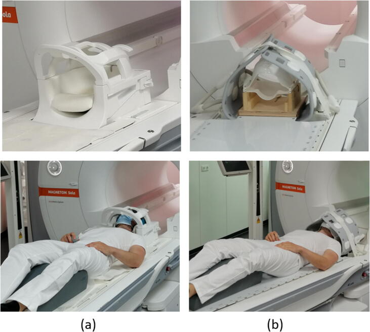
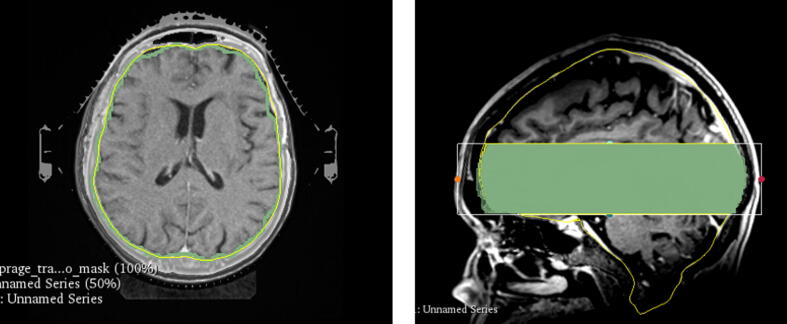
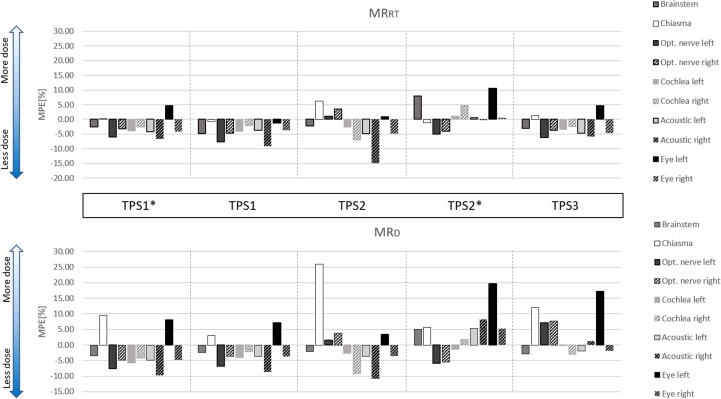
Methods and materials: MRI of 20 brain radiotherapy patients was acquired in two MRI acquisition setups (RT and diagnostic). Three different automatic registration tools provided by three treatment planning systems were used to rigidly register both MRIs and CT in addition to the clinical registration. Segmentation-based evaluation using Hausdorff Distance (HD)/Dice Similarity Coefficient and landmark-based evaluation were used as evaluation metrics. Dose-volume-histograms were evaluated for target volumes and various organs at risks.

Results: MRI acquisition in the RT setup provided a similar head extension as compared to the planning CT. The registration method had a more significant influence than the acquisition setup (Wilcoxon signed-rank test, p<0.05). When registering using a less optimal registration method, the RT setup improved the registration accuracy compared to the diagnostic setup (Difference: ΔMHD = 0.16 mm, ΔHDP95 = 0.64 mm, mean Euclidean distance (ΔmEuD) = 2.65 mm). Different registration methods and acquisition setups lead to the variation of the clinical DVH. Acquiring MRI in the RT setup can improve PTV and GTV coverage compared to the diagnostic setup.

Conclusions: Both MRI acquisition setup and registration method influence the MRI-CT registration accuracy in brain RT patients geometrically and dosimetrically. MR-simulation in the RT setup assures optimal registration accuracy if automatic registration is impaired, and therefore recommended for brain RT.

Keywords: Brain radiotherapy; Intracranial stereotactic radiotherapy; Magnetic-resonance imaging; Patient immobilization; Registration.


Conflict of interest statement

Declaration of Competing Interest Universitätsklinikum Erlangen, Strahlenklinik and Siemens Healthineers (Erlangen, Germany) have institutional research contracts not related to this specific work. Christoph Bert and Florian Putz act as speakers in training courses of Siemens Healthineers.

Figures

Figure 1
Figure 1
(a) The standard head coil (Head/Neck 20-channel coil) for MR-measurement in the diagnostic setup, and (b) the radiotherapy setup using an immobilization mask, a dedicated mask holder, an RT flat table top, and two 18-channel Ultraflex coils.
Figure 2
Figure 2
(a) A transversal view of the segmented brain of CT (yellow) and MRI (green), and (b) a sagittal view of the cropped brain segmentation used for segmentation-based evaluation. (For interpretation of the references to colour in this figure legend, the reader is referred to the web version of this article.)
Figure 3
Figure 3
Flow diagram of the RTSTRUCT transformation between different coordinate systems (CT, MRRT, MRD) for the dosimetry-based evaluation method. Different font colors/attributes for the registration (R) and transformation (T) refer to two clinical cases: red+underlined = if MRD was used for clinical plan and; black = if MRRT was used for the clinical plan. Shown in grey boxes are fixed processes independent of the MR used for the clinical case. (For interpretation of the references to colour in this figure legend, the reader is referred to the web version of this article.)
Figure 4
Figure 4
Rotation angle (roll-pitch-yaw) of MRI-CT rigid-registration, (a) MRI acquired in diagnostic setup, and (b) MRI acquired in radiotherapy setup.
Figure 5
Figure 5
The results of the segmentation-based registration accuracy evaluation: (a) Mean Hausdorff distance, (b) 95th percentile of Hausdorff Distance, (c) Dice Similarity Coefficient, and the results of the landmarks-based registration accuracy evaluation: (d) Mean Euclidean Distance of landmarks across 3 different treatment planning platforms (# with ROI) and 2 different MRI acquisition setups (RT: MRI acquisition in radiotherapy setup, D: MRI acquisition in diagnostic setup). Arrow direction points to better values. * on top of the graph indicates statistical significance between different setups within the same TPS, and (*) under the graph indicates statistical significance to the clinical registration (Wilcoxon signed-rank test, p<0.05).
Figure 6
Figure 6
Correlation matrix between mean Euclidean Distance (mEuD), mean Hausdorff Distance, 95th percentile of the Hausdorff Distance, and Dice Similarity Coefficient. Data points are linear fitted, where R is the correlation coefficient.
Figure 7
Figure 7
Box plots of dosimetric evaluation of MRI-CT registrations: (a) V80% of patients prescribed to 80% isodose (b) V95% of patients prescribed to 95% isodose, and (c) GTV coverage: across 3 different treatment planning platforms (# with ROI) and 2 different patient acquisition (RT: MRI acquisition in radiotherapy setup, D: MRI acquisition in diagnostic setup.). * on top of the graph indicates statistical significance between different setups within the same TPS, and (*) under the graph indicates statistical significance to the clinical registration (Wilcoxon signed-rank test, p < 0.05).
Figure 8
Figure 8
Mean percentage error (MPE) of Dmax and Dmean of OARs registered using various treatment planning systems (TPS) compared to clinical registration. (MRRT: MR acquisition in radiotherapy setup, MRD: MR acquisition in diagnostic setup.)

References

    1. Kocher M., et al. Stereotactic radiosurgery for treatment of brain metastases. A report of the DEGRO Working Group on Stereotactic Radiotherapy. Strahlenther Onkol. 2014;190(6):521–532. - PubMed
    1. Schell M.C., No R., et al. 054 -Stereotactic Radiosurgery. Am Assoc Phys Med. 1995
    1. Seung S.K., et al. American College of Radiology (ACR) and American Society for Radiation Oncology (ASTRO) Practice Guideline for the Performance of Stereotactic Radiosurgery (SRS) Am J Clin Oncol. 2013;36(3):310–315. - PMC - PubMed
    1. Guckenberger M., et al. Definition and quality requirements for stereotactic radiotherapy: consensus statement from the DEGRO/DGMP Working Group Stereotactic Radiotherapy and Radiosurgery. Strahlenther Onkol. 2020 - PMC - PubMed
    1. Salkeld A.L., et al. Changes in brain metastasis during radiosurgical planning. Int J Radiat Oncol Biol Phys. 2018;102(4):727–733. - PubMed

MeSH terms