

Automatic lesion segmentation using atrous convolutional deep neural networks in dermoscopic skin cancer images
- PMID: 35644612
- PMCID: PMC9148511
- DOI: 10.1186/s12880-022-00829-y
Automatic lesion segmentation using atrous convolutional deep neural networks in dermoscopic skin cancer images
Abstract
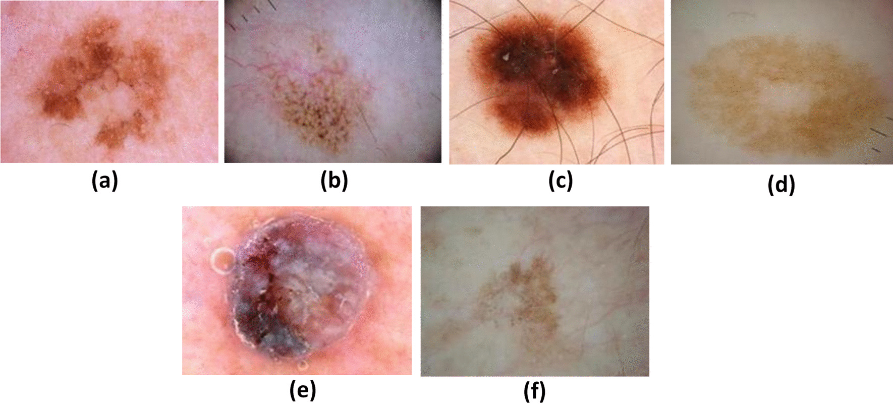
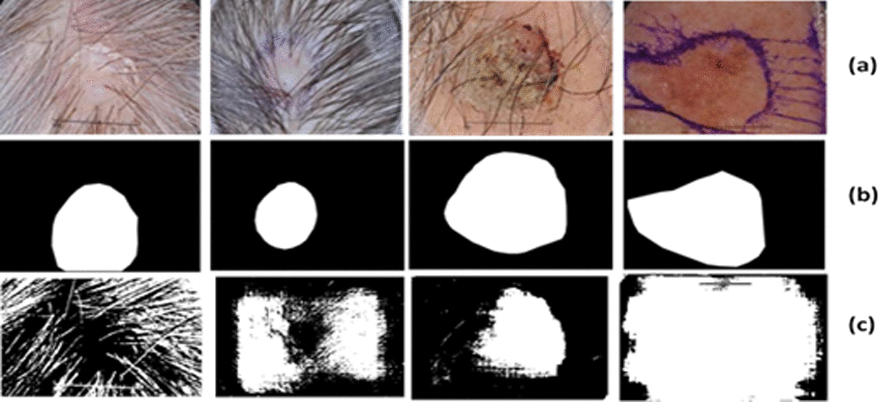
Background: Melanoma is the most dangerous and aggressive form among skin cancers, exhibiting a high mortality rate worldwide. Biopsy and histopathological analysis are standard procedures for skin cancer detection and prevention in clinical settings. A significant step in the diagnosis process is the deep understanding of the patterns, size, color, and structure of lesions based on images obtained through dermatoscopes for the infected area. However, the manual segmentation of the lesion region is time-consuming because the lesion evolves and changes its shape over time, making its prediction challenging. Moreover, it is challenging to predict melanoma at the initial stage as it closely resembles other skin cancer types that are not malignant as melanoma; thus, automatic segmentation techniques are required to design a computer-aided system for accurate and timely detection.
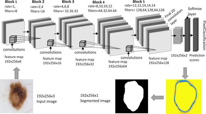
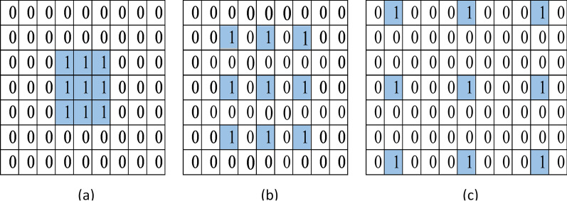
Methods: As deep learning approaches have gained significant attention in recent years due to their remarkable performance, therefore, in this work, we proposed a novel design of a convolutional neural network (CNN) framework based on atrous convolutions for automatic lesion segmentation. This architecture is built based on the concept of atrous/dilated convolutions which are effective for semantic segmentation. A deep neural network is designed from scratch employing several building blocks consisting of convolutional, batch normalization, leakyReLU layer, and fine-tuned hyperparameters contributing altogether towards higher performance.
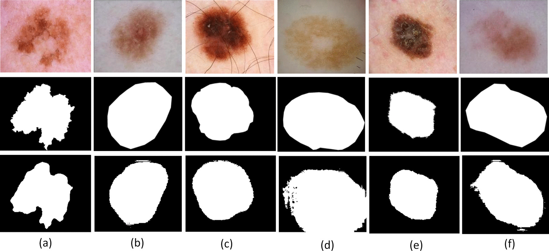
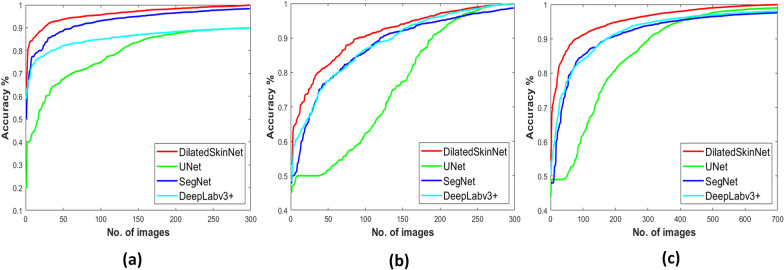
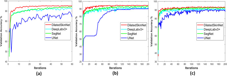
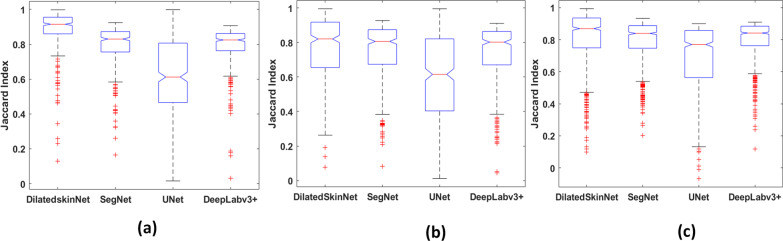
Conclusion: The network was tested on three benchmark datasets provided by International Skin Imaging Collaboration (ISIC), i.e., ISIC 2016, ISIC 2017, and ISIC 2018. The experimental results showed that the proposed network achieved an average Jaccard index of 90.4% on ISIC 2016, 81.8% on ISIC 2017, and 89.1% on ISIC 2018 datasets, respectively which is recorded as higher than the top three winners of the ISIC challenge and other state-of-the-art methods. Also, the model successfully extracts lesions from the whole image in one pass in less time, requiring no pre-processing step. The conclusions yielded that network is accurate in performing lesion segmentation on adopted datasets.
Keywords: CNN; Deep learning; Lesion segmentation; Skin cancer.
© 2022. The Author(s).
Conflict of interest statement
The authors declare no competing interest.
Figures
Similar articles
-
Melanoma segmentation using deep learning with test-time augmentations and conditional random fields.Sci Rep. 2022 Mar 10;12(1):3948. doi: 10.1038/s41598-022-07885-y. Sci Rep. 2022. PMID: 35273282 Free PMC article.
-
Efficient skin lesion segmentation using separable-Unet with stochastic weight averaging.Comput Methods Programs Biomed. 2019 Sep;178:289-301. doi: 10.1016/j.cmpb.2019.07.005. Epub 2019 Jul 8. Comput Methods Programs Biomed. 2019. PMID: 31416556
-
DSNet: Automatic dermoscopic skin lesion segmentation.Comput Biol Med. 2020 May;120:103738. doi: 10.1016/j.compbiomed.2020.103738. Epub 2020 Apr 2. Comput Biol Med. 2020. PMID: 32421644
-
Deep Learning Approaches Towards Skin Lesion Segmentation and Classification from Dermoscopic Images - A Review.Curr Med Imaging. 2020;16(5):513-533. doi: 10.2174/1573405615666190129120449. Curr Med Imaging. 2020. PMID: 32484086 Review.
-
Diagnosis and prognosis of melanoma from dermoscopy images using machine learning and deep learning: a systematic literature review.BMC Cancer. 2025 Jan 13;25(1):75. doi: 10.1186/s12885-024-13423-y. BMC Cancer. 2025. PMID: 39806282 Free PMC article.
Cited by
-
EMCAH-Net: an effective multi-scale context aggregation hybrid network for medical image segmentation.Quant Imaging Med Surg. 2025 Apr 1;15(4):3064-3083. doi: 10.21037/qims-24-1983. Epub 2025 Mar 28. Quant Imaging Med Surg. 2025. PMID: 40235751 Free PMC article.
-
Medical Image Segmentation with Learning Semantic and Global Contextual Representation.Diagnostics (Basel). 2022 Jun 25;12(7):1548. doi: 10.3390/diagnostics12071548. Diagnostics (Basel). 2022. PMID: 35885454 Free PMC article.
-
A Survey on Human Cancer Categorization Based on Deep Learning.Front Artif Intell. 2022 Jun 27;5:884749. doi: 10.3389/frai.2022.884749. eCollection 2022. Front Artif Intell. 2022. PMID: 35832207 Free PMC article. Review.
-
Enhanced skin cancer diagnosis using optimized CNN architecture and checkpoints for automated dermatological lesion classification.BMC Med Imaging. 2024 Aug 2;24(1):201. doi: 10.1186/s12880-024-01356-8. BMC Med Imaging. 2024. PMID: 39095688 Free PMC article.
-
YOLOSAMIC: A Hybrid Approach to Skin Cancer Segmentation with the Segment Anything Model and YOLOv8.Diagnostics (Basel). 2025 Feb 16;15(4):479. doi: 10.3390/diagnostics15040479. Diagnostics (Basel). 2025. PMID: 40002630 Free PMC article.
References
-
- Bogo F, Peruch F, Fortina A, Peserico E, Celebi M, Mendonca T, Marques J. Where’s the lesion? Variability in human and automated segmentation of dermoscopy images of melanocytic skin lesions. Boca Raton: CRC Press; 2015.
-
- Massey University of New Zealand: Environmental Health Indicators New Zealand. https://www.ehinz.ac.nz/indicators/uv-exposure/melanoma/. Accessed 10 July 2020.
-
- The Skin Cancer Foundation. Skin cancer facts and statistics. https://www.skincancer.org/skin-cancer-information/skin-cancer-facts/. Accessed Jan 2021.
MeSH terms
LinkOut - more resources
Full Text Sources
Medical
Research Materials
