Review
. 2022 Jul;82(5):408-427.
doi: 10.1002/dneu.22879. Epub 2022 May 29.

Evaluation of advances in cortical development using model systems

Affiliations
Review

Evaluation of advances in cortical development using model systems

Patricia R Nano et al. Dev Neurobiol. 2022 Jul.

Abstract

Compared with that of even the closest primates, the human cortex displays a high degree of specialization and expansion that largely emerges developmentally. Although decades of research in the mouse and other model systems has revealed core tenets of cortical development that are well preserved across mammalian species, small deviations in transcription factor expression, novel cell types in primates and/or humans, and unique cortical architecture distinguish the human cortex. Importantly, many of the genes and signaling pathways thought to drive human-specific cortical expansion also leave the brain vulnerable to disease, as the misregulation of these factors is highly correlated with neurodevelopmental and neuropsychiatric disorders. However, creating a comprehensive understanding of human-specific cognition and disease remains challenging. Here, we review key stages of cortical development and highlight known or possible differences between model systems and the developing human brain. By identifying the developmental trajectories that may facilitate uniquely human traits, we highlight open questions in need of approaches to examine these processes in a human context and reveal translatable insights into human developmental disorders.

Keywords: cortical development; human; model organisms.


Conflict of interest statement

CONFLICT OF INTEREST

The authors declare no conflicts of interest.

Figures
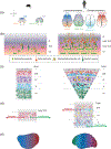
FIGURE 1
FIGURE 1
Model organisms, including mice, highlight principles of cortical development, and their modifications in the human cortex. (a) Many patterning mechanisms dissected in seminal mouse model experiments are conserved in the developing human cortex. However, humans also display unique expression patterns, such as the coexpression of SP8 and COUP-TF1 transcription factors or the graded expression of CNTNAP2. (b) Both mice and human neurogenesis in the neocortex rely on the production of newborn neurons from progenitors in the neurogenic ventricular zone (VZ) and subventricular zones (SVZ), followed by their migration through the intermediate zone (IZ) to the cortical surface. However, humans have progenitors with greater proliferative capacity, expanded subventricular zones, and a specialized progenitor cell type called the outer radial glia that enables cortical expansion. (c) In both mice and humans, newborn neurons migrate radially to generate a six-layered cortex, segregated largely by cell type. However, human cell types show differences in laminar identities, such as the presence of neurons homologous to mouse layer V neurons in the human layer III (light blue cells) and human-specific neuronal subtypes (yellow and magenta). The human cortex also displays an expansion in the upper layers. (d) Although studies to delineate the conservation of projection specification principles between humans and mice are still ongoing, primates have been shown to display stricter rules in the laminar segregation of feedback and feedforward projections. (e) Recent transcriptomic studies have shown that both human and mouse cortical areas are defined by graded differences in transcriptomic identity—variations that are exacerbated in the human cortex

Similar articles

Cited by

References

    1. Agmon A, Yang LT, O’Dowd DK, & Jones EG (1993). Organized growth of thalamocortical axons from the deep tier of terminations into layer IV of developing mouse barrel cortex. Journal of Neuroscience, 13(12), 5365–5382. - PMC - PubMed
    1. Alcamo EA, Chirivella L, Dautzenberg M, Dobreva G, Fariñas I, Grosschedl R, & McConnell SK (2008). Satb2 regulates callosal projection neuron identity in the developing cerebral cortex. Neuron, 57(3), 364–377. - PubMed
    1. Alexander L, Gaskin PL, Sawiak SJ, Fryer TD, Hong YT, Cockcroft GJ, Clarke HF, & Roberts AC (2019). Fractionating blunted reward processing characteristic of anhedonia by over-activating primate subgenual anterior cingulate cortex. Neuron, 101(2), 307–320.e306. - PMC - PubMed
    1. Alexander L, Wood CM, Gaskin PL, Sawiak SJ, Fryer TD, Hong YT, McIver L, Clarke HF, & Roberts AC (2020). Over-activation of primate subgenual cingulate cortex enhances the cardiovascular, behavioral and neural responses to threat. Nature Communications, 11(1), 5386. - PMC - PubMed
    1. Allman JM, Tetreault NA, Hakeem AY, Manaye KF, Semendeferi K, Erwin JM, Park S, Goubert V, & Hof PR (2010). The von Economo neurons in frontoinsular and anterior cingulate cortex in great apes and humans. Brain Structure and Function, 214(5), 495–517. - PubMed

Publication types