. 2022 Apr 20;7(2):49.
doi: 10.3390/biomimetics7020049.

Modification of Zirconia Implant Surfaces by Nd:YAG Laser Grooves: Does It Change Cell Behavior?

Affiliations

Modification of Zirconia Implant Surfaces by Nd:YAG Laser Grooves: Does It Change Cell Behavior?

Mariana Brito da Cruz et al. Biomimetics (Basel). .

Abstract

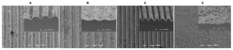
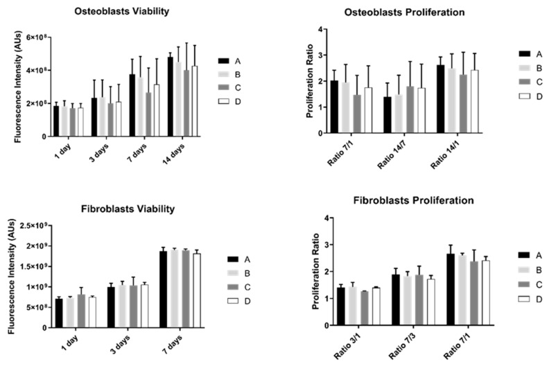
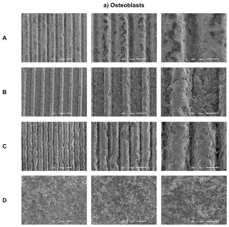
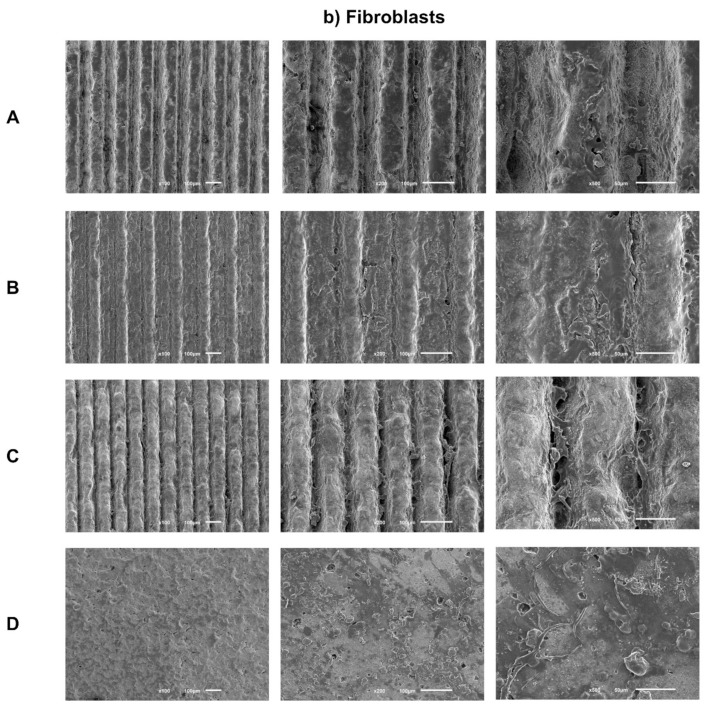
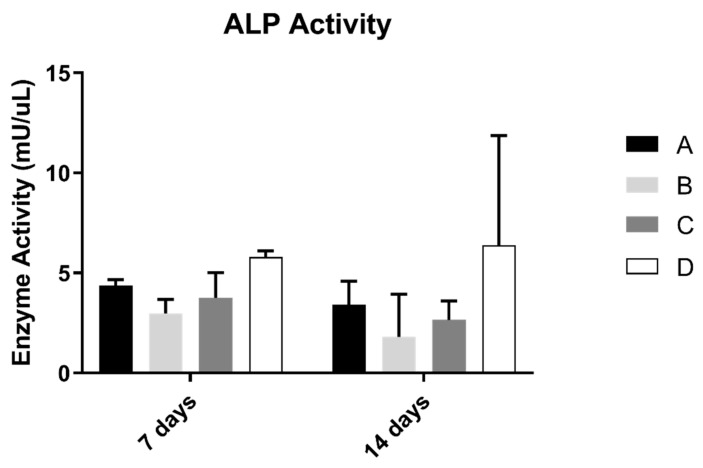
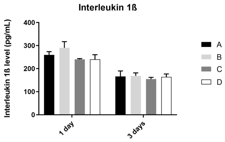
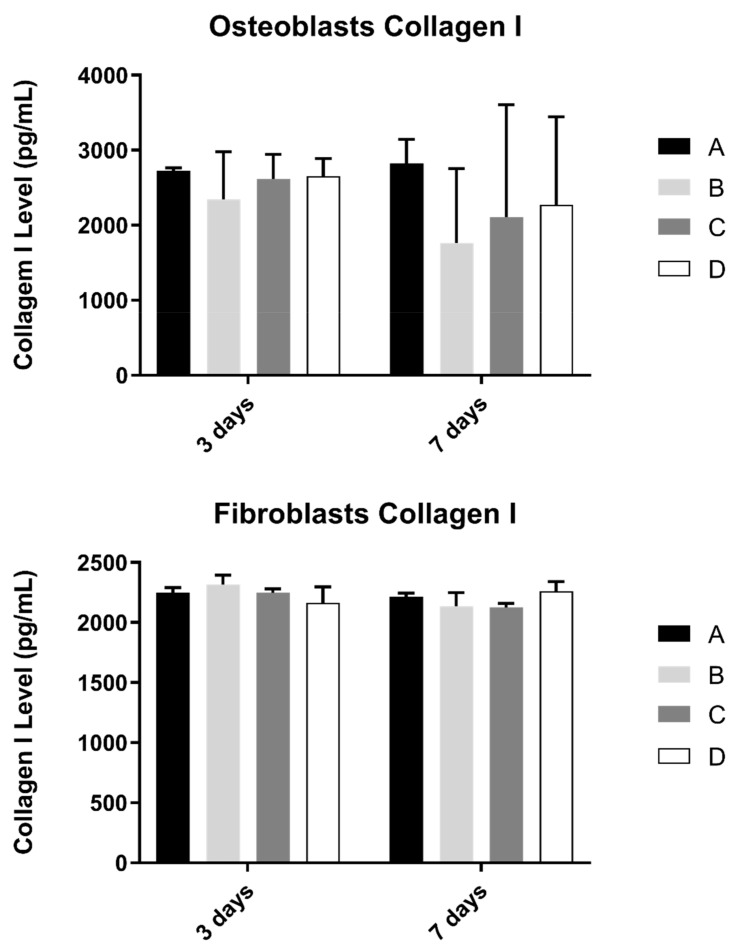
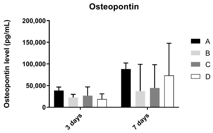
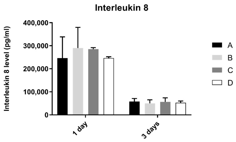
The aim of this study was to evaluate gingival fibroblasts and human osteoblasts' response to textured Nd:YAG laser microgrooves, with different dimensions, on zirconia implant surfaces. A total of 60 zirconia disks (8 mm in diameter and 2 mm in thickness) were produced and divided between four study groups (N = 15): three laser-textured (widths between 125.07 ± 5.29 μm and 45.36 ± 2.37 μm and depth values from 50.54 ± 2.48 μm to 23.01 ± 3.79 μm) and a control group without laser treatment. Human osteoblasts and gingival fibroblasts were cultured on these surfaces for 14 days. FEG-SEM (Field Emission Gun-Scanning Electron Microscope) images showed cellular adhesion at 24 h, with comparable morphology in all samples for both cell types. A similar cell spreading within the grooves and in the space between them was observed. Cell viability increased over time in all study groups; however, no differences were found between them. Additionally, proliferation, ALP (Alkaline phosphatase) activity, collagen type I, osteopontin and interleukin levels were not significantly different between any of the study groups for any of the cell types. Analysis of variance to compare parameters effect did not reveal statistically significant differences when comparing all groups in the different tests performed. The results obtained revealed similar cell behavior based on cell viability and differentiation on different microtopographic laser grooves, compared to a microtopography only established by sandblasting and acid-etching protocol, the reference surface treatment on zirconia dental implants.

Keywords: fibroblasts; laser; osteoblasts; zirconia.


Conflict of interest statement

The authors declare no conflict of interest.

Figures

Figure 1
Figure 1
The FEG-SEM micrographs taken after the surface treatment of all samples (100× magnification).
Figure 2
Figure 2
Graphs showing osteoblasts and fibroblasts’ viability as a mean of the registered fluorescence intensity of resorufin expressed in arbitrary units (N = 15) for each study group (as described in Table 1). Error bars represent a standard deviation. A Kruskal–Wallis, followed by Dunn’s test, was used for comparisons between study groups. Statistical significance was defined as: p < 0.05.
Figure 3
Figure 3
(a) Representative FEG-SEM micrographs of osteoblasts cultured on test samples and control surface after 1 day, with 100, 200, and 500× magnifications for each study group (as described in Table 1). (b) FEG-SEM micrographs of fibroblasts cultured on test samples and the control surface after 1 day, with 100, 200, and 500× magnifications for each study group (as described in Table 1).
Figure 3
Figure 3
(a) Representative FEG-SEM micrographs of osteoblasts cultured on test samples and control surface after 1 day, with 100, 200, and 500× magnifications for each study group (as described in Table 1). (b) FEG-SEM micrographs of fibroblasts cultured on test samples and the control surface after 1 day, with 100, 200, and 500× magnifications for each study group (as described in Table 1).
Figure 4
Figure 4
Mean alkaline phosphatase activity (mU/µL) measured in osteoblasts cultured for 7 and 14 days (N = 5), presented as the mean, for each study group (as described in Table 1). A Kruskal–Wallis test, followed by Dunn’s post-hoc tests, was used for comparison between study groups. Error bars represent the standard deviation. Statistical significance: p< 0.05.
Figure 5
Figure 5
Mean concentration of interleukin 1ß (pg/mL) measured in osteoblasts culture in all groups at 1 and 3 days (N = 4), presented as mean and standard deviation, for each study group (as described in Table 1). The Kruskal–Wallis test, followed by Dunn’s post-hoc tests, was used for comparison between study groups. Error bars represent standard deviation. Statistical significance: p< 0.05.
Figure 6
Figure 6
Collagen type I concentration (in pg/mL) obtained in osteoblasts and fibroblasts in all study groups at 3 and 7 days (N = 4), presented as mean and standard deviation (error bars), for each study group (as described in Table 1). The Kruskal–Wallis test, followed by Dunn’s post-hoc tests, was used for comparison between study groups. Statistical significance: p < 0.05.
Figure 7
Figure 7
Osteopontin concentration (in pg/mL) obtained in osteoblast culture at 3 and 7 days of cell culture (N = 4), presented as mean and standard deviation (error bars), for each study group (as described in Table 1). A Kruskal–Wallis test, followed by Dunn’s post-hoc tests, was used for comparison between study groups. Statistical significance: p < 0.05.
Figure 8
Figure 8
Interleukin 8 concentration (pg/mL) obtain in fibroblasts at 1 and 3 days (N = 4), presented as mean and standard deviation (error bars), for each study group (as described in Table 1). A Kruskal–Wallis test, followed by Dunn’s post-hoc tests, was used for comparison between study groups. Statistical significance: p < 0.05.

References

    1. Hwang J.W., Lee E.U., Lee J.S., Jung U.W., Lee I.S., Choi S.H. Dissolution behavior and early bone apposition of calcium phosphate-coated machined implants. J. Periodontal Implant Sci. 2013;43:291–300. doi: 10.5051/jpis.2013.43.6.291. - DOI - PMC - PubMed
    1. Guillaume B. Dental implants: A review. Morphologie. 2016;100:189–198. doi: 10.1016/j.morpho.2016.02.002. - DOI - PubMed
    1. Rupp F., Liang L., Geis-Gerstorfer J., Scheideler L., Hüttig F. Surface characteristics of dental implants: A review. Dent. Mater. 2018;34:40–57. doi: 10.1016/j.dental.2017.09.007. - DOI - PubMed
    1. Ivanovski S., Lee R. Comparison of peri-implant and periodontal marginal soft tissues in health and disease. Periodontology. 2018;76:116–130. doi: 10.1111/prd.12150. - DOI - PubMed
    1. Fischer N.G., Aparicio C. Junctional epithelium and hemidesmosomes: Tape and rivets for solving the “percutaneous device dilemma” in dental and other permanent implants. Bioact. Mater. 2022;18:178–198. doi: 10.1016/j.bioactmat.2022.03.019. - DOI - PMC - PubMed