. 2022 Jun;21(6):e13646.
doi: 10.1111/acel.13646. Epub 2022 May 30.

TNF-α/IFN-γ synergy amplifies senescence-associated inflammation and SARS-CoV-2 receptor expression via hyper-activated JAK/STAT1

Affiliations

TNF-α/IFN-γ synergy amplifies senescence-associated inflammation and SARS-CoV-2 receptor expression via hyper-activated JAK/STAT1

Renuka Kandhaya-Pillai et al. Aging Cell. 2022 Jun.

Abstract

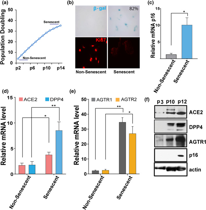
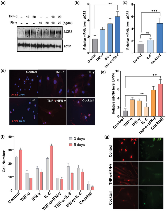
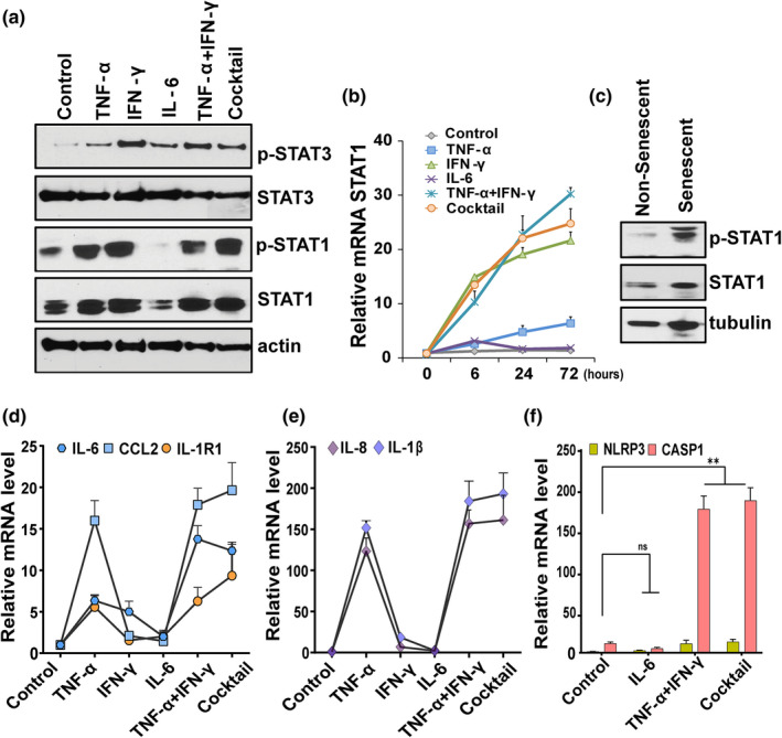
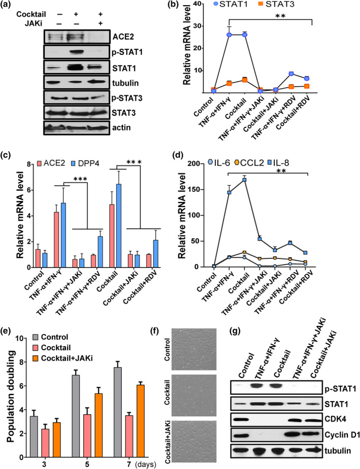
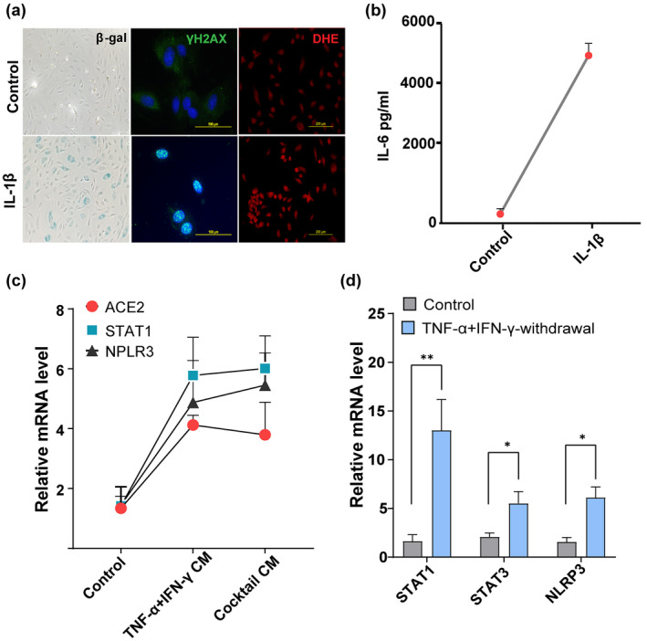
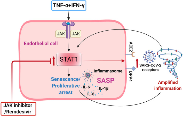
Older age and underlying conditions such as diabetes/obesity or immunosuppression are leading host risk factors for developing severe complications from COVID-19 infection. The pathogenesis of COVID-19-related cytokine storm, tissue damage, and fibrosis may be interconnected with fundamental aging processes, including dysregulated immune responses and cellular senescence. Here, we examined effects of key cytokines linked to cellular senescence on expression of SARS-CoV-2 viral entry receptors. We found exposure of human umbilical vein endothelial cells (HUVECs) to the inflammatory cytokines, TNF-α + IFN-γ or a cocktail of TNF-α + IFN-γ + IL-6, increased expression of ACE2/DPP4, accentuated the pro-inflammatory senescence-associated secretory phenotype (SASP), and decreased cellular proliferative capacity, consistent with progression towards a cellular senescence-like state. IL-6 by itself failed to induce substantial effects on viral entry receptors or SASP-related genes, while synergy between TNF-α and IFN-γ initiated a positive feedback loop via hyper-activation of the JAK/STAT1 pathway, causing SASP amplification. Breaking the interactive loop between senescence and cytokine secretion with JAK inhibitor ruxolitinib or antiviral drug remdesivir prevented hyper-inflammation, normalized SARS-CoV-2 entry receptor expression, and restored HUVECs proliferative capacity. This loop appears to underlie cytokine-mediated viral entry receptor activation and links with senescence and hyper-inflammation.

Keywords: ACE2; COVID-19; DPP4; JAK-STAT; SARS-COV-2 receptor; cytokines; inflammation; senescence.


Conflict of interest statement

The authors declare that they have no conflict of interest.

Figures

FIGURE 1
FIGURE 1
SARS‐CoV‐2 viral entry receptor expression is increased in senescent HUVECs. (a) Population doubling times of HUVECs. “Young” (passage 2–5) HUVECs were serially cultured until they entered proliferative arrest at passage 11–14 (Senescent). (b) Non‐senescent (passage 2) and senescent (passage 14) cells were stained for SA‐β‐gal activity (blue cells) and the proliferative marker Ki67 (in red). (c) Real‐time PCR analysis for the senescence marker p16 INK4a in non‐senescent and senescent cultures. (d, e) quantification of ACE2, DPP4, AGTR1, and AGTR2 mRNA in non‐senescent and senescent cells. GAPDH was used as an internal control. Error bars show mean ± SD, *p < 0.05, **p < 0.01, ***p < 0.001, Student's T‐test. f) Western blots for ACE2, DPP4, AGTR1, p16, and actin in non‐senescent (passage 3) and senescent cells (passage 10–12). All real‐time PCR reactions were conducted in triplicate. Data are representative of three (a–d) or two (e) independent experiments
FIGURE 2
FIGURE 2
TNF‐α/IFN‐γ synergistically increase ACE2 and DPP4 and inhibit proliferation. (a) Western blot for ACE2 in HUVECs exposed to TNF‐α, IFN‐γ, or both for 3 days at the indicated concentrations. (b, c) relative expression of ACE2 in cells stimulated with TNF‐α, IFN‐β, IL‐6 or TNFα+IFN‐γ individually or as a cocktail (TNF‐α + IFN‐γ + IL‐6). (d) Immunostaining for cell surface expressed‐ACE2 (red) and DAPI (blue) in cells exposed to the indicated cytokines. DAPI staining indicates nuclear DNA. (e) Real‐time expression of DPP4 in cells exposed to the indicated cytokines. (f) Graph represents cell number as % of control after 3 and 5 days of stimulation with cytokines as indicated. (g) Immunostaining for the proliferation indicator, Ki67 (red), and the cell cycle marker, BrdU (green), in cells exposed to cytokines for 5 days. Data are representative of two (a, d, g) or three (b, c, e, f) independent experiments. Error bars show mean ± SD, *p < 0.05, **p < 0.01, ***p < 0.001; ns: Non‐significant
FIGURE 3
FIGURE 3
TNF‐α/IFN‐γ synergy accentuates inflammation in a STAT‐dependent manner. (a) Western blot analysis of pSTAT1, STAT1, pSTAT3, STAT3, and actin in HUVECs stimulated with the indicated cytokines for 3 days. (b) Time course analysis of STAT1 mRNA in cells treated with cytokines for the indicated times. (c) Western blot analysis of pSTAT1 and non‐phosphorylated STAT1 in non‐senescent and senescent HUVECs. Tubulin served as the control. (d–f) HUVECs were exposed to different cytokines for 3 days as indicated and qPCR performed for the SASP components, IL‐6, CCL2, IL‐1R1, IL‐8, and IL‐1β, and NLPR3 and CASP1. Data are representative of two (a–c) or three (b–f) independent experiments. Error bars show mean ± SD. *p < 0.05, **p < 0.01, and ***p < 0.001 by Student's T‐test
FIGURE 4
FIGURE 4
Targeting the JAK/STAT pathway reduces expression of SARS‐CoV‐2 viral entry receptors and suppresses inflammation. HUVECs were exposed to the indicated cytokines or vehicle or pretreated with ruxolitinib (1 μM) (JAKi) or remdesivir (2 μM) (RDV) for 30 min prior to cytokine stimulation. (a) Western blot analysis of ACE2, pSTAT1, STAT1, pSTAT3, STAT3, actin, and tubulin in control or cocktail or cocktail with JAK inhibitor. (b–d) Analysis of STAT1, STAT3, ACE2, DPP4, IL‐6, CCL2, and IL‐8 mRNA in control or cells treated with cytokines as indicated. (e) Cellular proliferation of control cells or cells exposed cocktail alone or with JAK inhibitor (1 μM) for the number of days indicated. (f) Representative phase contrast microscopic images of control or cells treated with cocktail or with JAK inhibitor. (g) Western blot analysis of pSTAT1, STAT1, CDK4, and cyclin D1 in cells exposed to the combination of TNF‐α + IFN‐γ or the cocktail or with the JAK inhibitor, ruxolitinib. Data are representative of two (a), two (g), or three (b‐f) independent experiments. Error bars show mean ± SD. *p < 0.05, **p < 0.01, or ***p < 0.001
FIGURE 5
FIGURE 5
A positive feedback loop connects STAT and inflammation. (a) SA‐β‐gal, γ‐H2AX, and DHE (for ROS detection) staining in HUVECs stimulated with or without IL‐1β (20 ng/ml) for 48 h. (b) Quantification of IL‐6 production by ELISA in supernatants collected from cells cultured in the presence or absence of IL‐1β for 48 h. (c) Culture media from control cells or cells exposed to cytokines for 3 days were collected, centrifuged, filtered, and mixed with culture medium at a 1:2 ratio (culture supernatants: Culture medium). Cells were then treated with control conditioned medium or conditioned medium derived from cells exposed to cytokines for 24 h and analyzed for ACE2, STAT1, and NLRP3 mRNA. (d) Quantification of STAT1, STAT3, and NLRP3 mRNA in controls or 24 h after withdrawal of the cytokines, TNF‐α + IFN‐γ. data are representative of three (a–d) independent experiments. Error bars show mean ± SD. *p < 0.05, **p < 0.01, or ***p < 0.001
FIGURE 6
FIGURE 6
Schematic of TNF‐α + IFN‐γ‐mediated effect on endothelial cells. Exposure of HUVECs to TNF‐α + IFN‐γ increases SARS‐CoV‐2 receptor expression, accentuates the SASP, and induces senescence‐like proliferative arrest. Hyperactivated STAT1 coupled with the SASP and inflammasome creates a feedback loop, allowing sustained inflammation and spread of senescence. Targeting STAT1 with a JAK inhibitor (ruxolitinib), or the antiviral remdesivir, normalized ACE2/DPP4 expression and suppressed inflammation. Schematic sketch created with Biorender.com

References

    1. Acosta, J. C. , O'Loghlen, A. , Banito, A. , Guijarro, M. V. , Augert, A. , Raguz, S. , Fumagalli, M. , da Costa, M. , Brown, C. , Popov, N. , Takatsu, Y. , Melamed, J. , d'Adda di Fagagna, F. , Bernard, D. , Hernando, E. , & Gil, J. (2008). Chemokine signaling via the CXCR2 receptor reinforces senescence. Cell, 133(6), 1006–1018. 10.1016/j.cell.2008.03.038 - DOI - PubMed
    1. Bajaj, V. , Gadi, N. , Spihlman, A. P. , Wu, S. C. , Choi, C. H. , & Moulton, V. R. (2021). Aging, immunity, and COVID‐19: How age influences the host immune response to coronavirus infections? Frontiers in Physiology, 11(1793), 571416. 10.3389/fphys.2020.571416 - DOI - PMC - PubMed
    1. Baker, S. A. , Kwok, S. , Berry, G. J. , & Montine, T. J. (2021). Angiotensin‐converting enzyme 2 (ACE2) expression increases with age in patients requiring mechanical ventilation. PLoS One, 16(2), e0247060. 10.1371/journal.pone.0247060 - DOI - PMC - PubMed
    1. Bartleson, J. M. , Radenkovic, D. , Covarrubias, A. J. , Furman, D. , Winer, D. A. , & Verdin, E. (2021). SARS‐CoV‐2, COVID‐19 and the aging immune system. Nature Aging, 1(9), 769–782. 10.1038/s43587-021-00114-7 - DOI - PMC - PubMed
    1. Bonafe, M. , Prattichizzo, F. , Giuliani, A. , Storci, G. , Sabbatinelli, J. , & Olivieri, F. (2020). Inflamm‐aging: Why older men are the most susceptible to SARS‐CoV‐2 complicated outcomes. Cytokine & Growth Factor Reviews, 53, 33–37. 10.1016/j.cytogfr.2020.04.005 - DOI - PMC - PubMed

Publication types

MeSH terms