

Cone-beam Computed Tomographic Analysis of Deciduous Root Canals after Instrumentation with Different Filing Systems: An In Vitro Study
- PMID: 35645508
- PMCID: PMC9108822
- DOI: 10.5005/jp-journals-10005-2126
Cone-beam Computed Tomographic Analysis of Deciduous Root Canals after Instrumentation with Different Filing Systems: An In Vitro Study
Abstract
Aim and objective: To evaluate root canal transportation, centering ability ratio (CAR), remaining dentine thickness, dentinal cracks, and instrumentation time after instrumentation with different filing systems in root canals of primary teeth by cone beam computed tomography (CBCT) analysis.
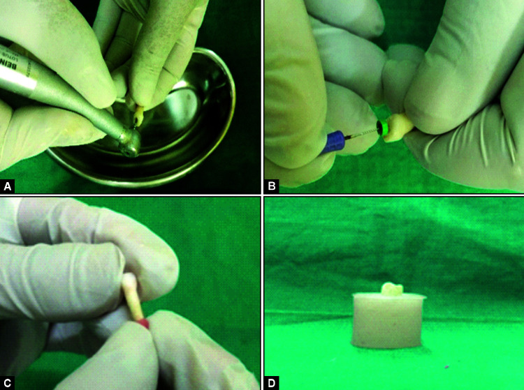
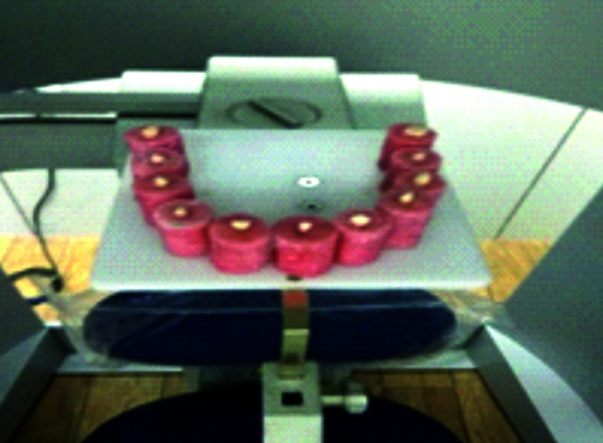
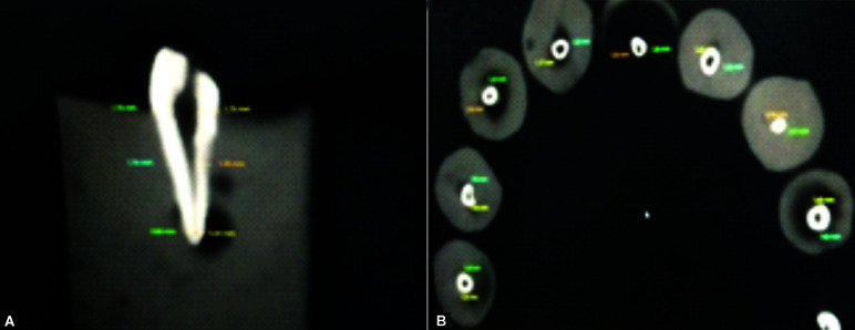
Materials and methods: Sixty prepared canals of primary teeth divided into 4 groups with 15 canals in each were prepared with NiTi K files, Proaper Next (PTN) files, OneShape (OS), and WaveOne (WO) files, respectively. Using CBCT scan, the pre- and postinstrumentation scan was done to obtain images at three levels (apical, middle, and cervical). The results obtained were statistically analyzed using SPSS 21 statistical software version.
Result: Significant statistical difference was found between different filing systems.
Conclusion: ProTaper Next files showed least canal transportation and the best centering ability was shown by OS file system. The NiTi K hand files preserved maximum remaining dentin thickness (RDT) and produced minimum dentin cracks. WO file system took least instrumentation time when compared to the other three filing systems.
Clinical significance: The use of rotary instruments in the pulpectomy of primary teeth represents a promising technique being advantageous for the pediatric patients by maintaining the original canal curvatures, showing greater ability to maintain dentin thickness, causing lesser dentin cracks, and reducing chair time thus favoring preparation of more conical root canals and better obturation.
How to cite this article: Singh P. Cone-beam Computed Tomographic Analysis of Deciduous Root Canals after Instrumentation with Different Filing Systems: An In Vitro Study. Int J Clin Pediatr Dent. 2022;15(S-1):S22-S29.
Keywords: Cone beam computed tomography; Nickel-titanium; Primary teeth; ProTaper; WaveOne.
Copyright © 2022; The Author(s).
Conflict of interest statement
Source of support: Nil Conflict of interest: None
Figures
References
-
- Prabhakar AR, Yavagal C, Dixit K, et al. Reciprocating vs rotary nstrumentation in pediatric endodontics: cone beam computed tomographic analysis of deciduous root canals using two single-file systems. Int J Clin Pediatr Dent. 2016;9(01):45–49. doi: 10.5005/jp-journals-10005-1332. - DOI - PMC - PubMed
-
- Pham KV, Phan TN. Evaluation of root canal preparation using two nickel-titanium instrument systems via cone-beam computed tomography. Saudi Endod J. 2019;9:210–215. doi: 10.4103/sej.sej_147_18. - DOI
LinkOut - more resources
Full Text Sources