. 2022 May 13:16:879634.
doi: 10.3389/fnsys.2022.879634. eCollection 2022.

Novel Cerebello-Amygdala Connections Provide Missing Link Between Cerebellum and Limbic System

Affiliations

Novel Cerebello-Amygdala Connections Provide Missing Link Between Cerebellum and Limbic System

Se Jung Jung et al. Front Syst Neurosci. .

Abstract

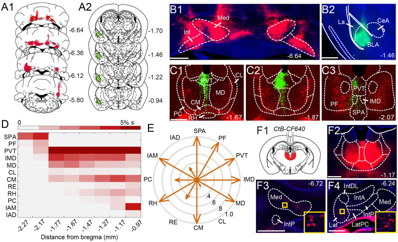
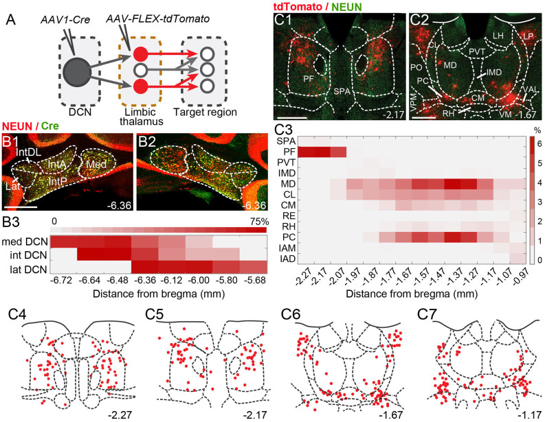
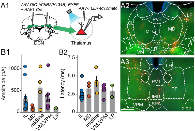
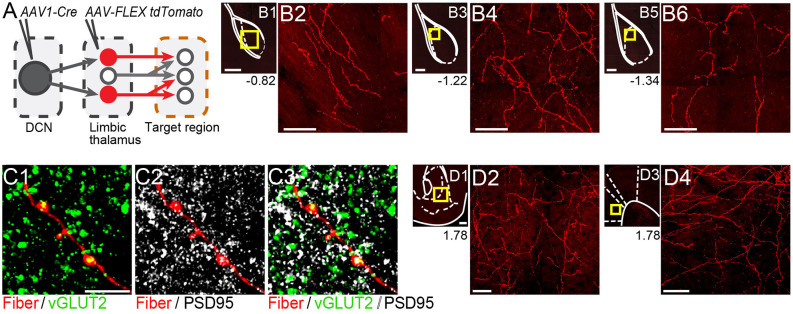
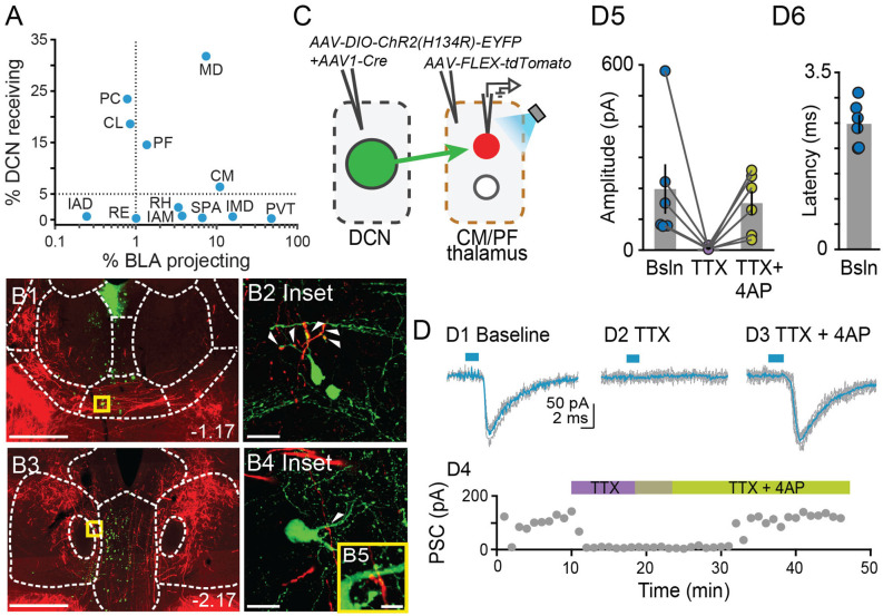
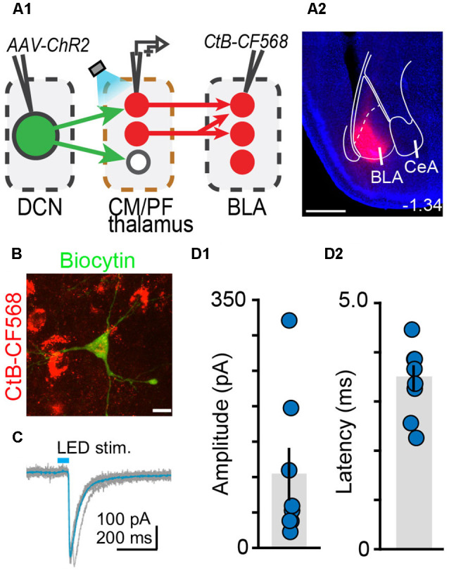
The cerebellum is emerging as a powerful regulator of cognitive and affective processing and memory in both humans and animals and has been implicated in affective disorders. How the cerebellum supports affective function remains poorly understood. The short-latency (just a few milliseconds) functional connections that were identified between the cerebellum and amygdala-a structure crucial for the processing of emotion and valence-more than four decades ago raise the exciting, yet untested, possibility that a cerebellum-amygdala pathway communicates information important for emotion. The major hurdle in rigorously testing this possibility is the lack of knowledge about the anatomy and functional connectivity of this pathway. Our initial anatomical tracing studies in mice excluded the existence of a direct monosynaptic connection between the cerebellum and amygdala. Using transneuronal tracing techniques, we have identified a novel disynaptic circuit between the cerebellar output nuclei and the basolateral amygdala. This circuit recruits the understudied intralaminar thalamus as a node. Using ex vivo optophysiology and super-resolution microscopy, we provide the first evidence for the functionality of the pathway, thus offering a missing mechanistic link between the cerebellum and amygdala. This discovery provides a connectivity blueprint between the cerebellum and a key structure of the limbic system. As such, it is the requisite first step toward obtaining new knowledge about cerebellar function in emotion, thus fundamentally advancing understanding of the neurobiology of emotion, which is perturbed in mental and autism spectrum disorders.

Keywords: anatomy; basolateral amydala; cerebellar nuclei; channelrhodopsin; circuit; electrophysiology; limbic; mouse.


Conflict of interest statement

The authors declare that the research was conducted in the absence of any commercial or financial relationships that could be construed as a potential conflict of interest.

Figures

Figure 1
Figure 1
Anatomical tracing uncovers putative disynaptic pathways from the cerebellum tobasolateral amygdala. (A) Injection sites for anterogradeviral tracer in DCN (A1, red) and retrograde viraltracer in BLA (A2, green). (B) Mosaic epifluorescence image of injection sites in DCN (B1) and BLA (B2). (C1–C3) Mosaic epifluorescence images of overlapping DCN axons (red) and BLA-projecting neurons (green) in limbic thalamus. (D) Relative distribution of BLA-projecting neurons across nuclei of the limbic thalamus, normalized to the total number of labeled neurons and averaged across experiments, as a function of distance from bregma. Antero-posterior coordinates for each nucleus are given in Table 1. (E) Quantification of overlap between DCN axons and BLA-projecting thalamic neurons. Arrow length in compass plot indicates proportion (0.0–1.0) of experiments with overlap in each thalamic nucleus. (F1,F2) Schematic and confocal image of injection site for retrograde tracer CtB CF-640 in limbic thalamus. (F3,F4) CtB-labeled projection neurons (red) in DCN at different distances from bregma. Insets show high-magnification images of areas in yellow squares. For all panels, numbers denote distance (in mm) from bregma. Blue: DAPI. Scale bars: 500 μm.
Figure 2
Figure 2
Theintralaminar and mediodorsal nuclei are major cerebellar postsynaptic targets in the limbic thalamus. (A) Schematic of experimental approach for disynaptic pathway tracing. (B1,B2) Example images of bilateral Cre expression in DCN. Red: immunofluorescence for NeuN neural marker; Green: anti-Cre immunoreactivity; Yellow: merge. (B3) Heatmap of Cre immunofluorescence in DCN, normalized to NeuN signal and averaged across experiments, as a function of distance (in mm) from bregma. (C1,C2) Example images of thalamic neurons conditionally expressing tdTomato (red) upon transneuronal transfer of Cre from cerebellar presynaptic axons. Green: NeuN immunofluorescence. (C3) Heatmap of the relative distribution of tdTomato+ neurons across thalamic nuclei, normalized to the total number of labeled neurons and averaged across experiments, as a function of distance from bregma. (C4–C7) Example registration of tdTomato+ neurons to the Allen mouse brain atlas. Numbers at the bottom denote distance (in mm) from bregma. Antero-posterior coordinates for each nucleus can be found in Table 1. Scale bars: 500 μm.
Figure 3
Figure 3
Electrophysiologicalvalidation of virally-identified cerebello-thalamic connectivity. (A1) Schematic of experimental approach for ex vivo optophysiology. (A2,A3) Epifluorescence images of anterior (A2) and posterior (A3) thalamic slices acutely prepared for recordings. DCN input-receiving neurons are tdTomato+. Scale bars: 500 μm. (B) Average (± SEM) amplitude (B1) and onset latency (B2) of ChR2-evoked synaptic currents as a function of recording location in the thalamus. Intralaminar (IL) group: CL, PC, CM, and PF; midline group: IMD and RH.
Figure 4
Figure 4
Thalamicneurons receiving cerebellar input form synapses in the basolateral amygdala and also target the nucleus accumbens and prelimbic cortex. (A) Schematic diagram of the experimental approach. Targets of tdTomato+ axons of thalamic neurons receiving cerebellar input were identified through imaging. (B) Mosaic confocal images of tdTomato+ axons along the anterior-posterior axis of the BLA. (C) High resolution airyscan confocal images of tdTomato+ axons in the BLA colocalizing with presynaptic (vGLUT2) (C1) and postsynaptic (PSD95) (C2) markers of excitatory synapses. Green: vGLUT2, gray: PSD95, yellow/white in (C3): overlay. (D) tdTomato+ axons in nucleus accumbens (D1,D2) and prelimbic cortex (D3,D4). Yellow squares in (B1,B3,B5,D1,D3) show zoom-in areas for (B2,B4,B6,D2,D4) images, respectively. Numbers at the bottom of images indicate the distance (in mm) from bregma. Scale bars: (B1,B3,B5,D1,D3): 200 μm; (B2,B4,B6,D2,D4): 50 μm; (C1–C3): 5 μm.
Figure 5
Figure 5
Centromedial and parafascicular neurons project to the basolateral amygdala andreceive functional monosynaptic input from the cerebellum.(A) Scatterplot of % neurons receiving DCN input vs. % neurons projecting to BLA, for limbic thalamus nuclei. (B1–B4) Airyscan confocal images of DCN axons (red) and BLA-projecting neurons (green) in the centromedial (CM; B1) and parafascicular (PF; B3) thalamic nuclei. (B2,B4,B5) Zoomed-in areas in yellow squares from (B1,B3). Scale bars: (B1,B3): 500 μm; (B2,B4): 20 μm; (B5): 5 μm. (C) Schematic diagram of ex vivo optophysiology approach to test for monosynaptic connections between DCN and CM/PF thalamic n. (D1–D3) Average ChR2-evoked synaptic current (teal), overlaid onto single trial responses (gray), at baseline (D1); upon addition of the action potential blocker tetrodotoxin (TTX, 1 μm; D2); after further addition of the potassium channel blocker 4-aminopyridine (4AP, 100 μm; D3). (D4) Time course of the wash-in experiment for the same example cell. (D5) Summary of effects on amplitude (mean ± SEM) of ChR2-evoked synaptic responses for the indicated conditions. Bsln: baseline. (D6) Average (± SEM) onset latency of ChR2-evoked responses at baseline.
Figure 6
Figure 6
The centromedial and parafascicular thalamus is a functional node of the cerebello-amygdala circuit. (A) Experimental approach (A1) and example CtB-CF568 injection in amygdala (A2). Blue: DAPI. Scale bar: 500 μm. (B) BLA-projecting neuron in centromedial (CM) thalamus retrogradely labeled with CtB CF-568 (red) is also labeled with biocytin (green) through the patch pipette. Scale bar: 10 μm. (C) Example ChR2-evoked synaptic response. Average trace (teal) overlaid onto single trials (gray). (D1,D2) Average (± SEM) amplitude (D1) and onset latency (D2) of ChR2-evoked synaptic currents at DCN-CM/PF synapses.

References

    1. Adamaszek M., D’Agata F., Ferrucci R., Habas C., Keulen S., Kirkby K. C., et al. . (2017). Consensus paper: cerebellum and emotion. Cerebellum 16, 552–576. 10.1007/s12311-016-0815-8 - DOI - PubMed
    1. Adolphs R. (2010). What does the amygdala contribute to social cognition? Ann. N Y Acad. Sci. 1191, 42–61. 10.1111/j.1749-6632.2010.05445.x - DOI - PMC - PubMed
    1. Ahmed N., Headley D. B., Paré D. (2021). Optogenetic study of central medial and paraventricular thalamic projections to the basolateral amygdala. J. Neurophysiol. 126, 1234–1247. 10.1152/jn.00253.2021 - DOI - PMC - PubMed
    1. Ahs F., Pissiota A., Michelgård A., Frans O., Furmark T., Appel L., et al. . (2009). Disentangling the web of fear: amygdala reactivity and functional connectivity in spider and snake phobia. Psychiatry Res. 172, 103–108. 10.1016/j.pscychresns.2008.11.004 - DOI - PubMed
    1. Amir A., Paré J., Smith Y., Paré D. (2019). Midline thalamic inputs to the amygdala: ultrastructure and synaptic targets. J. Comp. Neurol. 527, 942–956. 10.1002/cne.24557 - DOI - PMC - PubMed