. 2022 May 11:16:886634.
doi: 10.3389/fnbeh.2022.886634. eCollection 2022.

Chronic Ethanol Causes Persistent Increases in Alzheimer's Tau Pathology in Female 3xTg-AD Mice: A Potential Role for Lysosomal Impairment

Affiliations

Chronic Ethanol Causes Persistent Increases in Alzheimer's Tau Pathology in Female 3xTg-AD Mice: A Potential Role for Lysosomal Impairment

Autumn E Tucker et al. Front Behav Neurosci. .

Abstract

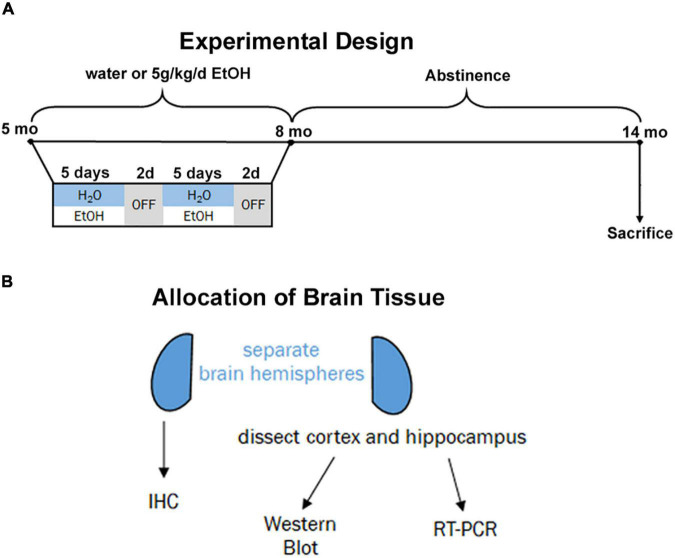
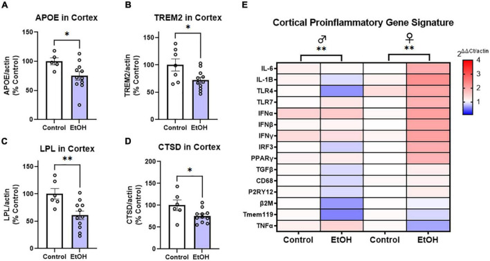
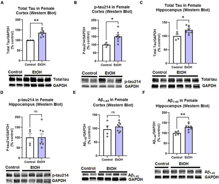
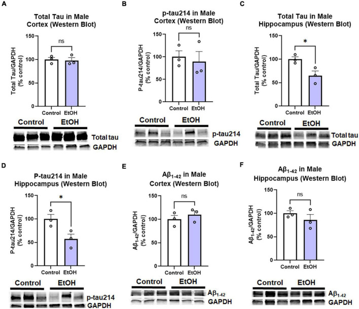
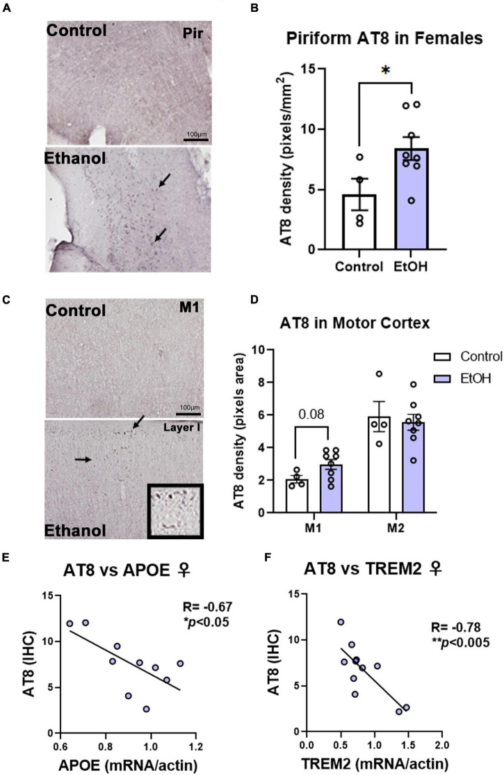
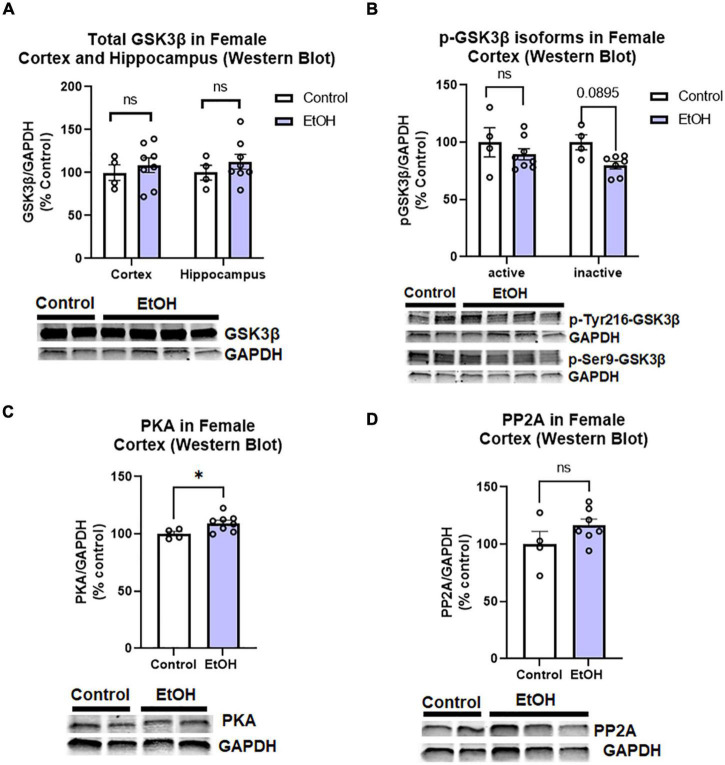
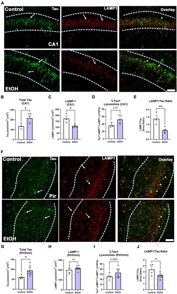
Epidemiological studies have found that heavy alcohol use is associated with increased risk for Alzheimer's disease (AD), with frequent drinking earlier in adulthood increasing risk. The increases in neuroinflammation featured in both heavy alcohol use and AD may be partially responsible for this link. However, it is unknown if abstinence mitigates this risk. We hypothesized that binge ethanol during mid adult life would persistently increase AD pathology even after prolonged abstinence. Male and female 3xTg-AD mice (APPSwe, tauP301, Psen1tm1Mpm) which feature progressive amyloid (Aβ) and tau pathology, received chronic binge ethanol (5g/kg/day, 5-days-on/2-days-off, i.g.) or water during adulthood (from 5.5 to 9 months of age), followed by abstinence and assessment at 14 months of age. The effects of ethanol on protective AD genes (e.g., APOE and TREM2) as well as proinflammatory genes were measured by PCR. Levels of pathologic tau and Aβ were measured by immunohistochemistry and western blot. Ethanol caused persistent reductions in protective AD genes: APOE (25% reduction, *p < 0.05), TREM2 (28%, *p < 0.05), LPL (40%, ** p < 0.01), and CTSD (24%, *p < 0.05) and promoted a proinflammatory gene signature in female, but not male cortex. Concurrently, ethanol increased total and hyperphosphorylated tau (AT8) in piriform cortex and hippocampus of females, but not males. Levels of AT8 were negatively correlated with APOE (R = -0.67, *p < 0.05) and TREM2 (R = -0.78, **p < 0.005) suggesting protective roles in pathogenesis. No differences were found in levels of main regulators of tau phosphorylation state (GSK3β, PKA, PP2A), suggesting ethanol disrupted clearance of tau. Therefore, we measured the effect of ethanol on lysosomes, which degrade tau, and lysosomal localization of tau using co-immunofluorescence. In females, ethanol caused a persistent reduction in mature LAMP1 lysosomes in CA1 of hippocampus (35%, *p < 0.05), along with a 60% increase in total tau (*p < 0.05). Thus, chronic binge ethanol during mid adult life causes a persistent enhancement of tau pathology in cortical and hippocampal brain regions of females. Persistent AD pathology was associated with an increased proinflammatory signature and a reduction of mature lysosomes. This implicates binge ethanol exposure with increased risk of AD pathologic progression in females.

Keywords: Alzheimer’s disease; abstinence; addiction; alcohol; neuro-inflammation; tau.


Conflict of interest statement

The authors declare that the research was conducted in the absence of any commercial or financial relationships that could be construed as a potential conflict of interest.

Figures

FIGURE 1
FIGURE 1
Experimental design and tissue allocation. (A) Adult 3xTg mice (5 mo, P168) received either water gavage (i.g.) or ethanol (5g/kg/d, i.g.) for 3 months (P168–P264), in a 5-days on 2-days off pattern. Mice were then left without intervention until sacrifice at 14 months (P432). (B) 1 hemisphere was drop-fixed in PFA for immunohistochemistry and 1 hemisphere dissected for cortex and hippocampus for both western blot and RT-PCR analyses.
FIGURE 2
FIGURE 2
Chronic binge ethanol persistently reduces key disease-associated microglial (DAM) genes and promotes a proinflammatory gene profile in females. Adult male and female 3xTg-AD mice received EtOH (5g/kg/d, i.g., 5d on 2d off) for 3 months (P168–P264), followed by prolonged abstinence until 14 months of age. Gene expression was measured by RT-PCR. (A–D) Expression of key protective DAM genes in cortex of both males and females (combined). Ethanol caused a significant reduction of (A) APOE, 25%, (B) TREM2, 28%, (C) LPL, 40%, and (D) CTSD, 24%. *p < 0.05, **p < 0.01, t-tests. (E) Heat map showing expression of pro-inflammatory genes in cortex. Ethanol persistently increased expression of several proinflammatory genes in females (11/13), with reductions in several genes found in males (8/13 genes). Gene Set Enrichment Analysis (GSEA) found significant enrichment of genes in both sexes (ethanol vs control), N = 3 males/group; 3 female control and 8 female ethanol. **p < 0.01.
FIGURE 3
FIGURE 3
Persistent effects of ethanol on tau and Aβ proteins in female 3xTg-AD mice. Adult female 3xTg-AD mice received either EtOH (5g/kg/d, i.g., 5d on 2d off, N = 8) or the same schedule of water (i.g., N = 4) for 3 months (P168–P264), followed by prolonged abstinence until 14 months. Pathologic tau and Aβ protein levels were measured in cortex and hippocampus by western blot. (A,B) Tau species in cortex. In the cortex EtOH caused a persistent increase in (A) total tau, 36% (N = 3 control, 8 ethanol, 1 outlier) and (B) phosphorylated tau214, p-tau214, 50%, *p < 0.05. (C,D) Tau in hippocampus. In the hippocampus, EtOH persistently increased levels of (C) total Tau, 23% increase, *p < 0.05 without altering (D) p-tau214, p = 0.9 levels. (E,F)1–42 levels. EtOH had no persistent impact on levels of Aβ1–42 protein in female cortex, p = 0.4 (E), though a (F) 30% increase was found in the hippocampus. *p < 0.05, **p < 0.01, t-test.
FIGURE 4
FIGURE 4
Persistent effects of ethanol on tau and Aβ proteins in male 3xTg-AD mice. Adult male 3xTg-AD mice received EtOH (5g/kg/d, i.g., 5d on 2d off, N = 3) or the same schedule of water (i.g., N = 3) for 3 months (P168–P264), followed by prolonged abstinence with assessment at 14 months. Pathologic tau and Aβ protein levels were measured in cortex and hippocampus by western blot. (A,B) Tau species in cortex. EtOH had no persistent effects on neither (A) total tau, p = 0.8 nor (B) phosphorylated tau214 (p-tau214) p = 0.7. (C,D) Tau in hippocampus. In the hippocampus, EtOH caused a persistent reduction in (C) total Tau, 35% reduction, *p < 0.05, and (D) p-tau214, 43% reduction, *p < 0.05. (E,F)1–42 levels. EtOH had no persistent impact on levels of Aβ1–42 in either male (E) cortex, p = 0.4 or (F) hippocampus, p = 0.3. *p < 0.05, t-test.
FIGURE 5
FIGURE 5
Chronic binge ethanol persistently increases phosphorylated tau AT8 staining in female cortex. Adult female 3xTg-AD mice received EtOH (5g/kg/d, i.g., 5d on 2d off) for 3 months (P168–P264), followed by prolonged abstinence at assessment at 14 months. Pathological phosphorylated tau (AT8) was assessed by IHC in piriform and motor cortex. (A) Representative images showing increased AT8 immunoreactivity (+IR) in piriform cortex caused by ethanol. Arrows denote AT8+IR cells. (B) Quantification of AT8 pixel density showed a ∼2-fold increase in AT8 in the piriform cortex after ethanol. *p < 0.05, t-test. (C) Images of M1 motor cortex showing low levels of AT8 in control mice, but slightly increased +IR was seen in several ethanol-treated mice. High magnification insert shows cells positive for AT8. (D) Quantification showed a trend toward a 33% increase in AT8 in M1 of ethanol-treated mice (p = 0.08, t-test). No differences were seen in AT8 +IR in M2 of the motor cortex, though higher baseline levels were seen than in M1. *p < 0.05, t-test. (E) AT8+IR was negatively correlated with cortical APOE mRNA levels. R = –0.67, *p < 0.05, Pearson’s correlation coefficient. (F) AT8 +IR was negatively correlated with gene expression of TREM2 in cortex. R = –0.78, N = 4 control, 8 ethanol. **p < 0.005.
FIGURE 6
FIGURE 6
Chronic binge ethanol has no persistent effect on major regulators of tau phosphorylation in female 3x-TgAD mice. Adult female 3xTg-AD mice received EtOH (5g/kg/d, i.g., 5d on 2d off) for 3 months (P168–P264), followed by prolonged abstinence until 14 months. Common tau-phosphorylating kinases (GSK3β and PKA) and the tau de-phosphorylating enzyme PP2A were measured by Western Blot. (A) Total GSK3β protein level in cortex was unchanged by ethanol. (B) Levels of active p-Tyr216 GSK3β were unchanged by ethanol and levels of the inactive p-Ser9-GSK3β showed a trend toward a 20% reduction, p = 0.09. (C) Ethanol caused a slight 5% increase in protein kinase A (PKA), *p < 0.05. (D) Ethanol had no effect on expression levels of PP2A. N = 4 control, 8 ethanol. *p < 0.05, t-test.
FIGURE 7
FIGURE 7
Chronic binge ethanol causes persistent increase in tau and loss of LAMP1 positive lysosomes in hippocampus of female 3xTg-AD mice. Adult female 3xTg-AD mice received EtOH (5g/kg/d, i.g., 5d on 2d off) for 3 months (P168–P264), followed by prolonged abstinence until 14 months. (A) Total tau and lysosomal LAMP1 involved in autophagy and tau clearance were measured by co-immunofluorescence (co-IF). Left-pointing arrows denote tau stain in cell body and processes. Right pointing arrows denote punctate LAMP-1 staining. Arrowheads denote co-localization. (B) Quantification of total tau in the CA1 region revealed a persistent increase by 60%, *p < 0.05, t-test and (C) quantification of lysosomal LAMP1 in the CA1 region indicated a persistent reduction by 35%, *p < 0.05. (D) Overlaying total tau and LAMP1 revealed a trend toward a 43% increase in the percent of tau-positive lysosomes in the CA1, p = 0.07, however, (E) there was also a sharp 59% decline in the amount of LAMP1 overlapping with total tau in the CA1, **p < 0.01. (F) Total tau and lysosomal LAMP1 involved in autophagy and tau clearance were measured by co-IF in piriform cortex (Pir). Left-pointing arrows denote tau stain in cell body and processes. Right pointing arrows denote punctate LAMP-1 staining. Arrowheads denote co-localization. (G) Ethanol caused a persistent increase in tau in the piriform cortex (Pir), 56%m*p < 0.05, t-test. (H) Ethanol had no effect on LAMP1 level in Pir. (I) Ethanol caused a trend toward an increase in the percent of Tau+ lysosomes p = 0.17. (J) Ethanol caused a persistent reduction in the ratio of LAMP1 to Tau. *p < 0.05, t-test. Scale bar = 50μm.

References

    1. Bala S., Marcos M., Gattu A., Catalano D., Szabo G. (2014). Acute binge drinking increases serum endotoxin and bacterial dna levels in healthy individuals. PLoS One 9:E96864. 10.1371/journal.pone.0096864 - DOI - PMC - PubMed
    1. Barnett A., David E., Rohlman A. R., Nikolova V. D., Moy S. S., Vetreno R., et al. (2022). Adolescent binge alcohol enhances early Alzheimer’s disease pathology in adulthood through proinflammatory neuroimmune activation. Front. Pharmacol. 2020:170. 10.3389/Fphar.2022.884170 - DOI - PMC - PubMed
    1. Barton E. A., Baker C., Leasure J. L. (2017). Investigation of sex differences in the microglial response to binge ethanol and exercise. Brain Sci. 7 1–14. 10.3390/brainsci7100139 - DOI - PMC - PubMed
    1. Behl T., Yadav H. N., Sharma P. L. (2021). Alcoholic neuropathy: involvement of multifaceted signalling mechanisms. Curr. Mol. Pharmacol. 14 2–10. 10.2174/1874467213666200512114943 - DOI - PubMed
    1. Belfiore R., Rodin A., Ferreira E., Velazquez R., Branca C., Caccamo A., et al. (2019). Temporal and regional progression of alzheimer’s disease-like pathology in 3xtg-ad mice. Aging Cell 18:E12873. 10.1111/acel.12873 - DOI - PMC - PubMed

LinkOut - more resources