. 2022 May 20:2022:2984789.
doi: 10.1155/2022/2984789. eCollection 2022.

Relative Perfusion Differences between Parathyroid Adenomas and the Thyroid on Multiphase 4DCT

Affiliations

Relative Perfusion Differences between Parathyroid Adenomas and the Thyroid on Multiphase 4DCT

Steven P M J Raeymaeckers et al. Int J Biomed Imaging. .

Abstract

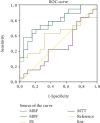
A multiphase 4DCT technique can be useful for the detection of parathyroid adenomas. Up to 16 different phases can be obtained without significant increase of exposure dose using wide beam axial scanning. This technique also allows for the calculation of perfusion parameters in suspected lesions. We present data on 19 patients with histologically proven parathyroid adenomas. We find a strong correlation between 2 perfusion parameters when comparing parathyroid adenomas and thyroid tissue: parathyroid adenomas show a 55% increase in blood flow (BF) (p < 0.001) and a 50% increase in blood volume (BV) (p < 0.001) as compared to normal thyroid tissue. The analysis of the ROC curve for the different perfusion parameters demonstrates a significantly high area under the curve for BF and BV, confirming these two perfusion parameters to be a possible discriminating tool to discern between parathyroid adenomas and thyroid tissue. These findings can help to discern parathyroid from thyroid tissue and may aid in the detection of parathyroid adenomas.


Conflict of interest statement

The authors declare that there is no conflict of interest regarding the publication of this paper.

Figures

Figure 1
Figure 1
Boxplot. BF normal thyroid tissue vs BF parathyroid adenomas.
Figure 2
Figure 2
(a) 4DCT. On the arterial phase, we find a large intrathyroidal parathyroid adenoma (arrow). (b) Parametric perfusion map, color-coded overlay of BF values. Note the elevated BF in the parathyroid adenoma as compared to the thyroid tissue.
Figure 3
Figure 3
Boxplot. BV normal thyroid tissue vs BV parathyroid adenomas.
Figure 4
Figure 4
(a) 4DCT. On the arterial phase, we find a small parathyroid adenoma (arrow) in close relation to the left common carotid artery. (b) Parametric perfusion map, color-coded overlay of BV values. Note the elevated BV in the parathyroid adenoma as compared to the thyroid tissue.
Figure 5
Figure 5
ROC analysis of the perfusion values in parathyroid adenomas compared to thyroid tissue.

References

    1. Sung J. Y. Parathyroid ultrasonography: the evolving role of the radiologist. Ultrasonography . 2015;34(4):268–274. doi: 10.14366/usg.14071. - DOI - PMC - PubMed
    1. Bunch P. M., Randolph G. W., Brooks J. A., George V., Cannon J., Kelly H. R. Parathyroid 4D CT: what the surgeon wants to know. Radiographics . 2020;40(5):1383–1394. doi: 10.1148/rg.2020190190. - DOI - PubMed
    1. Bartsch D., Nies C., Hasse C., Willuhn J., Rothmund M. Clinical and surgical aspects of double adenoma in patients with primary hyperparathyroidism. The British Journal of Surgery . 1995;82(7):926–929. doi: 10.1002/bjs.1800820723. - DOI - PubMed
    1. Rodgers S. E., Hunter G. J., Hamberg L. M., et al. Improved preoperative planning for directed parathyroidectomy with 4-dimensional computed tomography. Surgery . 2006;140(6):932–941. doi: 10.1016/j.surg.2006.07.028. - DOI - PubMed
    1. Raeymaeckers S., Tosi M., De Mey J. 4DCT scanning technique for primary hyperparathyroidism: a scoping review. Radiology research and practice . 2021;2021:12. doi: 10.1155/2021/6614406.6614406 - DOI - PMC - PubMed

LinkOut - more resources