Review
. 2022 May;12(5):2171-2192.
doi: 10.1016/j.apsb.2021.12.022. Epub 2022 Jan 4.

Development of small molecule extracellular signal-regulated kinases (ERKs) inhibitors for cancer therapy

Affiliations
Review

Development of small molecule extracellular signal-regulated kinases (ERKs) inhibitors for cancer therapy

Xiaoli Pan et al. Acta Pharm Sin B. 2022 May.

Abstract

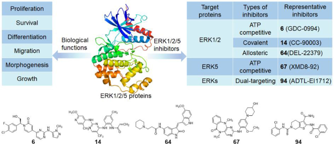
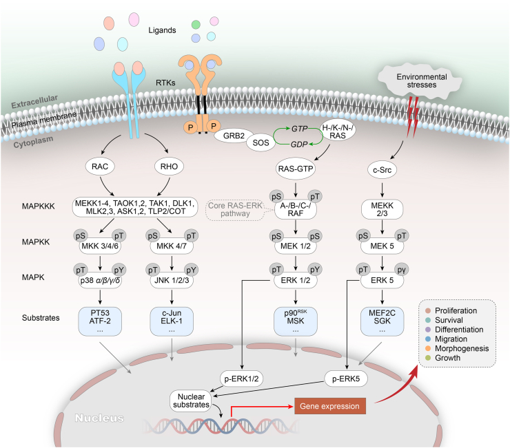
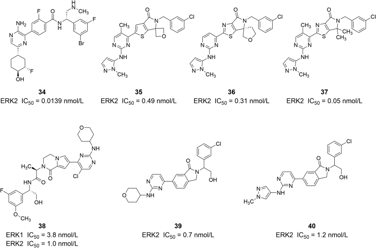
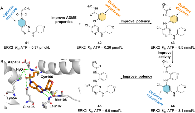
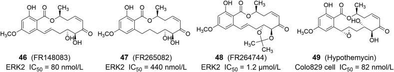
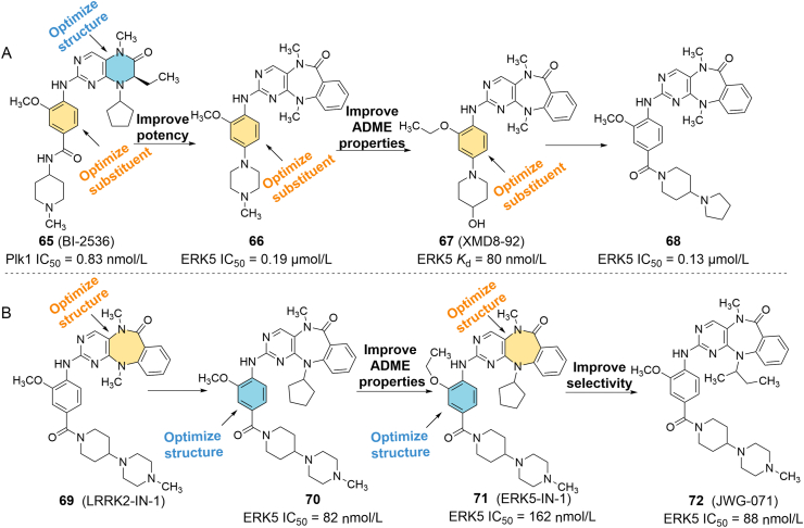
The mitogen-activated protein kinase (MAPK)/extracellular signal-regulated kinase 1/2 (ERK1/2) signaling pathway is widely activated by a variety of extracellular stimuli, and its dysregulation is associated with the proliferation, invasion, and migration of cancer cells. ERK1/2 is located at the distal end of this pathway and rarely undergoes mutations, making it an attractive target for anticancer drug development. Currently, an increasing number of ERK1/2 inhibitors have been designed and synthesized for antitumor therapy, among which representative compounds have entered clinical trials. When ERK1/2 signal transduction is eliminated, ERK5 may provide a bypass route to rescue proliferation, and weaken the potency of ERK1/2 inhibitors. Therefore, drug research targeting ERK5 or based on the compensatory mechanism of ERK5 for ERK1/2 opens up a new way for oncotherapy. This review provides an overview of the physiological and biological functions of ERKs, focuses on the structure-activity relationships of small molecule inhibitors targeting ERKs, with a view to providing guidance for future drug design and optimization, and discusses the potential therapeutic strategies to overcome drug resistance.

Keywords: Cancer; Extracellular signal-regulated kinase 1/2 inhibitors; Extracellular signal-regulated kinase 5 inhibitors; Inhibition; Mitogen-activated protein kinases; Selectivity.


Figures

Image 1
Graphical abstract
Figure 1
Figure 1
The MAPK cascades and the core RAS/ERK signaling pathway. Extracellular stimulus recruits the guanine nucleotide exchange factor SOS (Son of Sevenless) to induce RAS to release GDP and binds to a new GTP. Activated RAS promotes the dimerization and activation of the A-/B-/C-RAF, which further activates downstream signals MEK1/2. The kinase domain of activated MEK1/2 catalyzes the phosphorylation of tyrosine and threonine residues in the ERK1/2 activation segment to activate ERK1/2. Eventually activated ERK1/2 enters the cell nucleus to phosphorylate a series of transcription factors and modulate many critical aspects of cell physiology.
Figure 2
Figure 2
Schematic representation and crystal structures of ERK1 (PDB ID: 2ZOQ), ERK2 (PDB ID: 1ERK) and ERK5 (PDB ID: 4IC8). All MAPKs contain the Ser/Thr kinase domain between the N-terminal and C-terminal regions. Different additional domains are also present in some MAPKs, including a proline-rich domain 1 (PR1), proline-rich domain 2 (PR2) and nuclear localization sequence (NLS).
Figure 3
Figure 3
(A) Discovery and design procedure of the ATP competitive ERK1/2 inhibitor 6 (GDC-0994) by combing HTS strategy and medicinal chemistry. (B) X-ray structure of compound 3 (orange) bound to ERK2 (PDB ID: 4XJ0). Hydrogen-bonding interactions are illustrated with green dashed lines. (C) X-ray structure of 6 (orange) bound to ERK2 (PDB ID: 5K4I).
Figure 4
Figure 4
(A) Chemical structure and design strategy for compound 10. (B) X-ray crystal structure of 10 in complex with ERK2, green dotted lines indicate hydrogen bonds (PDB ID: 6DCG).
Figure 5
Figure 5
(A) Discovery of compound 23 from the lead compound 19 to improve its potency and affinity to ERK1/2. (B) Crystal structure of 19 bound to ERK2 (PDB ID: 2OJG). (C) Binding mode of compound 21 in complex with ERK2 (PDB ID: 2OJJ).
Figure 6
Figure 6
(A) X-ray crystal structure of 24 binding to the active site of ERK2. Hydrogen bonds were highlighted in green dashed lines (PDB ID: 1TVO). (B) Chemical structures of ATP competitive ERK inhibitors 2428 that discovered by adopting a CADD strategy.
Figure 7
Figure 7
(A) X-ray crystal structure of 32 in the active site of ERK2 with hydrogen bond interactions to the key residues highlighted in green dashed lines (PDB ID: 4QPA). (B) Chemical structures of ATP competitive ERK inhibitors 2933.
Figure 8
Figure 8
Chemical structures of ERK1/2 inhibitors 3440.
Figure 9
Figure 9
(A) Discovery and design procedure of the ERK1/2 covalent inhibitor 45 by combing HTS strategy and medicinal chemistry. (B) Cocrystal structure of compound 41 in complex with ERK2, green dotted lines indicate hydrogen bonds, red sphere indicate water molecule (PDB ID: 4ZZM).
Figure 10
Figure 10
Chemical structures of ERK1/2 covalent inhibitors 4649.
Figure 11
Figure 11
(A) Optimization of ERK5 inhibitor 67 with high activity and selectivity based on compound 65. (B) Optimization of ERK5 inhibitor 72 with high activity and selectivity based on compound 69.
Figure 12
Figure 12
Chemical structures of ERK5 inhibitors 7384.
Figure 13
Figure 13
Chemical structures of ERK1/2 dual-targeting inhibitors 8689.
Figure 14
Figure 14
(A) Design strategy used from the novel (7-aryl-1,5-naphthyridin-4-yl) ureas skeleton to the dual ERK2 and Aurora B kinases inhibitor 91. (B) Design strategy used from the leading compound 92 to the dual-target inhibitor of ERK1 and ERK5, 94, with high potency through a series of optimizations.

Similar articles

Cited by

References

    1. Schaeffer H.J., Weber M.J. Mitogen-activated protein kinases: specific messages from ubiquitous messengers. Mol Cell Biol. 1999;19:2435–2444. - PMC - PubMed
    1. Cargnello M., Roux P.P. Activation and function of the MAPKs and their substrates, the MAPK-activated protein kinases. Microbiol Mol Biol Rev. 2011;75:50–83. - PMC - PubMed
    1. Kim E.K., Choi E.J. Pathological roles of MAPK signaling pathways in human diseases. Biochim Biophys Acta. 2010;1802:396–405. - PubMed
    1. Lavoie H., Gagnon J., Therrien M. ERK signalling: a master regulator of cell behaviour, life and fate. Nat Rev Mol Cell Biol. 2020;21:607–632. - PubMed
    1. Coulombe P., Meloche S. Atypical mitogen-activated protein kinases: structure, regulation and functions. Biochim Biophys Acta. 2007;1773:1376–1387. - PubMed