. 2022 Apr 25;11(5):e929-e935.
doi: 10.1016/j.eats.2022.01.012. eCollection 2022 May.

Ultrasound-Assisted Arthroscopic All-Inside Repair Technique for Posterior Lateral Meniscus Tear

Affiliations

Ultrasound-Assisted Arthroscopic All-Inside Repair Technique for Posterior Lateral Meniscus Tear

Nobutake Ozeki et al. Arthrosc Tech. .

Abstract

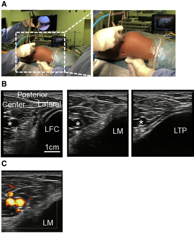
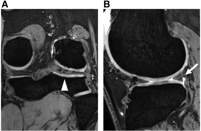
Arthroscopic repair of the posterior horn of the lateral meniscus (LM) from an anterolateral portal has a risk of popliteal artery injury. Here, we present an ultrasound-assisted, arthroscopic, all-inside repair technique for a posterior LM tear to reduce the risk of neurovascular injury. An ultrasound probe covered with a sterile sleeve is placed horizontally at the popliteal fossa by an assistant surgeon, and the popliteal artery and posterior LM are confirmed. From the anterolateral portal, an arthroscopic probe is inserted to push the posterior capsule of the lateral compartment, while an ultrasound image detects the tip of the probe. After the probe is confirmed not to be directed toward the popliteal artery, an all-inside suture device is introduced from the anterolateral portal. While the meniscus is penetrated, the surgeon can confirm by ultrasound images that the needle is directed away from the popliteal artery. The guide suture is pulled anteriorly to secure the anchors tightly, and an ultrasound confirms that the anchors are positioned behind the posterior portion of the LM. All sutures are secured under the assistance of ultrasound images, followed by arthroscopic confirmation of a properly secured LM by the all-inside repair technique.

Keywords: lateral meniscus tear; meniscal repair; ultrasound.


Figures

Fig 1
Fig 1
Preoperative MRI of the left knee joint (A: coronal planes; B: sagittal planes). (A) Disruption of the lateral meniscus (LM) is observed (arrowhead). (B) A gap is observed between the posterior LM and the capsule (arrow).
Fig 2
Fig 2
Arthroscopic images of the lateral meniscus (LM) injury in the left knee joint (viewed from an anteromedial portal). (A) The posterior portion of the LM shows fraying (asterisk) and disruption. (B) The posterior LM (asterisk) is pulled anteriorly over the center of the lateral compartment by probing. F, femoral condyle; T, tibial plateau.
Fig 3
Fig 3
Settings of ultrasound-assisted arthroscopic lateral meniscus repair in the left knee joint and ultrasound images of the posterior knee compartment. (A) Under an arthroscopic view from an anteromedial portal, a probe is inserted from the anterolateral portal with a figure 4 position. An ultrasound probe, covered with a sterile sleeve, is placed horizontally at the popliteal fossa by an assistant surgeon. (B) Ultrasound image of the posterior knee compartment showing the lateral femoral condyle (left panel), lateral meniscus (center panel), and lateral tibial plateau (right panel). The asterisk (∗) denotes popliteal artery. (C) Doppler image of the posterior knee compartment. LFC, lateral femoral condyle; LM, lateral meniscus; LTP, lateral tibial plateau.
Fig 4
Fig 4
Ultrasound-guided arthroscopic lateral meniscus repair in the left knee joint (arthroscopic view from an anteromedial portal and ultrasound image of the posterior lateral meniscus; LM). (A) All-inside suture device (JuggerStitch) is introduced from the anterolateral portal, and the meniscus (asterisk) is penetrated. (B) Ultrasound image shows the needle (yellow dashed line) avoiding the neurovascular structures (asterisk). (C) After two anchors are introduced at the posterior side of the LM (asterisk), (D) Ultrasound image confirms the anchors (arrowheads) are secured behind the posterior portion of the LM. Popliteal artery (asterisk). F, femoral condyle; T, tibial plateau.
Fig 5
Fig 5
Arthroscopic findings after meniscus repair under ultrasound guidance in the left knee joint (arthroscopic view from an anterolateral portal). (A) Arthroscopic examination shows the properly secured LM (asterisk) following the all inside repair technique. (B) Posterior LM (asterisk) is stabilized and is not pulled anteriorly over the center of the lateral compartment by probing.
Fig 6
Fig 6
Postoperative magnetic resonance image 3 months after arthroscopic meniscus repair under the guidance of ultrasound in the left knee joint. (A) Healing of the lateral meniscus (LM) is observed (arrowhead). (B) The gap between the posterior LM and the capsule has disappeared.

References

    1. Katano H., Koga H., Ozeki N., et al. Trends in isolated meniscus repair and meniscectomy in Japan, 2011-2016. J Orthop Sci. 2018;23:676–681. - PubMed
    1. Seil R., Becker R. Time for a paradigm change in meniscal repair: Save the meniscus. Knee Surg Sports Traumatol Arthrosc. 2016;24:1421–1423. - PubMed
    1. Ozeki N., Seil R., Krych A.J., Koga H. Surgical treatment of complex meniscus tear and disease: State of the art. J ISAKOS. 2021;6:35–45. - PubMed
    1. Sanada T., Iwaso H., Honda E., Yoshitomi H., Inagawa M. All-inside repair for radial tear at the posterior horn of the lateral meniscus: A figure-8 suture technique. Arthrosc Techn. 2021;10:e1973–e1977. - PMC - PubMed
    1. Lutz C., Dalmay F., Ehkirch F.P., et al. Meniscectomy versus meniscal repair: 10 years radiological and clinical results in vertical lesions in stable knee. Orthop Traumatol Surg. 2015;101:S327–S331. - PubMed

LinkOut - more resources