

Phenol-Soluble Modulins From Staphylococcus aureus Biofilms Form Complexes With DNA to Drive Autoimmunity
- PMID: 35646719
- PMCID: PMC9131096
- DOI: 10.3389/fcimb.2022.884065
Phenol-Soluble Modulins From Staphylococcus aureus Biofilms Form Complexes With DNA to Drive Autoimmunity
Abstract
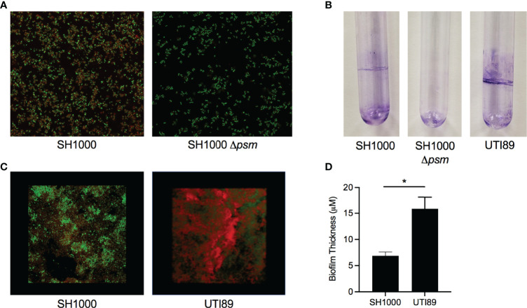
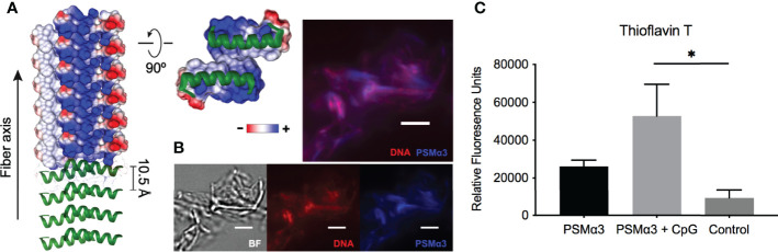
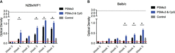
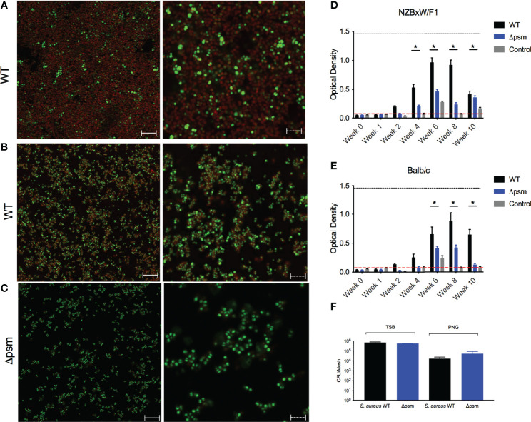
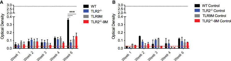
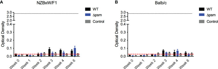
The bacterial amyloid curli, produced by Enterobacteriales including Salmonella species and Escherichia coli, is implicated in the pathogenesis of several complex autoimmune diseases. Curli binds to extracellular DNA, and these complexes drive autoimmunity via production of anti-double-stranded DNA autoantibodies. Here, we investigated immune activation by phenol-soluble modulins (PSMs), the amyloid proteins expressed by Staphylococcus species. We confirmed the amyloid nature of PSMs expressed by S. aureus using a novel specific amyloid stain, (E,E)-1-fluoro-2,5-bis(3-hydroxycarbonyl-4-hydroxy) styrylbenzene (FSB). Direct interaction of one of the S. aureus PSMs, PSMα3, with oligonucleotides promotes fibrillization of PSM amyloids and complex formation with bacterial DNA. Finally, utilizing a mouse model with an implanted mesh-associated S. aureus biofilm, we demonstrated that exposure to S. aureus biofilms for six weeks caused anti-double-stranded DNA autoantibody production in a PSM-dependent manner. Taken together, these results highlight how the presence of PSM-DNA complexes in S. aureus biofilms can induce autoimmune responses, and suggest an explanation for how bacterial infections trigger autoimmunity.
Keywords: PSM; Phenol Soluble Modulins; SLE; Staphycoccus aureus; autoimmune disease; biofilm; curli; mesh.
Copyright © 2022 Grando, Nicastro, Tursi, De Anda, Lee, Wong and Tükel.
Conflict of interest statement
The authors declare that the research was conducted in the absence of any commercial or financial relationships that could be construed as a potential conflict of interest.
Figures
Similar articles
-
Do amyloid structures formed by Staphylococcus aureus phenol-soluble modulins have a biological function?Int J Med Microbiol. 2018 Aug;308(6):675-682. doi: 10.1016/j.ijmm.2017.08.010. Epub 2017 Sep 1. Int J Med Microbiol. 2018. PMID: 28867522 Free PMC article.
-
Probing the drivers of Staphylococcus aureus biofilm protein amyloidogenesis and disrupting biofilms with engineered protein disaggregases.mBio. 2023 Aug 31;14(4):e0058723. doi: 10.1128/mbio.00587-23. Epub 2023 May 17. mBio. 2023. PMID: 37195208 Free PMC article.
-
Functional amyloids composed of phenol soluble modulins stabilize Staphylococcus aureus biofilms.PLoS Pathog. 2012;8(6):e1002744. doi: 10.1371/journal.ppat.1002744. Epub 2012 Jun 7. PLoS Pathog. 2012. PMID: 22685403 Free PMC article.
-
Phenol-soluble modulins and staphylococcal infection.Nat Rev Microbiol. 2013 Oct;11(10):667-73. doi: 10.1038/nrmicro3110. Epub 2013 Sep 10. Nat Rev Microbiol. 2013. PMID: 24018382 Free PMC article. Review.
-
Bacterial Amyloids: The Link between Bacterial Infections and Autoimmunity.Trends Microbiol. 2019 Nov;27(11):954-963. doi: 10.1016/j.tim.2019.07.002. Epub 2019 Aug 15. Trends Microbiol. 2019. PMID: 31422877 Free PMC article. Review.
Cited by
-
Amyloid-containing biofilms and autoimmunity.Curr Opin Struct Biol. 2022 Aug;75:102435. doi: 10.1016/j.sbi.2022.102435. Epub 2022 Jul 18. Curr Opin Struct Biol. 2022. PMID: 35863164 Free PMC article. Review.
-
YjbH contributes to Staphylococcus aureus skin pathology and immune response through Agr-mediated α-toxin regulation.Virulence. 2024 Dec;15(1):2399798. doi: 10.1080/21505594.2024.2399798. Epub 2024 Sep 9. Virulence. 2024. PMID: 39229975 Free PMC article.
-
Bioprospecting the Skin Microbiome: Advances in Therapeutics and Personal Care Products.Microorganisms. 2023 Jul 27;11(8):1899. doi: 10.3390/microorganisms11081899. Microorganisms. 2023. PMID: 37630459 Free PMC article. Review.
-
Extracellular G-quadruplexes and Z-DNA protect biofilms from DNase I, and G-quadruplexes form a DNAzyme with peroxidase activity.Nucleic Acids Res. 2024 Feb 28;52(4):1575-1590. doi: 10.1093/nar/gkae034. Nucleic Acids Res. 2024. PMID: 38296834 Free PMC article.
-
DNA at the center of mammalian innate immune recognition of bacterial biofilms.Trends Immunol. 2024 Feb;45(2):103-112. doi: 10.1016/j.it.2023.12.004. Epub 2024 Jan 27. Trends Immunol. 2024. PMID: 38281884 Free PMC article. Review.
References
-
- Bird A. P. (1987). CpG Islands as Gene Markers in the Vertebrate Nucleus. Trends Genet. 3, 342–347. doi: 10.1016/0168-9525(87)90294-0 - DOI
-
- Ceccarelli F., Perricone C., Olivieri G., Cipriano E., Spinelli F. R., Valesini G., et al. . (2019). Staphylococcus Aureus Nasal Carriage and Autoimmune Diseases: From Pathogenic Mechanisms to Disease Susceptibility and Phenotype. Int. J. Mol. Sci. 20, E5624. doi: 10.3390/ijms20225624 - DOI - PMC - PubMed
Publication types
MeSH terms
Substances
Grants and funding
LinkOut - more resources
Full Text Sources
Medical
Miscellaneous