. 2022 May 11:12:873536.
doi: 10.3389/fcimb.2022.873536. eCollection 2022.

The Regulatory Protein ChuP Connects Heme and Siderophore-Mediated Iron Acquisition Systems Required for Chromobacterium violaceum Virulence

Affiliations

The Regulatory Protein ChuP Connects Heme and Siderophore-Mediated Iron Acquisition Systems Required for Chromobacterium violaceum Virulence

Vinicius M de Lima et al. Front Cell Infect Microbiol. .

Abstract

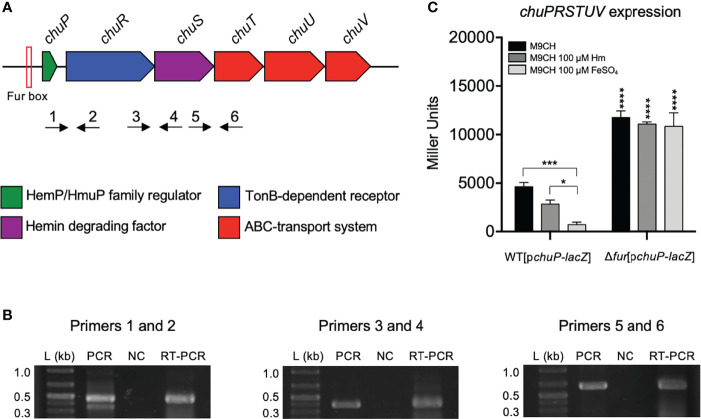
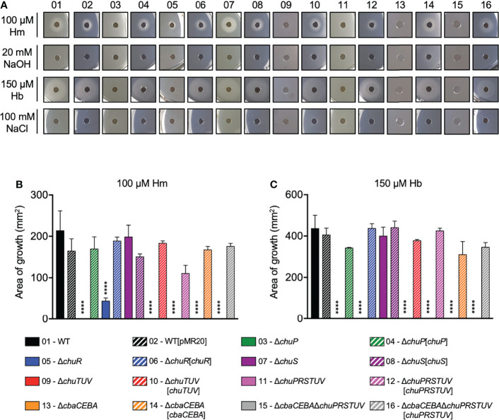
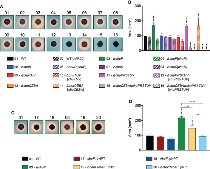
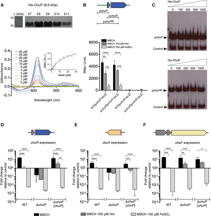
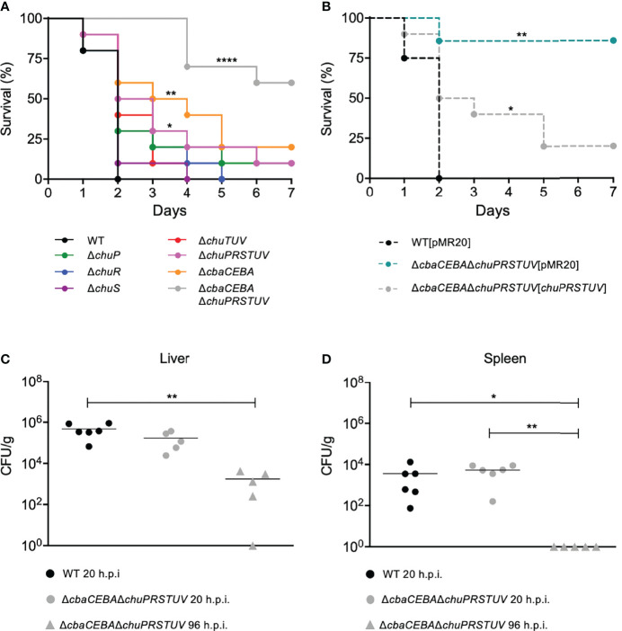
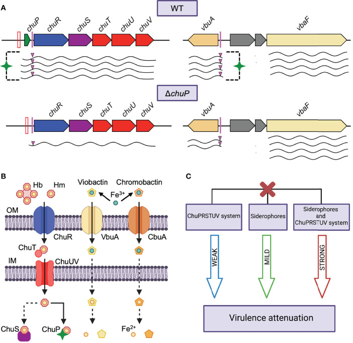
Chromobacterium violaceum is an environmental Gram-negative beta-proteobacterium that causes systemic infections in humans. C. violaceum uses siderophore-based iron acquisition systems to overcome the host-imposed iron limitation, but its capacity to use other iron sources is unknown. In this work, we characterized ChuPRSTUV as a heme utilization system employed by C. violaceum to explore an important iron reservoir in mammalian hosts, free heme and hemoproteins. We demonstrate that the chuPRSTUV genes comprise a Fur-repressed operon that is expressed under iron limitation. The chu operon potentially encodes a small regulatory protein (ChuP), an outer membrane TonB-dependent receptor (ChuR), a heme degradation enzyme (ChuS), and an inner membrane ABC transporter (ChuTUV). Our nutrition growth experiments using C. violaceum chu deletion mutants revealed that, with the exception of chuS, all genes of the chu operon are required for heme and hemoglobin utilization in C. violaceum. The mutant strains without chuP displayed increased siderophore halos on CAS plate assays. Significantly, we demonstrate that ChuP connects heme and siderophore utilization by acting as a positive regulator of chuR and vbuA, which encode the TonB-dependent receptors for the uptake of heme (ChuR) and the siderophore viobactin (VbuA). Our data favor a model of ChuP as a heme-binding post-transcriptional regulator. Moreover, our virulence data in a mice model of acute infection demonstrate that C. violaceum uses both heme and siderophore for iron acquisition during infection, with a preference for siderophores over the Chu heme utilization system.

Keywords: Chromobacterium violaceum; bacterial physiology; bacterial virulence; heme transporter; heme uptake; iron homeostasis; siderophores.


Conflict of interest statement

The authors declare that the research was conducted in the absence of any commercial or financial relationships that could be construed as a potential conflict of interest.

Figures

Figure 1
Figure 1
The chuPRSTUV genes compose an operon regulated by the iron levels and Fur. (A) Genomic organization of the chuPRSTUV genes in C. violaceum. A predicted Fur box is indicated. Numbered arrows indicate the primers used in RT-PCR (not scaled). (B) Confirmation of co-transcription of the chuPRSTUV genes. The RT-PCR reactions amplified fragments of 439 bp (Primers 1 and 2), 373 bp (Primers 3 and 4), and 662 bp (Primers 5 and 6). Conventional PCR was performed using genomic DNA (PCR) and RNA (NC) as controls. L, 1 Kb plus DNA Ladder (Thermo Scientific). (C) Promoter activity of the chu operon in response to iron and Fur. β-galactosidase assays were performed from the WT and Δfur strains harboring the chuP-lacZ fusion grown in M9CH medium and either untreated or treated with 100 μM Hm or 100 μM FeSO4. Data are from three biological replicates. ****p < 0.0001; ***p < 0.001; *p < 0.05; when not indicated, not significant. Two-way ANOVA followed by Dunnett’s multiple-comparison test.
Figure 2
Figure 2
The chu operon encodes a heme uptake system (ChuRTUV) and a regulatory protein (ChuP) required for heme and hemoglobin utilization. (A) Nutrition assay for Hm and Hb under DP-imposed iron deficiency. The indicated strains were embedded into M9CH medium supplemented with 125 μM DP. Aliquots of 100 μM Hm and 150 μM Hb were provided as iron sources, while 20 mM NaOH and 100 mM NaCl were used as negative controls. Growth halos around the discs indicate compound utilization. Representative images are shown. (B, C) Quantification of Hm and Hb-stimulated growth. The area of the growth halos stimulated by Hm (B) and Hb (C) was measured using Image J software by subtracting the area of the discs. Data are from three biological replicates. Mutant and complemented strains were compared to WT and WT[pMR20], respectively. ****p < 0.0001; when not indicated, not significant. One-way ANOVA followed by Tukey’s multiple-comparison test.
Figure 3
Figure 3
Deletion of chuP impacts the siderophore levels in C. violaceum. (A, B) Role of the chu operon on the siderophore levels in C. violaceum. Mutant strains without chuP showed increased siderophore halos. (C, D) The effect of ChuP occurs on the siderophore viobactin. For all indicated strains, the siderophore detection was performed by CAS assays on PSA-CAS plates. C. violaceum cultures were spotted onto the plate surface, and the orange halos indicating secreted siderophores were photographed (A, C) and measured (B, D), after incubation for 24 hours at 37°C, using Image J software. The area of the siderophore halos was calculated subtracting the area of bacterial growth. Data are from three biological replicates. Mutant and complemented strains (B) were compared to WT and WT[pMR20], respectively. Insertion mutants (D) were compared to the strains they derived from. **p < 0.01; ***p < 0.001; ****p < 0.0001; when not indicated, not significant. Vertical asterisks indicate comparisons with the WT strain. One-way ANOVA followed by Tukey’s multiple-comparison test.
Figure 4
Figure 4
ChuP is a heme-binding regulatory protein that controls chuR and vbuA expression at a post-transcriptional level. (A) ChuP binds heme. The His-ChuP protein was purified (top) and incubated (10 μM protein) with the indicated concentrations of Hm (bottom). The absorption spectra were measured from 300 nm to 600 nm on a SpectraMax i3 MiniMax Imaging Cytometer. The changes at 413 nm were used to calculate the ChuP-Hm affinity (insert). Data are shown as differential absorption spectra: the difference of values obtained from the sample cuvette (His-ChuP and Hm) against the reference cuvette (Hm). Data are from a single experiment of three independent replicates. L, protein ladder; E7 to E11, eluted fractions of purified ChuP. (B) ChuP does not regulate the promoter of the chu operon. The scheme (top) indicates the regions used for β-galactosidase or EMSA assays. β-galactosidase assays were performed from the WT and ΔchuP strains harboring chuP-lacZ and chuR-lacZ fusions grown in M9CH in the indicated conditions of iron availability. Data are from three biological replicates. **p < 0.01; ***p < 0.001; ****p < 0.0001. When not shown, n.s. (not significant). Two-way ANOVA followed by Dunnett’s multiple-comparison test. (C) ChuP does not bind to DNA probes covering from chuP to chuR. The indicated concentrations of His-ChuP were used in EMSA assays with the chu indicated probes. In both cases, the promoter region of CV_2599 (control) was used as an in-reaction unspecific negative control. (D-F) chuR and vbuA but not vbaF have HPRE sequences and are regulated by ChuP. The predicted HPREs are indicated as colored bars in the gene maps. Expression was evaluated by RT-qPCR. cDNA was reverse transcribed from RNA obtained from the WT, ΔchuP, and ΔchuP[chuP] strains grown in M9CH, and either untreated or treated with 100 μM Hm or 100 μM FeSO4. Expression of chuR, vbuA, and vbaF is shown as the fold change relative to the control condition (WT in M9CH 100 μM Hm). Data are from three biological replicates. ****p < 0.0001; ***p < 0.001; **p < 0.01; *p < 0.05; when not indicated, not significant. Vertical asterisks indicate comparisons with the WT strain at the same condition. One-way ANOVA followed by Tukey’s multiple-comparison test.
Figure 5
Figure 5
C violaceum requires siderophores and heme but prefers siderophores in a mice model of acute infection. (A, B) Survival curves of infected BALB/c mice. Animals (n = 8 for WT[pMR20]; n = 7 for ΔcbaCEBAΔchuPSRTUV[chuPRSTUV]; n = 10 for all other strains) were i.p. injected with 106 CFU of the indicated C. violaceum mutant (A) and complemented (B) strains. Animal survival was monitored daily for a week. *p < 0.05; **p < 0.01; ****p < 0.0001; when not shown, n.s. (not significant). Log-rank (Mantel-Cox) test. (C, D) Bacterial burden in organs. Animals were infected with 106 CFU of the indicated strains. After 20h or 96 h post-infection (h.p.i.), the liver (C) and the spleen (D) were collected, homogenized, serially diluted, and plated for CFU quantification. *p < 0.05; **p < 0.01; when not indicated, not significant. One-way ANOVA followed by Tukey’s (liver) or Dunnett’s (spleen) multiple-comparison tests.
Figure 6
Figure 6
Model of how C. violaceum connects iron acquisition by heme and siderophore during infection. (A) Regulatory function of ChuP over chuR and vbuA. (B) Iron acquisition systems in C. violaceum for the uptake of heme (this work) and the siderophores viobactin and chromobactin (Batista et al., 2019). (C) Interplay between the uptake of heme and siderophore in the C. violaceum virulence. We propose that ChuP links heme and siderophore utilization by acting as a positive regulator of chuR and vbuA, which encode TBDRs for the uptake of heme (ChuR) and the siderophore viobactin (VbuA). In addition to Fur derepression, the expression of chuR and vbuA depends on ChuP under iron deficiency, possibly by a post-transcriptional mechanism involving HPRE (HmuP-responsive elements) sequences found in the 5’-UTR of the chuR and vbuA transcripts. In the absence of ChuP, the abundance of chuR and vbuA transcripts decreases, causing a reduction in heme and viobactin utilization. Different levels of virulence attenuation occurred when the ChuPRSTUV system (weak), the siderophores (mild), or both (strong) were deleted, indicating that C. violaceum prefers siderophores over heme during infection but relies on heme in the absence of siderophores. Red bars, Fur boxes; Pink bars and triangles, predicted HPRE sequences; Green stars, ChuP protein; Waved lines, mRNAs; Red X, mutant strains; OM, Outer membrane; IM, Inner membrane; Dashed lines, unknown mechanisms.

Similar articles

Cited by

References

    1. Abdelhamed H., Ibrahim I., Baumgartner W., Lawrence M. L., Karsi A. (2018). The Virulence and Immune Protection of Edwardsiella Ictaluri HemR Mutants in Catfish. Fish. Shellfish. Immunol. 72, 153–160. doi: 10.1016/j.fsi.2017.10.041 - DOI - PubMed
    1. Amarelle V., Koziol U., Fabiano E. (2019). Highly Conserved Nucleotide Motifs Present in the 5’UTR of the Heme-Receptor Gene shmR are Required for HmuP-Dependent Expression of shmR in Ensifer Meliloti . Biometals 32, 273–291. doi: 10.1007/s10534-019-00184-6 - DOI - PubMed
    1. Amarelle V., Koziol U., Rosconi F., Noya F., O’Brian M. R., Fabiano E. (2010). A New Small Regulatory Protein, HmuP, Modulates Haemin Acquisition in Sinorhizobium Melioti . Microbiology 156, 1873–1882. doi: 10.1099/mic.0.037713-0 - DOI - PMC - PubMed
    1. Amarelle V., Rosconi F., Lázaro-Martinez J. M., Buldain G., Noya F., O’Brian M. R., et al. . (2016). HmuS and HmuQ of Ensifer/Sinorhizobium Meliloti Degrade Heme In Vitro and Participate in Heme Metabolism In Vivo . Biometals 29, 333–347. doi: 10.1007/s10534-016-9919-3 - DOI - PubMed
    1. Balhesteros H., Shipelskiy Y., Long N. J., Majumdar A., Katz B. B., Santos N. M., et al. . (2017). TonB-Dependent Heme/Hemoglobin Utilization in Caulobacter Crescentus HutA. J. Bacteriol. 199, e00723-16. doi: 10.1128/JB.00723-16 - DOI - PMC - PubMed

Publication types

Supplementary concepts

LinkOut - more resources