. 2022 May 11:12:824575.
doi: 10.3389/fcimb.2022.824575. eCollection 2022.

Effect of Angiogenesis and Lymphangiogenesis in Diesel Exhaust Particles Inhalation in Mouse Model of LPS Induced Acute Otitis Media

Affiliations

Effect of Angiogenesis and Lymphangiogenesis in Diesel Exhaust Particles Inhalation in Mouse Model of LPS Induced Acute Otitis Media

Byeong-Gon Kim et al. Front Cell Infect Microbiol. .

Abstract

Lymphangiogenesis and angiogenesis might have significant involvement in the pathogenesis of otitis media with effusion. This study investigated the effect of diesel exhaust particles (DEP) on inflammation and lymphangiogenesis in a mouse model of acute otitis media (AOM). BALB/c mice were injected with LPS and exposed to 100 µg/m3 DEP. The mice were divided into four groups: control (no stimulation), AOM, AOM + DEP, and DEP + AOM. The effects of DEP inhalation pre- and post-DEP induction were estimated based on measurements of the auditory brainstem response, mRNA levels of lymphangiogenesis-related genes and cytokines, and histology of the middle ear. Cell viability of human middle ear epithelial cells decreased in a dose-response manner at 24 and 48 hours post-DEP exposure. DEP alone did not induce AOM. AOM-induced mice with pre- or post-DEP exposure showed thickened middle ear mucosa and increased expression of TNF-α and IL1-β mRNA levels compared to the control group, but increased serum IL-1β levels were not found in the AOM + Post DEP. The mRNA expression of TLR4, VEGFA, VEGFAC, and VEGFR3 was increased by pre-AOM DEP exposure. The expression of VEFGA protein was stronger in the AOM + Post DEP group than in any other group. The expression of CD31 and CD45 markers in the mouse middle ear tissue was higher in the Pre DEP + AOM group than in the AOM group. This result implies that pre-exposure to DEP more strongly increases inflammation and lymphangiogenesis in a mouse model of acute otitis media.

Keywords: angiogenesis; diesel exhaust particles; lipopolysaccharides; lymphangiogenesis; otitis media.


Conflict of interest statement

The authors declare that the research was conducted in the absence of any commercial or financial relationships that could be construed as a potential conflict of interest.

Figures

Figure 1
Figure 1
Viability of LPS- and DEP-exposed HMEECs assessed by the CCK-8 assay and study design of the in vitro model. LPS and DEP exposure decreased cell viability in HMEECs. *p < 0.05 compared with the 24 hr and 48 hr normal control groups. LPS, lipopolysaccharide; DEP, diesel exhaust particles; HMEECs, human middle ear epithelial cells.
Figure 2
Figure 2
Study design and DEP inhalation in a mouse model of acute otitis media induced by transtympanic LPS injection. (A) HMEECs were treated with a cytotoxic dose of LPS (1 µg/mL) and DEP (80 µg/mL) for 24 hr and 48 hr, or both in different sequences or simultaneously as indicated. (B) Experimental protocol for DEP exposure in a mouse model of AOM (n = 13 in each group) (DEP 100 µg/m3; 10 µL emulsion of 1 mg/mL LPS). (C) The DEP were observed in transmission electron microscopy images inside eustachian tube cells connected to tissues only among mice that inhaled 100 µg/m3 DEP for 1 hr a day for 5 days a week (N, nucleus; scale bar, 1 µm; magnification ×30,000). LPS, lipopolysaccharide; DEP, diesel exhaust particles; HMEECs, human middle ear epithelial cells; AOM, acute otitis media.
Figure 3
Figure 3
Acute otitis media symptoms and H&E staining analysis of the middle ear in LPS + DEP exposure mouse. (A) Otoscope images of the tympanic membrane before sacrifice. (B) H&E staining of middle ear pathology. (C) The thickness of the middle ear epithelium is higher in the DEP + LPS exposure group. (D) The symptom score is higher in the acute otitis media group. (E–G) Auditory brainstem response threshold changes in the mouse model. The same location shown in each slide was chosen (H&E, magnification ×400). *p < 0.05, compared with the control group; ***p < 0.05 compared to the AOM group. LPS, lipopolysaccharide; DEP, diesel exhaust particles; H&E, hematoxylin and eosin; AOM, acute otitis media.
Figure 4
Figure 4
Analysis of cytokine expression in LPS + DEP-exposed mice and HMEECs. The qRT-PCR analysis of mRNA levels of (A) IL-1β and (B) TNF-α in LPS in HMEECs. The mRNA levels of (C) IL-1β and (D) TNF-α a mouse model of acute otitis media. The cytokines in the serum of mice were assessed via ELISA kits, including (E) IL-1β, (F) TNF-α. (G) Immunohistochemical staining of mouse middle ear tissue using antibodies against CD45 (Microvessel; black arrow, magnification ×400). (H) Quantitation of the CD45 staining intensity. *p < 0.05, compared with the control group; ***p < 0.05 compared to the LPS 24 hr and AOM group. LPS, lipopolysaccharide; DEP, diesel exhaust particles; HMEECs, human middle ear epithelial cells; AOM, acute otitis media.
Figure 5
Figure 5
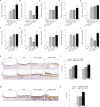
Analysis of the expression of angiogenesis and lymphangiogenesis-related genes in LPS + DEP-exposed mice and HMEECs. The results show the mRNA levels of (A) TLR4 and (B) VEGFA, (C) VEGFC, (D) VEGFR1, and (E) VEGFR3 in LPS-exposed HMEECs. The mRNA levels of (F) TLR4 and (G) VEGFA, (H) VEGFC, (I) VEGFR1, (J) VEGFR3 in LPS in a mouse model of AOM. Immunohistochemical (IHC) staining of mouse middle ear tissue using antibodies against (K) VEGFA, VEGFC, and (M) CD31 (IHC, magnification ×400). The same location shown in each slide was chosen (IHC, magnification ×400). (L, N) Quantitation of the CD45 staining intensity. *p < 0.05, compared with the control group; ***p < 0.05 compared to the LPS 24 hr and AOM group. LPS, lipopolysaccharide; DEP, diesel exhaust particles; HMEECs, human middle ear epithelial cells; AOM, acute otitis media.
Figure 6
Figure 6
Summary of the pathway of diesel exhaust particle-induced angiogenesis and lymphangiogenesis in acute otitis media.

Similar articles

References

    1. Becker S., Soukup J. M. (1999). Exposure to Urban Air Particulates Alters the Macrophage-Mediated Inflammatory Response to Respiratory Viral Infection. J. Toxicol. Environ. Health A 57 (7), 445–457. doi: 10.1080/009841099157539 - DOI - PubMed
    1. Brauer M., Gehring U., Brunekreef B., de Jongste J., Gerritsen J., Rovers M., et al. . (2006). Traffic-Related Air Pollution and Otitis Media. Environ. Health Perspect. 114 (9), 1414–1418. doi: 10.1289/ehp.9089 - DOI - PMC - PubMed
    1. Cheeseman M. T., Tyrer H. E., Williams D., Hough T. A., Pathak P., Romero M. R., et al. . (2011). HIF-VEGF Pathways are Critical for Chronic Otitis Media in Junbo and Jeff Mouse Mutants. PloS Genet. 7 (10), e1002336. doi: 10.1371/journal.pgen.1002336 - DOI - PMC - PubMed
    1. Chun Y. M., Moon S. K., Lee H. Y., Webster P., Brackmann D. E., Rhim J. S., et al. . (2002). Immortalization of Normal Adult Human Middle Ear Epithelial Cells Using a Retrovirus Containing the E6/E7 Genes of Human Papillomavirus Type 16. Ann. Otol. Rhinol. Laryngol 111, 507–517. doi: 10.1177/000348940211100606 - DOI - PubMed
    1. Clifford S., Mazaheri M., Salimi F., Ezz W. N., Yeganeh B., Low-Choy S., et al. . (2018). Effects of Exposure to Ambient Ultrafine Particles on Respiratory Health and Systemic Inflammation in Children. Environ. Int. 114, 167–180. doi: 10.1016/j.envint.2018.02.019 - DOI - PubMed

Publication types