Review
. 2022 May 12:10:886382.
doi: 10.3389/fchem.2022.886382. eCollection 2022.

Deciphering the Structure and Formation of Amyloids in Neurodegenerative Diseases With Chemical Biology Tools

Affiliations
Review

Deciphering the Structure and Formation of Amyloids in Neurodegenerative Diseases With Chemical Biology Tools

Isabelle Landrieu et al. Front Chem. .

Abstract

Protein aggregation into highly ordered, regularly repeated cross-β sheet structures called amyloid fibrils is closely associated to human disorders such as neurodegenerative diseases including Alzheimer's and Parkinson's diseases, or systemic diseases like type II diabetes. Yet, in some cases, such as the HET-s prion, amyloids have biological functions. High-resolution structures of amyloids fibrils from cryo-electron microscopy have very recently highlighted their ultrastructural organization and polymorphisms. However, the molecular mechanisms and the role of co-factors (posttranslational modifications, non-proteinaceous components and other proteins) acting on the fibril formation are still poorly understood. Whether amyloid fibrils play a toxic or protective role in the pathogenesis of neurodegenerative diseases remains to be elucidated. Furthermore, such aberrant protein-protein interactions challenge the search of small-molecule drugs or immunotherapy approaches targeting amyloid formation. In this review, we describe how chemical biology tools contribute to new insights on the mode of action of amyloidogenic proteins and peptides, defining their structural signature and aggregation pathways by capturing their molecular details and conformational heterogeneity. Challenging the imagination of scientists, this constantly expanding field provides crucial tools to unravel mechanistic detail of amyloid formation such as semisynthetic proteins and small-molecule sensors of conformational changes and/or aggregation. Protein engineering methods and bioorthogonal chemistry for the introduction of protein chemical modifications are additional fruitful strategies to tackle the challenge of understanding amyloid formation.

Keywords: aggregation; amyloid fibril; fluorescent probes; nanobody; native chemical ligation; neurodegenerative diseases; posttranslational modifications; protein semisynthesis.


Conflict of interest statement

The authors declare that the research was conducted in the absence of any commercial or financial relationships that could be construed as a potential conflict of interest.

Figures

FIGURE 1
FIGURE 1
Amyloid proteins involved in neurodegenerative diseases (tau, Aβ, α-synuclein, TDP-43 and Huntingtin), and their amyloid folds and polymorphs. Scheme of protein sequences highlighting domains, and eventually isoforms are presented (left). For tau protein, some prominent pathological phospho-epitopes (AT8, AT180, PHF-1) associated to tauopathies are indicated. N1, N2, N-terminal inserts; R1-R4, microtubule-binding repeats 1 to 4; NAC, non-amyloid-β component; NTD, N-terminal domain; NLS, nuclear localization signal; RRM, RNA Recognition Motifs; LCD, low complexity domain; N17, 17-residue N-terminal region; PRD, proline-rich domain; polyQ, polyglutamine; HEAT, HEAT repeats. The solution NMR structure of micelle-bound α-synuclein is depicted. Some PTM sites studied by chemical ligation and/or chemical mutagenesis are indicated. The cryo-EM structures of representative fibrils from individual brains (grey structures) and synthetic fibrils (blue structures) highlight structural discrepancies pointing to the role of trans-acting cofactors and PTMs of amyloid proteins in the amyloidogenic process. For a more comprehensive description of the multiple tau folds associated to diverse tauopathies and their classification, refer to (Shi et al., 2021). AD, Alzheimer’s disease; CBD, corticobasal degeneration; PiD, Pick’s disease; MSA, multiple system atrophy; ALS with FTLD, amyotrophic lateral sclerosis with frontotemporal lobar degeneration.
FIGURE 2
FIGURE 2
Investigating the role of PTMs in amyloidogenesis by protein semisynthesis and/or chemical mutagenesis. Amyloid proteins are extracted from patient brains and purified as insoluble material, then the PTM patterns of amyloid fibrils are deciphered by complementary biophysical and biochemical tools (right panel). The precise role of site-specific PTMs is unraveled by encoding individual PTM or pathological epitope using chemical biology tools. This allows investigating the effect of these specific chemical modifications on protein conformation and function, on oligomer and amyloid assembly, stability and properties (left panel). The synthetic fibril morphology and cryo-EM atomic structures could thus be compared to bona fide fibrils from patient brains, here illustrated with pY39 α-synuclein synthetic fibrils (PDB ID: 6LIT) and α-synuclein fibrils from MSA (PDB ID: 6XYO), providing important information about the role of cofactors in amyloid aggregation.
FIGURE 3
FIGURE 3
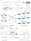
Expressed protein ligation (EPL) strategy for site-specific modification of proteins (A) and segmental isotope labeling for NMR studies (B). (A) The synthetic peptide (blue) incorporating a site-specific modification (yellow) is obtained by SPPS and ligated to a recombinant protein fragment (orange) expressed in a heterologous system which can eventually be isotopically labeled for NMR study. The recombinant protein can be expressed as intein fusion protein (intein in green) with a CBD tag (pink) for purification on chitin beads. The reaction with sodium 2-mercaptoethane sulfonate (MESNa) and a synthetic peptide with a N-terminal cysteine leads to a semisynthetic protein. (B) In the NMR 1H-15N HSQC spectrum, only the 15N-labeled region of the protein is visible, therefore the synthetic region bearing the modification is invisible (orange spectrum). This strategy called segmental isotopic labeling allows a reduction of NMR signals in the spectrum relative to the full-length, uniformly labeled protein (black) without PTM (black spectrum) or with enzymatically installed PTMs (yellow spectrum). It is noteworthy that modifications of protein resonances observed locally for the modified residues and its neighbors in the primary sequence in the uniformly labeled protein cannot be observed in the semisynthetic protein with segmental labeling due to the absence of isotopic labeling in the region of PTM. However, this modification may have a long-range structural impact on residues of the 15N-labeled fragment due to conformational proximity that can be detected through perturbations of signals in the isotopically labeled region.
FIGURE 4
FIGURE 4
Chemical biology strategies of protein ubiquitination. (A) Posttranslational chemical mutagenesis and EPL approaches involve both the preparation of a C-terminal activated ubiquitin from a ubiquitin-intein fusion protein that provides a reactive C-terminal thioester. (B) Posttranslational chemical mutagenesis exemplified for K23 ubiquitination of α-synuclein requires first the thiol activation of a cysteine residue obtained by site-directed mutagenesis at the PTM site. The activated thiol is further involved in a reaction with ubiquitin-aminoethanethiol (ubiquitin-AET) leading to a bis-thio-acetone (BTA) analog of the ubiquitin-lysine isopeptide bond (Lewis et al., 2016). Alternatively, a ubiquitin-AET activated by 2,2′-dithiobis(5-nitropyridine) (ubiquitin-AET-DTNP) can react with the cysteine thiol in a disulfide-directed ubiquitination (Moon et al., 2020). (C) The EPL strategy involves first, the synthesis of full-length α-synuclein by EPL of two fragments, a N-terminal peptide (fragment 1–18) bearing a protected δ-mercaptolysine at position K6 for further ubiquitin linkage and a C-terminal recombinantly expressed fragment with a N-terminal cysteine. After NCL and thiol deprotection of the δ-mercaptolysine, EPL with the recombinantly expressed ubiquitin with C-terminal thioester and subsequent desulfurization provide the full-length, native α-synuclein with a native ubiquitin-lysine isopeptide bond (Hejjaoui et al., 2011). Chemical reactions are indicated by blue arrows and enzymatic reactions by red arrows.
FIGURE 5
FIGURE 5
EPL strategy applied to the semisynthesis of full-length tau-S400-O-GlcNAc protein (Schwagerus et al., 2016). The C-terminal synthetic fragment incorporating the S400-O-GlcNAc modification and a biotin selectively linked to K438 residue for purification is obtained by NCL of two synthetic peptides, followed by desulfurization to restore the native A426 at the ligation site and deprotection of the GlcNAc moiety. The resulting C-terminal S400-O-GlcNAc peptide is then ligated by EPL with the N-terminal fragment recombinantly expressed as intein-GST fusion purified on glutathione affinity chromatography. The ligation product is isolated from the unreacted recombinant fragment through streptavidin-biotin affinity chromatography and traceless released by photocleavage of the biotin linker. The final product is the full-length protein tau-S400-O-GlcNAc with a A390C mutation at the EPL ligation site that cannot be recovered by desulfurization due to the presence of two native cysteine in tau sequence.
FIGURE 6
FIGURE 6
Posttranslational chemical mutagenesis. (A) General scheme of the posttranslational chemical mutagenesis approach for incorporation of PTM mimics by Michael addition of PTM-thiol derivatives on dehydroalanine (Dha) alkene function. The latter is obtained from serine/threonine/lysine-to-cysteine mutation at the PTM site and β-elimination of the bis-alkylated thiol function of cysteine. Addition of thiophosphate, S-GlcNAc, N-acetylcysteamine and captamine generates mimetics of phosphorylation, O-GlcNAcylation, N-acetylation and N,N-dimethylation, respectively. The thio-ether bond formation leads to a racemization of amino acid Cα. (B) This approach is illustrated by S356 phosphorylation and K311 acetylation in tau protein for the investigation of the PTM effect on loss-of-function in microtubule polymerization (PDB ID: 7PQP) (Brotzakis et al., 2021; Lindstedt et al., 2021). Chemical reactions are indicated by blue arrows and enzymatic reactions by red arrows.
FIGURE 7
FIGURE 7
Posttranslational chemical mutagenesis, genetic code expansion and chemo-enzymatic reactions for incorporation of O-GlcNAc mimics and derivatives for O-GlcNAc detection/purification. Chemical reactions are indicated by blue arrows and enzymatic reactions by red arrows. Some examples of the manifold approaches for the incorporation of S-GlcNAc and O-GlcNAc derivatives are given. An extensive review of O-GlcNAc installation and detection can be found in (Saha et al., 2021). Site-specific O-GlcNAc mimics can be incorporated directly on the cysteine thiol function after Ser/Thr into Cys site-directed mutagenesis or using the gene-editing tool CRISPR-Cas9. This allows for instance the enzymatic installation of S-GlcNAc mimics with OGT leading to a stable GlcNAc derivative since hydrolysis by OGA is not possible. Alternatively, the engineered cysteine residue may also be an intermediate to provide a bioorthogonal reactive dehydroalanine (Dha) derivative after thiol bis-alkylation and subsequent β-elimination. The Dha handle is amenable to a Michael addition with a GlcNAc-thiol derivative for installation of a S-GlcNAc derivative. A third approach involved the incorporation of unnatural amino acids (UAAs) bearing a reactive handle amenable to “click” chemistry, e.g., acetophenone or propargyl-lysine, using the amber codon suppression strategy of genetic code expansion for the site-specific O-GlcNAc modifications. Coupling of the O-GlcNAc moiety is performed by azide-alkyne [3 + 2] cycloaddition. For detection or enrichment of O-GlcNAcylated proteins, labeling of O-GlcNAc is performed as a first step either (i) by “metabolic labeling” through oligosaccharide engineering that introduces chemical handles (azide, alkyne,…) directly on the GlcNAc group for subsequent reaction (via the biosynthesis of UDP-GlcNAc derivatives, depicted in the box) or (ii) by “chemoenzymatic labeling” through an enzymatic reaction of the O-GlcNAc group with a mutant of β1,4-galactosyltransferase (GalT-Y289L) that forms a glycosidic bond with a galactose derivative (GalNAz). In both strategies, the second step is the subsequent conjugation of reactive probes (e.g., fluorescent probes, mass tag, biotin…) for downstream enrichment or detection. The latter processes through a reaction of “click” chemistry making use of azide bioorthogonal reactivity through the copper(I)-catalyzed (CuAAC) or strain-promoted azide-alkyne cycloaddition (SPAAC).
FIGURE 8
FIGURE 8
Detection of amyloids by fluorescence labeling (A–D) or functionalized gold nanoparticles (E). (A) Structures of extrinsic fluorescent probes used for amyloid detection. (B) Aggregation-induced emission (AIE) molecules exemplified by Thioflavin-T (ThT) emit a fluorescent signal upon binding into hydrophobic pockets of amyloid β-sheet structures due to restriction of intramolecular rotation (PDB ID: 3MYZ). The structure of [11C]-Pittsburgh Compound B used for PET imaging is based on ThT structure. (C) Some examples of fluorescent molecules based on BODIPY or tetraphenylethylene (TPE) scaffolds are depicted as well as a near-infrared (near-IR) fluorescent probe and the oligothiophene p-FTAA (pentameric form of formyl thiophene acetic acid). (D) An example of fluorescent SNAP-tagging is depicted in a strategy called “AggTag” for amyloid detection. The engineering of a SNAP-tag amyloid protein allows coupling of an AggFluor probe through the selective reaction of a SNAP tag cysteine residue with O-benzylguanine derivatives. The subsequent aggregation of amyloid-forming proteins turns on fluorescence of the AggFluor probe. Various AggFluor probes has been designed to detect oligomers and amyloid fibrils, or amyloid fibrils selectively (Liu et al., 2018; Wolstenholme et al., 2020). (E) Functionalization of gold nanoparticles (gold-thiol polymer) with a mixture of 11-mercapto-1-undecanesulfonate: 1-octanethiol (MUS:OT) enables the selective detection of amyloid fibrils and polymorphism by cryo-EM imaging (Cendrowska et al., 2020).
FIGURE 9
FIGURE 9
Nitroxide spin labeling and FRET fluorescent pair labeling for distance measurements by NMR, EPR and FRET exemplified in tau protein. (A) Nitroxide spin labels such as MTSL (S-(1-oxyl-2,2,5,5-tetramethyl-2,5-dihydro-1H-pyrrol-3-yl)methyl methanesulfonothioate) can be introduced on thiol function(s) of engineered cysteine residues obtained by site-directed mutagenesis for NMR experiments. Single spin label is used in paramagnetic relaxation enhancement (PRE) experiments. Double spin labeling of two distinct cysteine residues is used for paramagnetic relaxation interference (PRI) experiments (not shown in the figure). The same labeling strategy with single or double paramagnetic probes can be used in EPR experiments (not shown in the figure). Single spin labels are introduced at different positions along the protein sequence as illustrated by A72C and A239C labeling. Proximal and long-range perturbations of NMR signals (distance >10 Å up to 25 Å) from the spin label is indicated by red areas (Bibow et al., 2011). The PRE phenomenon is characterized by a selected decrease of peak intensities in the 1H-15N HSQC experiment acquired on the 15N-uniformly labeled protein and quantified by the Ipara/Idia ratio corresponding to the intensities measured in the paramagnetic specie (Ipara) and the intensities measured in the diamagnetic specie (Idia) in which the nitroxide is reduced by vitamin C. Such a spatial proximity to the probe of remote residues in the protein sequence suggests an overall folding of tau protein in its native monomeric form such as the paperclip-like conformation. (B) FRET measurement of distances requires both the mutagenesis of a hydrophobic amino acid into tryptophan as FRET donor probe (red spot) and coupling of IAEDANS as FRET acceptor probe (blue spot) to either a native (C322) or engineered cysteine (T17C) of tau protein. Different combinations of positions of FRET donor and acceptor probes allowed to define the paperclip conformation of native monomeric tau where both N- and C-termini are in close proximity of the central MTBR functional domain (Jeganathan et al., 2006; Jeganathan et al., 2012).
FIGURE 10
FIGURE 10
Structures of PET radiotracers for amyloid imaging of Aβ deposits (A) or tau inclusions (B). The cryo-EM structure of the PM-PBB3 (APN-1607) compound bound to PHF-tau amyloid structure shows selected interactions in the C-shaped cavities with R349, Q351 and K353 amino acids of the tau MTBR R4 repeats (PDB ID: 7NRV).
FIGURE 11
FIGURE 11
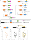
Chemical biology tools for controlling and deciphering the amyloid aggregation pathways. (A) Fibrils made of the mirror PHF6* hexapeptide of tau consisting of D-enantiomeric amino acids was used in mirror image phage display to screen L-peptides that bind to mirror PHF6* fibrils. The mirror peptides of the selected L-peptides were synthesized and evaluated as inhibitors of full-length tau aggregation exhibiting protease stability and reduced immunogenicity (Dammers et al., 2016; Malhis et al., 2021). (B) The strategy of “click” peptides or “switch” peptides is used to control protein aggregation and decipher the molecular elements responsible of amyloid aggregation (left panel). A molecular switch element (S) is under control of a protecting group “P” (green dots) that prevent aggregation (Soff) and is cleaved by an appropriate trigger factor (pH, enzyme, reducing agents, light, …). Upon deprotection, the spontaneous O→N or S→N acyl shift within the molecular switch element restores the native peptide bond between both fragments at its N- and C-terminal sides. As a result, the structural induction unit σ (grey fragment) is linked through a native peptide bond to the remaining part of the protein (blue fragment). If the deprotection and subsequent acyl shift trigger the amyloid aggregation of the protein of interest (Son), σ is identified as an aggregation hot spot as exemplified by the C-terminal region of Aβ42 peptide (right panel). In this case, a first molecular switch element (S1) at S26 under control of pH is removed without triggering any fibril formation. The enzymatic cleavage of the second switch element (S2) by the dipeptidyl-peptidase at S37 enables the fibrillization of native Aβ42 peptide supporting a role of the C-terminus in conformational changes and aggregation. (C) The photo-induced cross-linking of unmodified protein (PICUP) strategy is used to generate protein oligomers by light irradiation of a photocatalyst, the tris(bipyridyl)ruthenium(II), in presence of ammonium persulfate. Metastable oligomers that form transiently along the amyloid aggregation pathway, as shown by polyacrylamide gel electrophoresis under denaturing conditions, can thus be captured for further structural and functional investigations. (D) The use of fluoroproline derivatives, 4-cis-fluoro, 4-trans-fluoro or 4,4-difluoro-proline, in controlling amyloid protein conformational changes and aggregation rely on the alteration of the cis/trans conformational exchange rate and equilibrium of the peptidyl-prolyl bond with the 4-cis-fluoroproline the most favorable to the cis amide bond conformation. Furthermore, fluoroproline can be involved as probes in 19F-NMR studies of conformational exchange in peptides and proteins as shown for a model peptide of β2m, Ac-FH(F2-P32)SD-NH2. The 19F-NMR spectrum of the β2m peptide containing a 4,4-difluoroproline was reproduced from reference (Torbeev and Hilvert, 2013). The corresponding sequence is shown as sticks in the structure of β2m amyloid fibrils (PDB ID: 6GK3) with P32 (magenta) in trans conformation.
FIGURE 12
FIGURE 12
Modulation of O-GlcNAc modification of proteins by proximity-induced protein glycosylation using nanobody-OGT protein engineering. (A) Schematic representation of the O-GlcNAc dynamics regulated by single OGT and OGA enzymes, and modulation of OGA activity by an OGA inhibitor such as Thiamet G contributing to overall increased O-GlcNAc levels. (B) As OGT is the unique O-GlcNAc transferase in human, it has been suggested that it may act with regulatory subunits that address OGT to specific substrates depending on external stimuli and increase protein-specific O-GlcNAc levels. Treatment with Thiamet-G by increasing overall O-GlcNAc levels may contribute to increase protein-specific O-GlcNAc level. (C) Modulation of protein O-GlcNAc levels in cell or animal models. Condition 1, external stimuli and signal transduction lead to the expression of protein-specific regulatory subunits of OGT that increase its activity on selected proteins; condition 2, overall increase of protein O-GlcNAc levels upon treatment with Thiamet-G; condition 3, selected increase of O-GlcNAcylation of a target substrate by a nanobody fused to OGT targeting a specific protein tag. (D) This latter method (C, condition 3) was further extended to O-GlcNAcylation of endogenous α-synuclein using a nanobody-OGT targeting α-synuclein EPEA C-terminal sequence (Ramirez et al., 2020). EPEA-(4-TPR)-OGT refers to a truncated form of OGT containing 4 tetratricopeptide repeats fused to a nanobody targeting the EPEA sequence of α-synuclein. The TPR truncation of OGT limits the overall increase of O-GlcNAc levels while OGT coupling to a nanobody targeting the EPEA sequence increases the selectivity for α-synuclein. This approach leads to a selective elevation of α-synuclein O-GlcNAc levels.

References

    1. Abdulbagi M., Wang L., Siddig O., Di B., Li B. (2021). D-amino Acids and D-Amino Acid-Containing Peptides: Potential Disease Biomarkers and Therapeutic Targets? Biomolecules 11, 1716. 10.3390/biom11111716 - DOI - PMC - PubMed
    1. Abeywardana T., Lin Y. H., Rott R., Engelender S., Pratt M. R. (2013). Site-Specific Differences in Proteasome-dependent Degradation of Monoubiquitinated α-Synuclein. Chem. Biol. 20, 1207–1213. 10.1016/j.chembiol.2013.09.009 - DOI - PMC - PubMed
    1. Abraha A., Ghoshal N., Gamblin T. C., Cryns V., Berry R. W., Kuret J., et al. (2000). C-terminal Inhibition of Tau Assembly In Vitro and in Alzheimer's Disease. J. Cell Sci. 113, 3737–3745. 10.1242/jcs.113.21.3737 - DOI - PubMed
    1. Agard N. J., Prescher J. A., Bertozzi C. R. (2004). A Strain-Promoted [3 + 2] Azide−Alkyne Cycloaddition for Covalent Modification of Biomolecules in Living Systems. J. Am. Chem. Soc. 126, 15046–15047. 10.1021/ja044996f - DOI - PubMed
    1. Agouridas V., El Mahdi O., Diemer V., Cargoët M., Monbaliu J.-C. M., Melnyk O. (2019). Native Chemical Ligation and Extended Methods: Mechanisms, Catalysis, Scope, and Limitations. Chem. Rev. 119, 7328–7443. 10.1021/acs.chemrev.8b00712 - DOI - PubMed

LinkOut - more resources