Review
. 2022 May 13:10:903994.
doi: 10.3389/fcell.2022.903994. eCollection 2022.

When PIP2 Meets p53: Nuclear Phosphoinositide Signaling in the DNA Damage Response

Affiliations
Review

When PIP2 Meets p53: Nuclear Phosphoinositide Signaling in the DNA Damage Response

Yu-Hsiu Wang et al. Front Cell Dev Biol. .

Abstract

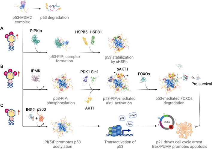
The mechanisms that maintain genome stability are critical for preventing tumor progression. In the past decades, many strategies were developed for cancer treatment to disrupt the DNA repair machinery or alter repair pathway selection. Evidence indicates that alterations in nuclear phosphoinositide lipids occur rapidly in response to genotoxic stresses. This implies that nuclear phosphoinositides are an upstream element involved in DNA damage signaling. Phosphoinositides constitute a new signaling interface for DNA repair pathway selection and hence a new opportunity for developing cancer treatment strategies. However, our understanding of the underlying mechanisms by which nuclear phosphoinositides regulate DNA damage repair, and particularly the dynamics of those processes, is rather limited. This is partly because there are a limited number of techniques that can monitor changes in the location and/or abundance of nuclear phosphoinositide lipids in real time and in live cells. This review summarizes our current knowledge regarding the roles of nuclear phosphoinositides in DNA damage response with an emphasis on the dynamics of these processes. Based upon recent findings, there is a novel model for p53's role with nuclear phosphoinositides in DNA damage response that provides new targets for synthetic lethality of tumors.

Keywords: DNA damage response (DDR); DNA repair; nuclear phosphoinositide signaling; p53; repair pathway choice; stress response.


Conflict of interest statement

The authors declare that the research was conducted in the absence of any commercial or financial relationships that could be construed as a potential conflict of interest.

Figures

FIGURE 1
FIGURE 1
Nuclear phosphoinositide metabolism and involved enzymes with implications in DNA damage response and/or tumor suppression. Representative proteins/isoforms are shown as full length structures predicted by AlphaFold. Studies indicate that PIPKIα, PIPKIIβ, and PTEN might exist as dimers. Whether dimerization is required for their kinase activities and in response to DNA damage requires further investigation (Heinrich et al., 2015; Hu et al., 2015; Droubi et al., 2016). Red dotted circle labels the catalytic site of each protein. The letters in the parentheses under each enzyme correspond to the pathways indicated in the center panel. Letters in gray represent a minor pathway. Note that PI(3,5)P2 is not detected in nucleus.
FIGURE 2
FIGURE 2
Nuclear PI(4,5)P2 sequestering by NLS-tagged PLCδPH domain suppresses the recruitment of ATR/ATRIP complex following UVA laser microirradiation, and the accumulation of its downstream effector, phospho-S345 Chk1. The recruitment of ATM, DNA-PKcs and other DNA repair proteins listed here are not affected. The data is adapted with permission from (Wang et al., 2017).
FIGURE 3
FIGURE 3
The roles of nuclear phosphoinositides and the regulation of their kinases and phosphatases in response to DNA damage at different time scales. Figures created with BioRender.com.
FIGURE 4
FIGURE 4
(A) PI(4,5)P2-centric view of nuclear phosphoinositide homeostasis in a resting state and (B) how the regulatory steps are altered in response to genotoxic stress. Figures created with BioRender.com.
FIGURE 5
FIGURE 5
Differential regulation of p53 by different nuclear phosphoinositide species in response to DNA damage. (A) PI(4,5)P2 drives complex formation with p53 and promotes p53 stabilization by recruiting small heat shock proteins (sHSPs). (B) IPMK phosphorylates p53-PI(4,5)P2 complex, which serves as a signaling hub for Akt activation and subsequent phosphorylation and degradation of FOXOs. (C) PI(5)P promotes chromatin association of ING2. It drives p300-mediated acetylation and transactivation of p53. Figures created with BioRender.com.
FIGURE 6
FIGURE 6
PARP-dependent rapid recruitment of p53 directs repair pathway choice. (A) Chemical structure of ADP-ribose and the illustration of auto-PARylated PARP. (B) p53 interacts with auto-PARylated PARP through its C-terminal polybasic stretch. (C) Auto-PARylated PARP drives the accumulation of p53, which promotes the recruitment of 53BP1 and DDB1 and therefore NHEJ and NER, respectively. (D) The recruitment of p53, as well as the subsequent recruitment of 53BP1 and DDB1, is suppressed by a PARP inhibitor. (E) Mutations that impair p53 rapid recruitment also suppress recruitment of 53BP1 and DDB1. (F) Early recruitment of p53 tips the balance of repair pathway selection in favor of the NHEJ and NER repair pathway in a transcription-independent fashion. Figures created with BioRender.com.

Similar articles

Cited by

References

    1. Alexandrova E. M., Yallowitz A. R., Li D., Xu S., Schulz R., Proia D. A., et al. (2015). Improving Survival by Exploiting Tumour Dependence on Stabilized Mutant P53 for Treatment. Nature 523, 352–356. 10.1038/nature14430 - DOI - PMC - PubMed
    1. Alvarez-Venegas R., Sadder M., Hlavacka A., Baluška F., Xia Y., Lu G., et al. (2006). The Arabidopsis Homolog of Trithorax, ATX1, Binds Phosphatidylinositol 5-phosphate, and the Two Regulate a Common Set of Target Genes. Proc. Natl. Acad. Sci. U.S.A. 103, 6049–6054. 10.1073/pnas.0600944103 - DOI - PMC - PubMed
    1. Andrin C., McDonald D., Attwood K. M., Rodrigue A., Ghosh S., Mirzayans R., et al. (2012). A Requirement for Polymerized Actin in DNA Double-Strand Break Repair. Nucleus 3, 384–395. 10.4161/nucl.21055 - DOI - PubMed
    1. Arrigo A.-P., Gibert B. (2014). HspB1, HspB5 and HspB4 in Human Cancers: Potent Oncogenic Role of Some of Their Client Proteins. Cancers 6, 333–365. 10.3390/cancers6010333 - DOI - PMC - PubMed
    1. Aubrey B. J., Kelly G. L., Janic A., Herold M. J., Strasser A. (2018). How Does P53 Induce Apoptosis and How Does This Relate to P53-Mediated Tumour Suppression? Cell Death Differ. 25, 104–113. 10.1038/cdd.2017.169 - DOI - PMC - PubMed