Review
. 2022 May 11:9:854554.
doi: 10.3389/fcvm.2022.854554. eCollection 2022.

Optical Coherence Tomography: An Eye Into the Coronary Artery

Affiliations
Review

Optical Coherence Tomography: An Eye Into the Coronary Artery

Ankush Gupta et al. Front Cardiovasc Med. .

Abstract

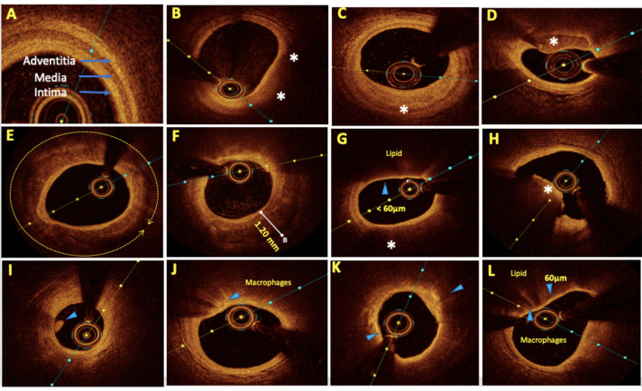
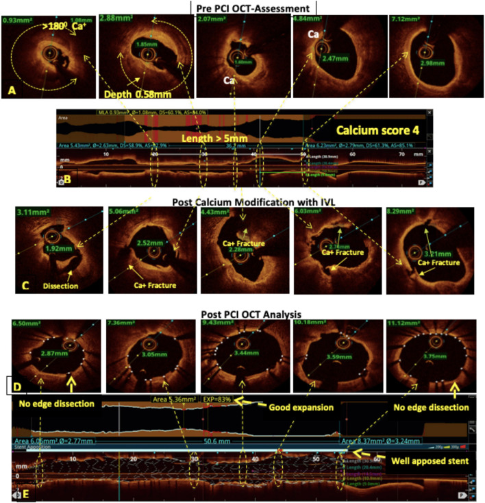
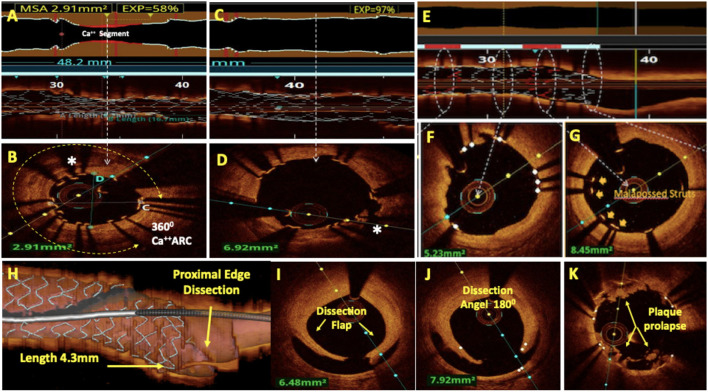
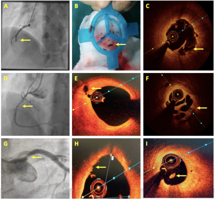
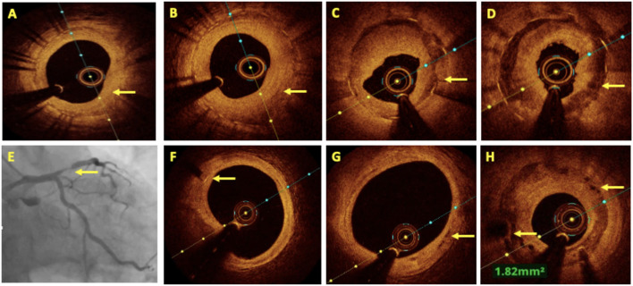
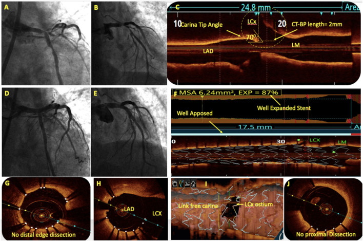
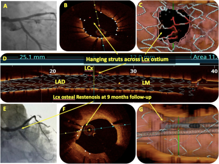
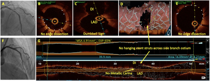
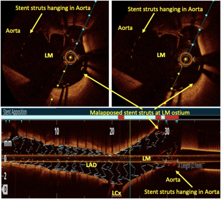
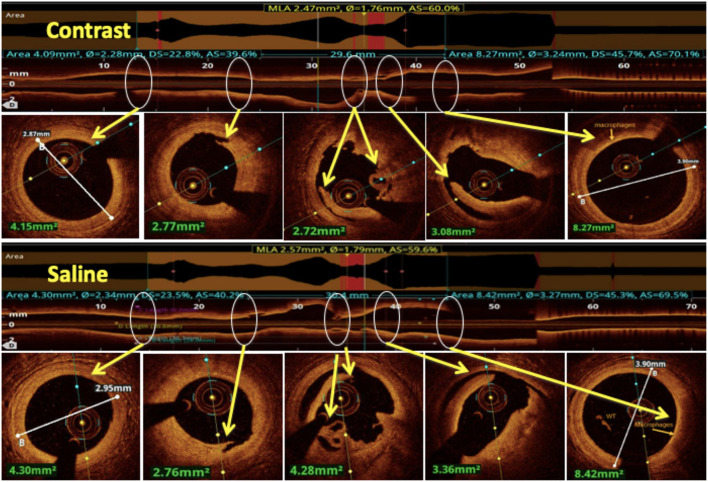
Optical coherence tomography (OCT) is slowly but surely gaining a foothold in the hands of interventional cardiologists. Intraluminal and transmural contents of the coronary arteries are no longer elusive to the cardiologist's probing eye. Although the graduation of an interventionalist in imaging techniques right from naked eye angiographies to ultrasound-based coronary sonographies to the modern light-based OCT has been slow, with the increasing regularity of complex coronary cases in practice, such a transition is inevitable. Although intravascular ultrasound (IVUS) due to its robust clinical data has been the preferred imaging modality in recent years, OCT provides a distinct upgrade over it in many imaging and procedural aspects. Better image resolution, accurate estimation of the calcified lesion, and better evaluation of acute and chronic stent failure are the distinct advantages of OCT over IVUS. Despite the obvious imaging advantages of OCT, its clinical impact remains subdued. However, upcoming newer trials and data have been encouraging for expanding the use of OCT to wider indications in clinical utility. During percutaneous coronary intervention (PCI), OCT provides the detailed information (dissection, tissue prolapse, thrombi, and incomplete stent apposition) required for optimal stent deployment, which is the key to successfully reducing the major adverse cardiovascular event (MACE) and stent-related morbidities. The increasing use of OCT in complex bifurcation stenting involving the left main (LM) is being studied. Also, the traditional pitfalls of OCT, such as additional contrast load for image acquisition and stenting involving the ostial and proximal LM, have also been overcome recently. In this review, we discuss the interpretation of OCT images and its clinical impact on the outcome of procedures along with current barriers to its use and newer paradigms in which OCT is starting to become a promising tool for the interventionalist and what can be expected for the immediate future in the imaging world.

Keywords: IVUS; OCT; OCT in ACS; OCT in bifurcation angioplasty; OCT in left main bifurcation angioplasty; calcified lesion modification; plaque morphology; saline OCT.


Conflict of interest statement

The authors declare that the research was conducted in the absence of any commercial or financial relationships that could be construed as a potential conflict of interest.

Figures

Figure 1
Figure 1
Representative optical coherence tomography (OCT) images with various plaque morphologies. (A) Normal coronary, (B) lipid-rich plaque (LRP), (C) fibrotic plaque, (D) calcific nodule, (E) near 360° arc of calcific plaque, (F) deep calcium deposition, (G) thin cap fibroatheroma (TCFA), (H) intraluminal red thrombus, (I) intraluminal white thrombus, (J) bright spots or bands at the boundary between the fibrous cap and lipid core suggestive of macrophages, (K) bright signal-rich cholesterol crystals, and (L) vulnerable plaque formed by a large lipid pool covered by TCFA with macrophage infiltration. Areas of interest were highlighted by arrows and asterisks.
Figure 2
Figure 2
OCT-guided management of calcified left anterior descending (LAD). (A,B) Pre-percutaneous coronary intervention (PCI) assessment showed a heavily calcified LAD with a calcium arc >180°, a calcium thickness of 0.58 mm, and a length of >5 mm resulting in a calcium score of 4. (C) Fractures in the calcium after modification with intravascular lithotripsy (IVL). (D,E) Post-PCI assessment showed 83% stent expansion, no edge dissection, and a well-apposed stent.
Figure 3
Figure 3
Post-PCI assessment by OCT. (A) Lumenogram and L mode showed under-expanded stent (UES) with minimum stent area (MSA) of 2.91 mm2 and 58% stent expansion. (B) Cross-sectional image at MSA revealed 360° arc of calcium as a cause of UES. (C) Lumenogram and L mode showed 97% stent expansion after the treatment of UES with ultra-high-pressure balloon dilatation. (D) A cross-sectional image shows the fracture of the calcium arc resulting in the resolution of the UES. (E) A rendered stent view shows well-apposed stent struts (white) and malapposed stent struts (red) corresponding to the white and red bars in the apposition indicator. Apposition (F) and mal-apposition (G) of the stent struts can also be well-appreciated on respective cross-sectional images. (H–J): Major proximal stent edge dissection is seen with the dissection flap extending to media, dissection angle of 180° and 4.3 mm dissection length on three-dimensional (3D) reconstruction. OCT with its better axial resolution enables clearer and more frequent visualization of in-stent tissue prolapse (K).
Figure 4
Figure 4
OCT in acute coronary syndrome. (A–C) A case of acute inferior wall STEMI with mid dominant right coronary artery (RCA) thrombotic occlusion. Thrombus aspiration followed by OCT revealed plaque rupture. (D–F) A 4-day old case of inferior wall STEMI showing Mid-RCA haziness. OCT revealed proximal RCA plaque rupture and mid-RCA recanalized thrombus in a Swiss-cheese pattern. (G–I) A case of NSTEMI having a separate origin of LAD and LCx with a significant stenosis in the proximal LAD. OCT showed luminal irregularities, intact thick fibrous cap, and intraluminal white thrombus suggestive of plaque erosion. Areas of interest are highlighted with arrows.
Figure 5
Figure 5
OCT in in-stent restenosis (ISR). (A) Homogenous neointimal hyperplasia (NIH) in bare metal stent (BMS) restenosis. (B) Heterogenous NIH in a drug-eluting stent (DES) ISR. (C,D) Showed neoatherosclerosis as a cause of ISR in a well-expanded stent with well-demarcated calcium and fibrotic ingrowth. (E–H) Coronary angiography showing proximal LAD bioresorbable vascular scaffolds (BVS) ISR after 2 years of implantation. (F) The OCT image shows a distal marker of BVS, (G) almost completely absorbed BVS struts in the middle and distal part of the stent, and (H) BVS ISR at the proximal segment with peri-strut low intensity areas.
Figure 6
Figure 6
OCT in bifurcation angioplasty. (A,B) Coronary angiography showed a significant left main (LM) bifurcation lesion (Medina 1.1.1). (C) Pre-PCI OCT showed the carina tip (CT) angle of 70° and CT to bifurcation point (CT-BP length) of 2 mm suggestive of bifurcation lesion suitable for provisional stenting without the risk of side branch (SB) compromise. (D) An OCT-guided LM-LAD cross-over stenting was done followed by the proximal optimization technique (POT) with balloons of appropriate size. (E) Post-PCI angiography showed TIMI III flow in left coronary system without LCx compromise. Post-PCI OCT showed well-apposed stent with 87% expansion (F), without proximal or distal edge dissection (G,J) and minimal stent struts across LCx ostium (H). 3D reconstruction showed minimal inappropriate stent apposition across LCx ostium with link-free carina (I).
Figure 7
Figure 7
OCT in bifurcation angioplasty. (A) Showed the final angiogram picture after LM bifurcation angioplasty with two stents using the DK crush technique. The angiographic result for LCx ostium was satisfactory but OCT pullback of the main branch (MB) after final kissing balloon inflation (KBI) showed hanging stent struts (red) across LCx ostium on the cross-sectional view (B), on 3D reconstruction (C), and on the rendered stent view (D). The same patient presented with crescendo angina after 9 months, and angiography showed LCx ostial ISR (E). OCT pullback of MB showed complete endothelization of hanging stent struts across LCx as a cause of ostial ISR (F,G).
Figure 8
Figure 8
OCT in bifurcation angioplasty. Coronary angiography showed a significant LAD/D1 stenosis (A) with Medina class (1, 1, 1). Patient underwent bifurcation angioplasty with two stents using the DK crush technique (F). OCT run of LAD after final KBI showed no edge dissection (B,E) and dumbbell sign (C). No hanging stent struts seen across D1 ostium on 3D reconstruction (D) and on L mode (G). No neo-metallic carina seen at LAD/D1 bifurcation on the rendered stent view (G).
Figure 9
Figure 9
Left main ostial imaging with OCT. OCT pullback from LAD to LM after provisional LM ostial to LAD crossover angioplasty with one wire in the aorta. Hanging stent struts in the aorta (suggestive of LM ostial coverage) can be clearly seen along with some malapposed struts both in the cross-sectional and rendered stent view.
Figure 10
Figure 10
Comparison between saline and contrast OCT. The upper panel shows pre-PCI run of RCA using contrast as a flushing media and in the lower panel the same vessel is imaged using saline as a flushing media for OCT and compared for image quality. All the lesion morphologies (including plaque rupture, dissections, recanalized thrombus, and macrophages) seen in contrast OCT are clearly seen with saline OCT (marked by yellow arrows at the same level).

Similar articles

Cited by

References

    1. Topol EJ, Nissen SE. Our preoccupation with coronary luminology. The dissociation between clinical and angiographic findings in ischemic heart disease. Circulation. (1995) 92:2333–42. 10.1161/01.CIR.92.8.2333 - DOI - PubMed
    1. Zir LM, Miller SW, Dinsmore RE, Gilbert JP, Harthorne JW. Interobserver variability in coronary angiography. Circulation. (1976) 53:627–32. 10.1161/01.CIR.53.4.627 - DOI - PubMed
    1. White CW, Wright CB, Doty DB, Hiratza LF, Eastham CL, Harrison DG, et al. . Does visual interpretation of the coronary arteriogram predict the physiologic importance of a coronary stenosis? N Engl J Med. (1984) 310:819–24. 10.1056/NEJM198403293101304 - DOI - PubMed
    1. Mintz GS. Clinical utility of intravascular imaging and physiology in coronary artery disease. J Am Coll Cardiol. (2014) 64:207–22. 10.1016/j.jacc.2014.01.015 - DOI - PubMed
    1. Neumann F-J, Sousa-Uva M, Ahlsson A, Alfonso F, Banning AP, Benedetto U, et al. . 2018 ESC/EACTS guidelines on myocardial revascularization. Euro Heart J. (2019) 40:87–165. 10.1093/eurheartj/ehy855 - DOI - PubMed