. 2022 May 13:9:868600.
doi: 10.3389/fcvm.2022.868600. eCollection 2022.

Therapeutic Effect of Pericytes for Diabetic Wound Healing

Affiliations

Therapeutic Effect of Pericytes for Diabetic Wound Healing

Kyeong Mi Kim et al. Front Cardiovasc Med. .

Abstract

Objective: Numerous attempts have been made to devise treatments for ischemic foot ulcer (IFU), which is one of the most severe and fatal consequences of diabetes mellitus (DM). Pericytes, which are perivascular multipotent cells, are of interest as a treatment option for IFU because they play a critical role in forming and repairing various tissues. In this study, we want to clarify the angiogenic potential of pericytes in DM-induced wounds.

Methods: We evaluated pericyte stimulation capability for tube formation, angiogenesis, and wound healing (cell migration) in human umbilical vein endothelial cells (HUVECs) with in-vivo and in-vitro models of high glucose conditions.

Results: When HUVECs were co-cultured with pericytes, their tube-forming capacity and cell migration were enhanced. Our diabetic mouse model showed that pericytes promote wound healing via increased vascularization.

Conclusion: The findings of this study indicate that pericytes may enhance wound healing in high glucose conditions, consequently making pericyte transplantation suitable for treating IFUs.

Keywords: angiogenesis; diabetes mellitus; ischemic foot ulcer; pericyte; wound healing.


Conflict of interest statement

The authors declare that the research was conducted in the absence of any commercial or financial relationships that could be construed as a potential conflict of interest.

Figures
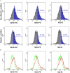
Figure 1
Figure 1
The expression of pericyte markers by flow cytometry was reduced in the diabetic condition. (A) Analysis of pericyte surface markers in the non-diabetic and diabetic conditions using flow cytometry [CD146-fluorescein isothiocyanate (FITC), Nestin-phycoerythrin (PE), and NG2-PE]. The results provided here are representative of those obtained in three independent studies. In the diabetic condition, Nestin and NG2-PE were seen to decrease by more than half. (B) Overlay of diabetic condition (green line) and non-diabetic (red line) condition flow cytometry. The diabetic condition showed the three pericyte markers to be decreased.
Figure 2
Figure 2
The group with pericytes showed improved tube formation and cell migration ability in the high glucose condition. (A) The in-vitro vascular, tube formation assay showed effects of exposure on FITC-tagged human umbilical vein endothelial cell (HUVEC) tube formation with and without PE-tagged human pericyte cells. The optimal observation time for tube formation was 8 h after seeding. In high glucose conditions, HUVECs with pericytes (10:1 ratio) showed active tube formation even compared to the without pericyte group. (B) The number of pieces in the assay was measured. (**P < 0.01 and ***P < 0.001 according to the Student's t-test). HUVEC tube formation capacity showed no significant difference in low glucose conditions. However, in high glucose conditions, the group with pericytes had a statistically significant increase in tube formation. (C) Photomicrographs were taken at 0, 12, and 24 h following initiation of the wound. HUVEC migration, often termed as wound closure ability, was more remarkable in the group with pericytes in both the high and low glucose conditions. (D) The quantitative approach applied to the wound closure assay, showing a histogram calculated based on the group breath ratio of the control group and the group with pericytes.
Figure 3
Figure 3
The diabetes mellitus (DM)-with-pericyte group exhibited substantial wound healing in a mouse wound model. (A) Illustration of injection of collagen type 1 scaffold + 5 × 10⁵ pericytes in the mouse wound. (B) Streptozotocin (STZ)-induced diabetic mice on blood glucose confirmed successful DM induction (****P < 0.0001). DM-induced mice (n = 10), control (n = 10). (C) Incisions with a diameter of 4 mm on either side of the midline were produced. To avoid wound contraction, the wounds were circumscribed with donut-shaped silicone splints. As observed on day 7, wounds of the DM-with-pericyte group healed, whereas those of the DM-without-pericyte and control groups did not heal. (D) The Y-axis of the graph represents the wound size, when measured on days 0, 2, 7, and 14. It was verified that all the groups, except the DM-without-pericyte group, had wound closure by the time of sacrifice on day 14 and approximately 3.6 mm of the wound of the DM-without-pericyte group remained. The pericyte and DM-with-pericyte groups showed the same wound size on days 0, 2, 7, and 14. (E) Quantification of wound size at sacrifice time points (**P < 0.01). In comparison to the DM-without-pericyte group, the DM-with-pericyte group exhibited substantial wound healing.
Figure 4
Figure 4
The DM-with-pericyte group showed successful repair of the collagen layer and higher blood vessel formation capacity compared to the DM-without-pericyte group. (A) H&E staining. (B) Masson's trichrome staining for wound analysis at 14 days. The black square indicates the regenerated dermal collagen layer. Scale bars = 200 μm. (C) CD31 and Ki67 immunofluorescence of wounds at 14 days. In diabetes wounds, new vessels were significantly less visible than in normal mice. However, when diabetic mice were injected with pericytes, new vessels were observed more often. (D) α-smooth muscle actin (α-SMA), collagen 1 (Col 1), and Nestin immunohistochemistry of wounds at 14 day. Scale bar = 50 μm.

References

    1. Fard AS, Esmaelzadeh M, Larijani B. Assessment and treatment of diabetic foot ulcer. Int J Clin Pract. (2007) 61:1931–8. 10.1111/j.1742-1241.2007.01534.x - DOI - PubMed
    1. Saeedi P, Petersohn I, Salpea P, Malanda B, Karuranga S, Unwin N, et al. . Global and regional diabetes prevalence estimates for 2019 and projections for 2030 and 2045: results from the international diabetes federation diabetes atlas. Diabetes res clin pract. (2019) 157:107843. 10.1016/j.diabres.2019.107843 - DOI - PubMed
    1. Yazdanpanah L, Nasiri M, Adarvishi S. Literature review on the management of diabetic foot ulcer. World J Diabetes. (2015) 6:37–53. 10.4239/wjd.v6.i1.37 - DOI - PMC - PubMed
    1. Ikura K, Hanai K, Oka S, Watanabe M, Oda Y, Hamada M, et al. . Brachial-ankle pulse wave velocity, but not ankle-brachial index, predicts all-cause mortality in patients with diabetes after lower extremity amputation. J Diabetes Invest. (2017) 8:250–3. 10.1111/jdi.12554 - DOI - PMC - PubMed
    1. Martins-Mendes D, Monteiro-Soares M, Boyko EJ, Ribeiro M, Barata P, Lima J, et al. . The independent contribution of diabetic foot ulcer on lower extremity amputation and mortality risk. J Diabetes Complications. (2014) 28:632–8. 10.1016/j.jdiacomp.2014.04.011 - DOI - PMC - PubMed

LinkOut - more resources