Review
. 2022 May 12:9:887671.
doi: 10.3389/fvets.2022.887671. eCollection 2022.

Comparative Review of Brucellosis in Small Domestic Ruminants

Affiliations
Review

Comparative Review of Brucellosis in Small Domestic Ruminants

Carlos Alberto Rossetti et al. Front Vet Sci. .

Abstract

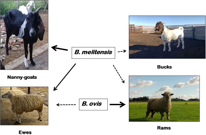
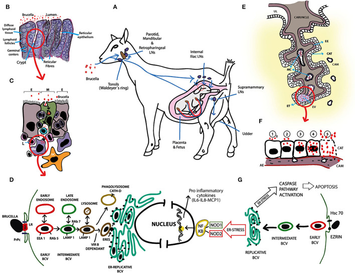
Brucella melitensis and Brucella ovis are the primary etiological agents of brucellosis in small domestic ruminants. B. melitensis was first isolated in 1887 by David Bruce in Malta Island from spleens of four soldiers, while B. ovis was originally isolated in Australia and New Zealand in early 1950's from ovine abortion and rams epididymitis. Today, both agents are distributed worldwide: B. melitensis remains endemic and associated with an extensive negative impact on the productivity of flocks in -some regions, and B. ovis is still present in most sheep-raising regions in the world. Despite being species of the same bacterial genus, B. melitensis and B. ovis have extensive differences in their cultural and biochemical characteristics (smooth vs. rough colonial phases, serum and CO2 dependence for in vitro growth, carbohydrate metabolism), host preference (female goat and sheep vs. rams), the outcome of infection (abortion vs. epididymitis), and their zoonotic potential. Some of these differences can be explained at the bacterial genomic level, but the role of the host genome in promoting or preventing interaction with pathogens is largely unknown. Diagnostic techniques and measures to prevent and control brucellosis in small ruminants vary, with B. melitensis having more available tools for detection and prevention than B. ovis. This review summarizes and analyzes current available information on: (1) the similarities and differences between these two etiological agents of brucellosis in small ruminants, (2) the outcomes after their interaction with different preferred hosts and current diagnostic methodologies, (3) the prevention and control measures, and (4) alerting animal producers about the disease and raise awareness in the research community for future innovative activities.

Keywords: Brucella melitensis; Brucella ovis; genomics; goats; pathogenesis; sheep.


Conflict of interest statement

The authors declare that the research was conducted in the absence of any commercial or financial relationships that could be construed as a potential conflict of interest.

Figures

Figure 1
Figure 1
Susceptibility of domestic small ruminants to B. melitensis and B. ovis infection. B. melitensis cause abortion, stillbirths and the birth of weak offspring in goats and sheep (solid arrow); and less commonly, epididymo-orchitis in bucks and rams (dashed arrow). Moreover, B. ovis affect rams almost exclusively (solid arrow), causing epididymitis and occasionally abortions in ewes and an increase in perinatal death (dashed arrow). In addition to sheep and goats, B. melitensis has a wide range of natural susceptible host species, including humans. Other than domestic sheep, B. ovis has only been isolated from naturally-acquired infections of farmed red deer (Cervus elaphus), but experimental infections have been established in goats, Rocky Mountain bighorn sheep (Ovis canadensis canadensis) and white-tailed deer (Odocoileus virginianus).
Figure 2
Figure 2
Pathogenesis of B. melitensis infection in small ruminants. (A) B. melitensis infects small ruminants mainly via the alimentary tract. Once in the oral cavity, B. melitensis enters through the mucosa or the oro- and nasopharyngeal tonsils (Waldeyer's ring) and colonizes, proliferates and persists in the lymph nodes (LNs) of the head (i.e., mandibular, parotid, and lateral and medial retropharyngeal LNs). Failure to eliminate B. melitensis on this primary line of defense results in escape of the bacteria through the efferent lymphatic vessels to the distal LN or via blood to the general circulation. In pregnant females, B. melitensis colonizes placenta and induces abortion or stillbirth. Brucella melitensis also has affinity for lactating udder. Significant numbers of B. melitensis are excreted in vaginal discharges, aborted fetus, placenta and milk. (B) Illustration of histological structure of tonsils. B. melitensis is first seen in the lumen of the oral cavity. Subsequently, B. melitensis colonizes the crypts and invade through reticular epithelium. (C) Reticular epithelium is composed of scattered M cells (M; green), lymphoid cells and the epithelial cell types (E). M cells endocytose B. melitensis from the lumen, after which transcytosis and basolateral release occurs. Immediately, the agent is up taken by resident antigen presenting cells, i.e., macrophages (MØ) and dendritic cells (DC), which in turn activate the immune response in the underlying lymphoid follicles. MØs and DCs traffic B. melitensis to other sites in the body or back through the reticular epithelium to the mucosal surface. (D) Schematic representation of B. melitensis intracellular trafficking in macrophages and evasion of killing. Following lipid rafts (LR)—and the prion protein (PrPc)—mediated internalization, B. melitensis is contained in an early vacuole (BCV: Brucella containing vacuole). This early vacuole sequentially interacts with cellular organelles (early and late endosomes, and lysosomes) and transiently acquires different markers (EEA1, Rab5, LAMP1, Rab7) in a VirB dependent-mechanism regulated by the pathogen effector proteins. To reach the replicative niche, BCV-LAMP1+ interacts with ER exit sites (ERES), and generates an ER-derived organelle permissive for B. melitensis replication (ER-Replicative BCV). Vacuoles containing VirB-deficient B. melitensis undergo phagolysosomal degradation. Spontaneous rough mutant generation might help parental pathogen release from infected MØ through lytic or non-lytic mechanisms, and the process repeats in other professional phagocytic cells. (E) Diagram of ruminant placentome with enlargement of one caruncular septa. Brucella invades placenta via maternal capillaries (MC). Extravasated (EV) maternal blood at the tip of the caruncular septa, along with B. melitensis, is phagocytosed by erythrophagocytic trophoblasts (ET). From these cells, B. melitensis spread infection to adjacent chorioallantoic trophoblasts (CAT). UL: Uterine lumen, EE: Endometrial epithelium, CAM: Chorioallantoic membrane. (F) Schematization of a complete cycle of B. melitensis-infected chorioallantoic trophoblast (CAT). 1: B. melitensis (B, red circles) adheres to the plasmatic membrane of CAT; 2: the bacteria are internalized and initial intracellular replication occurs; 3–4: Massive intracellular multiplication of the agent; 5: Apoptosis of CAT and release of huge number of B. melitensis. The cycle of endocytosis, intracellular replication and programmed cell death continues. CAM, Chorioallantoic membrane; AE, Allantoic ephitelium. (G) Schematic representation of B. melitensis intracellular trafficking in chorioallantioic trophoblasts. Trophoblasts invasion of B. melitensis is mediated by heat shock cognate protein 70 (Hsc70) and Ezrin. From this point until reaching of the replicative niche, intracellular trafficking is similar than that reported in macrophages (i.e., BCV goes from early BCV -red- to intermediate BCV -green- and to replicative BCV). Intracellular presence of Brucella induces ER stress that triggers production of the pro-inflammatory cytokines in a nucleotide-binding oligomerization domain (NOD) 1/2—dependent manner via activation of NF-kb pathway, and activates caspase pathway leading to chorioallantoic trophoblast apoptosis. These molecular responses of B. melitensis-infected trophoblasts may contribute to better understanding the pathogenesis of placentitis and abortion in small ruminant brucellosis.

References

    1. Rajendhran J. Genomic insights into Brucella. Infect Genet Evol J Mol Epidemiol Evol Genet Infect Dis. (2021) 87:104635. 10.1016/j.meegid.2020.104635 - DOI - PubMed
    1. Alton GG. Brucella Melitensis. Anim Brucell CRC. Boca Raton, FL: Press Inc. Nielsen K, Duncan JR, editors (1990). p. 383–409.
    1. Corbel J. Brucellosis in Humans and Animals. WHO , editor. Geneva: WHO; (2006). p. 89.
    1. Rossetti CA, Arenas-Gamboa AM, Maurizio E. Caprine brucellosis: a historically neglected disease with significant impact on public health. PLoS Negl Trop Dis. (2017) 11:e0005692. 10.1371/journal.pntd.0005692 - DOI - PMC - PubMed
    1. Blasco J. Brucella ovis. In: Nielsen K, Duncan JR, editors. Animal Brucellosis. Boca Raton, FL: CRC Press; (1990). p. 351–78.

LinkOut - more resources