

Utility of Diagnostic Ultrasound in the Assessment of Patellar Instability
- PMID: 35647210
- PMCID: PMC9134436
- DOI: 10.1177/23259671221098748
Utility of Diagnostic Ultrasound in the Assessment of Patellar Instability
Abstract
Background: The use of imaging to diagnose patellofemoral instability is often limited by the inability to dynamically load the joint during assessment. Therefore, the diagnosis is typically based on physical examination using the glide test to assess and quantify lateral patellar translation. However, precise quantification with this technique remains difficult.
Purpose: To quantify patellar position using ultrasound imaging under dynamic loading conditions to distinguish between knees with and without medial patellofemoral complex (MPFC) injury.
Study design: Controlled laboratory study.
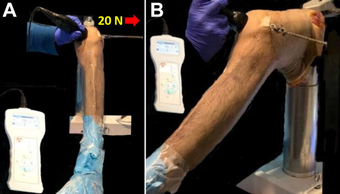
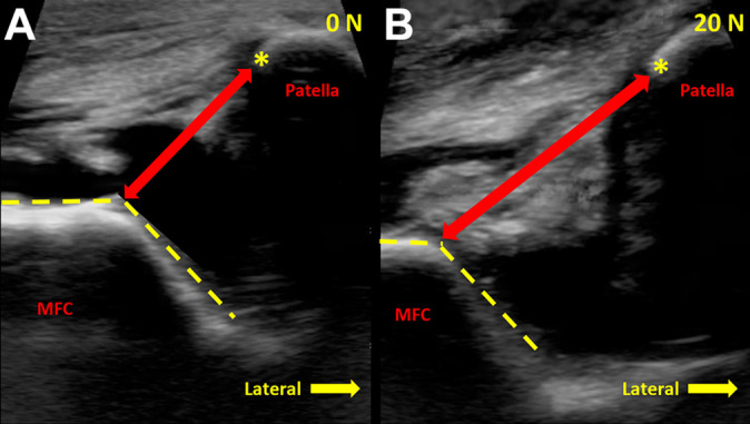
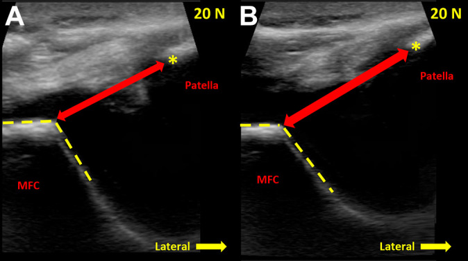
Methods: In 10 cadaveric knees, the medial patellofemoral distance was measured to quantify patellar position from 0° to 40° of knee flexion at 10° increments. Knees were evaluated at each flexion angle under unloaded conditions and with 20 N of laterally directed force on the patella to mimic the glide test. Patellar position measurements were made on ultrasound images obtained before and after MPFC transection and compared for significant differences. To determine the ability of medial patellofemoral measurements to differentiate between MPFC-intact and MPFC-deficient states, area under the receiver operating characteristic (ROC) curve analysis and the Delong test were used. The optimal cutoff value to distinguish between the deficient and intact states was determined using the Youden J statistic.
Results: A significant increase in medial patellofemoral distance was observed in the MPFC-deficient state as compared with the intact state at all flexion angles (P = .005 to P < .001). When compared with the intact state, MPFC deficiency increased medial patellofemoral distance by 32.8% (6 mm) at 20° of knee flexion under 20-N load. Based on ROC analysis and the J statistic, the optimal threshold for identifying MPFC injury was 19.2 mm of medial patellofemoral distance at 20° of flexion under dynamic loading conditions (area under the ROC curve = 0.93, sensitivity = 77.8%, specificity = 100%, accuracy = 88.9%).
Conclusion: Using dynamic ultrasound assessment, we found that medial patellofemoral distance significantly increases with disruption of the MPFC.
Clinical relevance: Dynamic ultrasound measurements can be used to accurately detect the presence of complete MPFC injury.
Keywords: dynamic imaging; knee; medial patellofemoral complex; medial patellofemoral ligament; patellar dislocation; patellar instability; patellofemoral; stress imaging; ultrasound.
© The Author(s) 2022.
Conflict of interest statement
One or more of the authors has declared the following potential conflict of interest or source of funding: C.W.D. has received education payments from Arthrex; consulting fees from Cartiva, Stryker, Wright Medical, and Zimmer Biomet; speaking fees from Wright Medical; and royalties from Extremity Medical. M.J.T. has received grant support from DJO and education payments from Kairos Surgical and Supreme Orthopedic Systems. AOSSM checks author disclosures against the Open Payments Database (OPD). AOSSM has not conducted an independent investigation on the OPD and disclaims any liability or responsibility relating thereto.
Figures
References
-
- Camp CL, Stuart MJ, Krych AJ, et al. CT and MRI measurements of tibial tubercle–trochlear groove distances are not equivalent in patients with patellar instability. Am J Sports Med. 2013;41(8):1835–1840. - PubMed
-
- Christian DR, Redondo ML, Cancienne JM, et al. Differential contributions of the quadriceps and patellar attachments of the proximal medial patellar restraints to resisting lateral patellar translation. Arthroscopy. 2020;36(6):1670–1676. - PubMed
-
- d’Entremont AG, Nordmeyer-Massner JA, Bos C, Wilson DR, Pruessmann KP. Do dynamic-based MR knee kinematics methods produce the same results as static methods? Magn Reson Med. 2013;69(6):1634–1644. - PubMed
LinkOut - more resources
Full Text Sources
