. 2022 May 25;8(5):546-561.
doi: 10.1021/acscentsci.1c01556. Epub 2022 Apr 27.

Durable Surfaces from Film-Forming Silver Assemblies for Long-Term Zero Bacterial Adhesion without Toxicity

Affiliations

Durable Surfaces from Film-Forming Silver Assemblies for Long-Term Zero Bacterial Adhesion without Toxicity

Hossein Yazdani-Ahmadabadi et al. ACS Cent Sci. .

Abstract

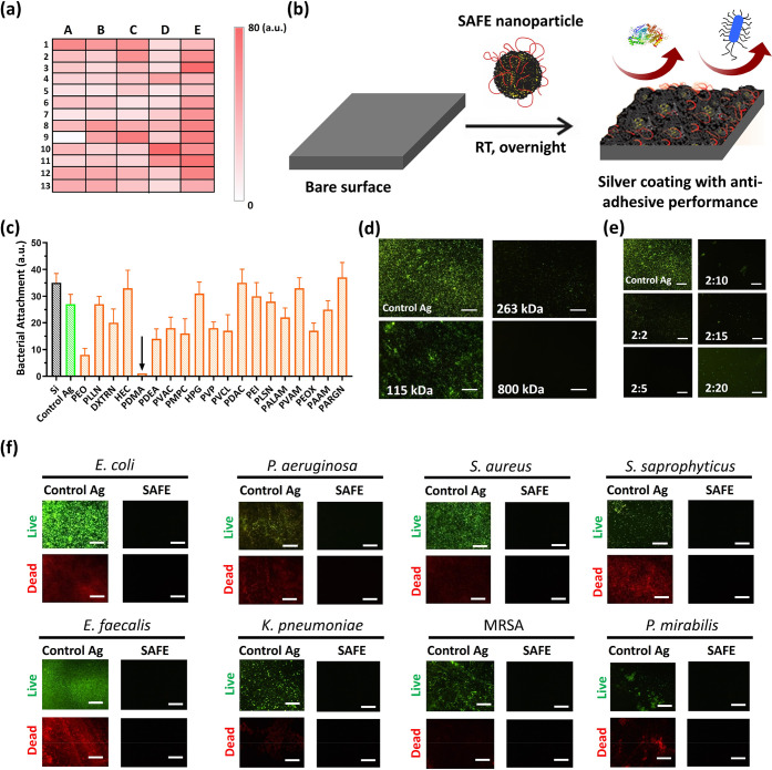
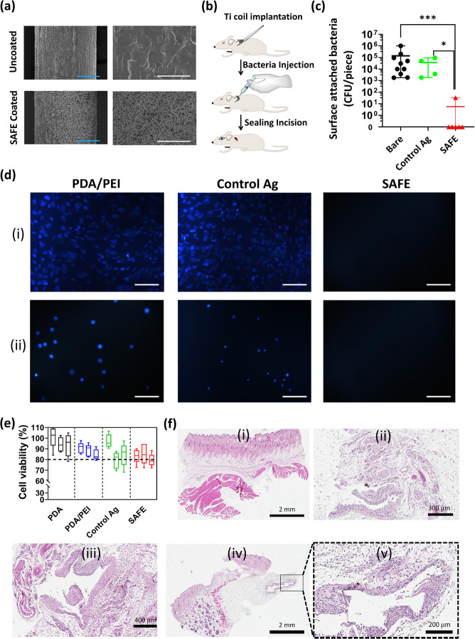
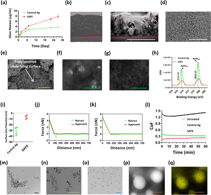
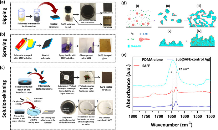
The long-term prevention of biofilm formation on the surface of indwelling medical devices remains a challenge. Silver has been reutilized in recent years for combating biofilm formation due to its indisputable bactericidal potency; however, the toxicity, low stability, and short-term activity of the current silver coatings have limited their use. Here, we report the development of silver-based film-forming antibacterial engineered (SAFE) assemblies for the generation of durable lubricous antibiofilm surface long-term activity without silver toxicity that was applicable to diverse materials via a highly scalable dip/spray/solution-skinning process. The SAFE coating was obtained through a large-scale screening, resulting in effective incorporation of silver nanoparticles (∼10 nm) into a stable nonsticky coating with high surface hierarchy and coverage, which guaranteed sustained silver release. The lead coating showed zero bacterial adhesion over a 1 month experiment in the presence of a high load of diverse bacteria, including difficult-to-kill and stone-forming strains. The SAFE coating showed high biocompatibility and excellent antibiofilm activity in vivo.


Conflict of interest statement

The authors declare the following competing financial interest(s): The University of British Columbia has filed for patent protection on the technology described here. H.Y.-A., K.Y., D.L., and J.N.K. are named as inventors on a PCT patent application submitted. The rest of the authors declare no competing interests.

Figures

Figure 1
Figure 1
High-throughput screening and identification of SAFE composition. (a) Heat map of the high-throughput screening results from the bacterial adhesion assay (E. coli, initial concentration of 1 × 10⁶ CFU/mL in LB, 24 h) (see also Table S1 for coating compositions corresponding to the heat map units). The color intensity indicates the bacterial load attached to the surface (white, no bacteria; intense red, high bacterial load). (b) Cartoon showing the synthesis of the SAFE coating with antiadhesive performance via a one-step dip-coating protocol at room temperature. (c) Relative bacterial attachment to the surface of coatings based on different UAPs incubated with E. coli (initial concentration of 1 × 10⁶ CFU/mL in LB) for 7 days. The black downward arrow is used to highlight the excellent bacterial adhesion prevention of the PDMA-containing coating. (d) Fluorescence images of biofilm formation by E. coli (initial concentration of 1 × 10⁶ CFU/mL in LB, 7 days) on the surface of coatings formed on the basis of different molecular weights of PDMA. (e) Fluorescence images of biofilm formation by E. coli (initial concentration of 1 × 10⁶ CFU/mL in LB, 7 days) on the surface of coatings formed on the basis of different DA:PDMA mass ratios. (f) Fluorescence images (green, live bacteria; red, dead bacteria) showing biofilm formation on the surface of the “control Ag” coating and the SAFE coating after 4 weeks of coincubation with diverse bacterial strains (initail concentration: 1 × 10⁶ CFU/mL). The scale bar is 100 μm.
Figure 2
Figure 2
Long-term antibacterial activity of the SAFE coating. (a) Concentration of the planktonic bacteria present in the LB medium after coincubation of the coated polyurethane (PU) substrates (two controls including the PDA/PEI control and the “control Ag” coating along with the SAFE coating) with diverse bacterial strains (initial concentration of 1 × 10⁶ CFU/mL in LB). The PDA/PEI control composition contains DA (2 mg/mL) and PEI (1.5 mg/mL). The downward arrows are used to highlight the prevention of planktonic bacterial growth (green, “control Ag”; blue, SAFE). (b) Fluorescence images (green, live bacteria; red, dead bacteria) showing the biofilm formation on the surface of the “control Ag” and the SAFE coating on PU substrates exposed to a stream of S. saprophyticus fluid (>1 × 10⁹ CFU/mL, LB, 5 mL/min) for 28 days. The scale bar is 100 μm.
Figure 3
Figure 3
In vivo activity and biocompatibility of SAFE coating. (a) SEM images of the uncoated Ti wire and the SAFE-coated Ti wire at two different magnifications including 0.35 k (left) and 5 k (right). The blue and white scale bars are 100 and 10 μm, respectively. (b) Cartoon showing the insertion of the Ti implant under the skin on the back of the rat in the subcutaneous pocket. (c) Number of bacterial colonies attached to the surface of uncoated (n = 9), “control Ag” (n = 4), and SAFE coated (n = 6) Ti implants after 7 days of implantation in the subcutaneous pockets of rats. * indicates a P value ≤0.05, ** indicates a P value ≤0.01, and *** indicates a P value ≤0.001. (d) Fluorescence microscopy images of cell adhesion on the surface of the “control Ag” coating and the SAFE coating following 24 h incubation with (i) fibroblast and (ii) bladder cells (T24) at 37 °C. (e) Viability (%) of cells (T24 bladder cells) grown for 24 h in the media (RPMI, 10% FBS, 1% penicillin/streptomycin) incubated with different coatings, including PDA, PDA/PEI, “control Ag” and SAFE coatings (n = 5) at 12 h (left box), 24 h (middle box), and 48 h (right box). (f) Optical microscopy images of the H&E-stained section of (i) healthy skin tissue and skin tissues in vicinity of the (ii) uncoated Ti implant, (iii) “control Ag”-coated Ti implant, and (iv, v) SAFE-coated Ti implant.
Figure 4
Figure 4
SAFE characterization. (a) Silver release profile for the “control Ag” coating and the SAFE coating over 28 days of incubation with water. SEM images of the FIB-created cross-section of (b) epoxy-embedded and (c) dehydrated SAFE coating on the silicon wafer. The purple and white scale bars are 4 and 5 μm, respectively. SEM images of the SAFE coating taken at two different magnifications: (d) 2 k and (e) 50 k. The yellow and white scale bars are 1 and 30 μm, respectively. The white arrow points out the full coverage of the underlying surface with the SAFE coating. BSE-SEM images of the (f) “control Ag” coating and (g) SAFE coating. The green scale bar is 400 nm. (h) High-resolution XPS spectra of silver for the “control Ag” coating and the SAFE coating. (i) Surface ζ potential of the “control Ag” coating (n = 4) and the SAFE coating (n = 4). Atomic force microscopy force–distance curves of (j) the “control Ag” coating and (k) the SAFE coating. (l) CoF of the coated glass against the PDMS ball (5 mm, 2 N) under wet conditions (water was used as the lubricant). The experiment was repeated three times, and the data presented are the average of the data collected from all three explements (n = 3). (m) TEM image of the solution-borne SAFE assemblies embedded with silver. The black scale bar is 30 nm. (n) Bright-field SEM image of the FIB-created cross section of the epoxy-embedded SAFE coating on a silicon wafer. The green scale bar is 400 nm. (o) TEM image of the reconstituted SAFE assemblies. The blue scale bar is 50 nm. (p) STEM dark field image and (q) silver mapping of the individual silver nanoparticle incorporated into the SAFE assembly/coating. The orange scale bar is 10 nm.
Figure 5
Figure 5
SAFE film formation. Schematic along with digital images showing different coating methods including (a) dipping, (b) spraying, and (c) solution-skinning. (d) Schematic shows different steps of the SAFE film formation based on the mechanism we proposed utilizing the SAFE assemblies and coating characterization: (i) a substrate exposed to a solution containing DA, PEI, silver nitrate, and PDMA at t = 0; (ii) the formation of irregularly shaped assemblies embedded with silver nanoparticles; (iii) the random deposition of assemblies forming a structurally loose film; (iv) reorganization of the film structure upon dehydration; (v) formation of the reorganized assemblies with a highly integrated and dense structure. (e) ATR-FTIR spectrum of the SAFE coating, spectrum of PDMA alone, and the spectrum resulting from the subtraction of SAFE spectrum from “control Ag” spectrum, denoted Sub (SAFE – “control Ag”).

References

    1. Onaizi S. A.; Leong S. S. J. Tethering Antimicrobial Peptides: Current Status and Potential Challenges. Biotechnol. Adv. 2011, 29 (1), 67–74. 10.1016/j.biotechadv.2010.08.012. - DOI - PubMed
    1. Raphel J.; Holodniy M.; Goodman S. B.; Heilshorn S. C. Multifunctional Coatings to Simultaneously Promote Osseointegration and Prevent Infection of Orthopaedic Implants. Biomaterials 2016, 84, 301–314. 10.1016/j.biomaterials.2016.01.016. - DOI - PMC - PubMed
    1. Yu K.; Lo J. C. Y.; Mei Y.; Haney E. F.; Siren E.; Kalathottukaren M. T.; Hancock R. E. W.; Lange D.; Kizhakkedathu J. N. Toward Infection-Resistant Surfaces: Achieving High Antimicrobial Peptide Potency by Modulating the Functionality of Polymer Brush and Peptide. ACS Appl. Mater. Interfaces 2015, 7 (51), 28591–28605. 10.1021/acsami.5b10074. - DOI - PubMed
    1. Yong Y.; Qiao M.; Chiu A.; Fuchs S.; Liu Q.; Pardo Y.; Worobo R.; Liu Z.; Ma M. Conformal Hydrogel Coatings on Catheters to Reduce Biofouling. Langmuir 2019, 35 (5), 1927–1934. 10.1021/acs.langmuir.8b03074. - DOI - PubMed
    1. Zhou C.; Wu Y.; Thappeta K. R. V.; Subramanian J. T. L.; Pranantyo D.; Kang E. T.; Duan H.; Kline K.; Chan-Park M. B. In Vivo Anti-Biofilm and Anti-Bacterial Non-Leachable Coating Thermally Polymerized on Cylindrical Catheter. ACS Appl. Mater. Interfaces 2017, 9 (41), 36269–36280. 10.1021/acsami.7b07053. - DOI - PubMed