. 2022 May 11:3:100107.
doi: 10.1016/j.crphar.2022.100107. eCollection 2022.

Low-dose melittin is safe for intravitreal administration and ameliorates inflammation in an experimental model of uveitis

Affiliations

Low-dose melittin is safe for intravitreal administration and ameliorates inflammation in an experimental model of uveitis

Brenda Fernanda Moreira Castro et al. Curr Res Pharmacol Drug Discov. .

Abstract

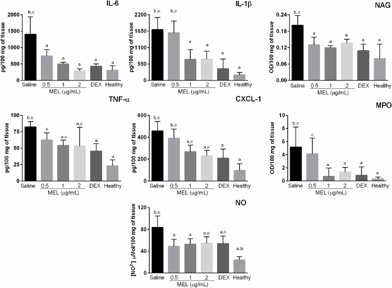
Uveitis is a group of sight-threatening ocular inflammatory disorders, whose mainstay of therapy is associated with severe adverse events, prompting the investigation of alternative treatments. The peptide melittin (MEL) is the major component of Apis mellifera bee venom and presents anti-inflammatory and antiangiogenic activities, with possible application in ophthalmology. This work aims to investigate the potential of intravitreal MEL in the treatment of ocular diseases involving inflammatory processes, especially uveitis. Safety of MEL was assessed in retinal cells, chick embryo chorioallantoic membranes, and rats. MEL at concentrations safe for intravitreal administration showed an antiangiogenic activity in the chorioallantoic membrane model comparable to bevacizumab, used as positive control. A protective anti-inflammatory effect in retinal cells stimulated with lipopolysaccharide (LPS) was also observed, without toxic effects. Finally, rats with bacille Calmette-Guerin- (BCG) induced uveitis treated with intravitreal MEL showed attenuated disease progression and improvement of clinical, morphological, and functional parameters, in addition to decreased levels of proinflammatory mediators in the posterior segment of the eye. These effects were comparable to the response observed with corticosteroid treatment. Therefore, MEL presents adequate safety profile for intraocular administration and has therapeutic potential as an anti-inflammatory and antiangiogenic agent for ocular diseases.

Keywords: Antiangiogenic; Eye; Inflammation; Melittin; Peptide; Uveitis.


Conflict of interest statement

The authors declare that they have no known competing financial interests or personal relationships that could have appeared to influence the work reported in this paper.

Figures

Image 1
Graphical abstract
Fig. 1
Fig. 1
Scheme of uveitis induction and intravitreal treatments. Rats were intraperitoneally inoculated with BCG on days 1 and 8 and the disease was induced on day 15 with an intravitreal injection of BCG. On day 18 the animals received an intravitreal injection of the following treatments: melittin 0.5, 1 and 2 ​μg/mL; dexamethasone sodium phosphate 4 ​mg/mL and saline. The healthy group was not submitted to any intervention and all animals were euthanized on day 25. BCG: bacille Calmette Guerin; MEL: melittin; DEX: dexamethasone sodium phosphate.
Fig. 2
Fig. 2
Effect of melittin on the viability of ARPE-19 ​cells and the vascular network of chicken embryos chorioallantoic membrane. (A) MTT assay performed on ARPE-19 ​cells 24, 48, and 72h after exposure to melittin 0.5–3.0 ​μg/mL showed that concentrations up to 1.5 ​μg/mL did not affect cells viability at all time-points evaluated. (B) Representative stereomicrographs showing no signs of vascular response on chick embryos chorioallantoic membranes after administration of melittin 0.1–3.0 ​μg/mL and saline (negative control), while the positive control (0,1 ​M NaOH) presented expected reactions. (C) Cumulative irritation scores and ocular irritation classification for melittin and controls according to HET-CAM assay, n ​= ​6/group. Data are mean ​± ​SD, n ​= ​3; ∗p ​< ​0.05 compared with control. a p ​< ​0.05 for 24 vs 72h; b p ​< ​0.05 for 48 vs 72h. IS: irritation score; MEL: melittin.
Fig. 3
Fig. 3
Melittin 0.5, 1, and 2 ​μg/mL are safe for intravitreal administration. (A) The IOP of melittin-treated animals measured at baseline, 6 and 14 days after the intravitreal injections was not significantly different from the vehicle group at all time-points. (B) Eye fundus images representative of vehicle, naïve, and melittin-treated animals showing preserved retinal vasculature and optic nerve head, as well as the absence of vitreous opacity in all groups, after 6 and 14 days of the intravitreal injections. (C) Representative photomicrographs of eyes enucleated 15 days after the intravitreal injection of melittin 0.5, 1, and 2 ​μg/mL or vehicle, evidencing an organized retina with preserved morphology and with no signs of inflammation or degeneration. (D) ERG examinations recorded 7 and 15 days after intravitreal melittin or vehicle showing preserved dark- and light-adapted parameters, except for a transient shorter implicit-time in the light-adapted exam of animals treated with MEL 2 ​μg/mL. (E) Similar mean ERG curves were observed for all groups evaluated 7 and 15 days after treatment. Data are mean ​± ​SD, n ​= ​4/group; ∗p ​< ​0.05 vs vehicle-treated animals; MEL: Melittin; GCL: ganglion cell layer; IPL: inner plexiform layer; INL: inner nuclear layer; OPL: outer plexiform layer; ONL: outer nuclear layer; PRL: photoreceptor layer; RPE: retinal pigment epithelium cells. Bar ​= ​50 ​μm.
Fig. 4
Fig. 4
Melittin conjugated with FITC penetrates through the retina and reaches the outer retina. Fluorescence photomicrographs representative of rats' retinas after 2 and 8h of intravitreal injection of melittin conjugated with FITC, FITC solution, or vehicle. MEL-FITC group showing fluorescent signals in the PRL and RPE after 2 and 8h, with additional signals in the GCL after 8h, while the FITC group exhibited fluorescence in PRL, RPE, and GCL after 2h with no evident signals after 8h; n ​= ​2/group. MEL-FITC: melittin conjugated with FITC; FITC: fluorescein isothiocyanate; GCL: ganglion cell layer; INL: inner nuclear layer; ONL: outer nuclear layer; PRL: photoreceptor layer; RPE: retinal pigment epithelium cells. Bar ​= ​50 ​μm.
Fig. 5
Fig. 5
Melittin inhibits angiogenesis in the chorioallantoic membrane. Chick embryos chorioallantoic membranes were treated with melittin 0.5, 1, and 2 ​μg/mL, bevacizumab 5 ​mg/mL, or saline on the 5th and 6th day after fertilization and photographed on the following day. (A) Melittin at all concentrations significantly reduced the vascularized area of the CAM in comparison to saline-treated group. (B) Representative photomicrographs of the CAM, taken 1 day after the last administration of each treatment. MEL: melittin. Data are mean ​± ​SD, n ​= ​12/group; ∗p ​< ​0.05 vs saline group; #p ​< ​0.05 vs bevacizumab.
Fig. 6
Fig. 6
Melittin reduces LPS-induced release of pro-inflammatory cytokines in ARPE-19 ​cells. Pre-treatment with melittin 1 and 2 ​μg/mL for 1h prior stimulation with LPS 10 ​μg/mL for 24h decreased IL-6, IL-8, and nitrite levels in the supernatant, determined by flow cytometry and Griess assay, respectively. Melittin 2 ​μg/mL did not stimulate de production of the proinflammatory markers measured in this assay in ARPE-19 ​cells. Data are mean ​± ​SD, n ​= ​4/group; a p ​< ​0.05 vs untreated LPS-activated cells; b p ​< ​0.05 vs control cells; MEL: melittin; LPS: lipopolysaccharide; NO: nitric oxide.
Fig. 7
Fig. 7
Intravitreal melittin improves retinal function in rats with BCG-induced uveitis. Rats inoculated with two weekly doses of BCG received 7 days later an intravitreal injection of the same antigen and were treated with intravitreal melittin 0.5, 1, or 2 ​μg/mL, dexamethasone, or saline. (A) ERG examinations performed at baseline, 3 days after BCG intravitreal injection, and 7 days after treatment showed a significantly reduced retinal function after disease induction for all groups with uveitis when compared to healthy animals and different responses after each treatment. (B) Rats treated with MEL showed significantly higher a and b-wave amplitudes compared to vehicle-treated group, with values similar to dexamethasone-treated animals, in light- and dark-adapted exams. (C) Mean ERG curves showing improved responses with treatment with MEL and dexamethasone. Data are mean ​± ​SD, n ​= ​4/group. a p ​< ​0.05 vs vehicle; b p ​< ​0.05 vs dexamethasone; c p ​< ​0.05 vs healthy group. Dashed lines represent mean values from the healthy group. MEL: melittin; DEX: dexamethasone.
Fig. 8
Fig. 8
Intravitreal melittin ameliorates clinical manifestation of uveitis in rats. Representative images were obtained with a slit-lamp, 6 days after intravitreal treatment with melittin, dexamethasone, or saline in rats with BCG-induced uveitis. Clinical signs of ocular inflammation, including iris congestion (arrows), posterior subcapsular cataract (asterisks), and synechiae (arrowheads) were observed in saline-treated animals. MEL 0.5 improved conjunctival hyperemia, although mild iris congestion and posterior subcapsular cataract were still present. An attenuated inflammatory reaction was observed in animals treated with MEL 1, 2 ​μg/mL and dexamethasone, whose anterior segment presented a normal aspect, except for mild iris congestion. Healthy animals exhibited eyes with a normal aspect; n ​= ​6 per group.
Fig. 9
Fig. 9
Intravitreal melittin attenuates morphological changes in rats with uveitis. Representative H&E photomicrographs of eyes enucleated 7 days after intravitreal treatment with melittin, dexamethasone, or saline in rats with BCG-induced uveitis. Severe retinal folds (asterisks) and disorganized retinal structure were observed in the saline-treated group, in addition to damaged photoreceptors layer (arrowhead) and inflammatory infiltrate (arrows) involving the ciliary body region and vitreous cavity. MEL 0.5-treated group showed a less intense inflammatory response in the ciliary body region, the retina slightly more organized but with an intense inflammatory infiltrate. Treatment with MEL 1, 2, and DEX attenuated the ocular inflammatory reaction, with animals exhibiting improved retinal structure and mild inflammatory infiltrate in the ciliary body region. No abnormal histological findings were observed on healthy animals; n ​= ​3 per group.
Fig. 10
Fig. 10
Treatment with intravitreal melittin decreases levels of pro-inflammatory markers in the posterior segment of rats with uveitis. Eyes were enucleated 7 days after treatment with melittin, dexamethasone, or saline, and cytokines/chemokine levels were determined by ELISA while nitrite concentration was assessed by the Griess reagent assay and NAG and MPO activity by a colorimetric reaction. Treatment with DEX, MEL 1 and 2 ​μg/mL was able to decrease the levels of all markers in comparison to the vehicle-treated group, whereas MEL 0.5 did not significantly affect CXCL-1, TNF-α, and MPO levels. Data are mean ​± ​SD, n ​= ​4/group. a p ​< ​0.05 vs vehicle; b p ​< ​0.05 vs dexamethasone; c p ​< ​0.05 vs healthy group. MEL: melittin; DEX: dexamethasone; MPO: myeloperoxidase; NAG: N-acetylglucosaminidase; NO: nitric oxide.

Similar articles

Cited by

References

    1. Ahmed C.M., Ildefonso C.J., Johnson H.M., Lewin A.S. A C-terminal peptide from type I interferon protects the retina in a mouse model of autoimmune uveitis. PLoS One. 2020;15(2):1–20. doi: 10.1371/journal.pone.0227524. - DOI - PMC - PubMed
    1. An H., Kim J., Kim W., Han S., Park K. The protective effect of melittin on renal fibrosis in an animal model of unilateral ureteral obstruction. Molecules. 2016;21(1137) doi: 10.3390/molecules21091137. - DOI - PMC - PubMed
    1. An H., Kim J., Kim W., Gwon M., Gu H.M., Jeon M.J., et al. Therapeutic effects of bee venom and its major component, melittin, on atopic dermatitis in vivo and in vitro. Br. J. Pharmacol. 2018;175:4310–4324. doi: 10.1111/bph.14487. - DOI - PMC - PubMed
    1. Arafat S.N., Suelves A.M., Spurr-Michaud S., James C., Foster S., Claes H.D., et al. Neutrophil collagenase, gelatinase and myeloperoxidase in tears of Stevens-Johnson syndrome and ocular cicatricial pemphigoid patients. Ophthalmology. 2014;121(1):79–87. doi: 10.1016/j.ophtha.2013.06.049.Neutrophil. - DOI - PMC - PubMed
    1. Arjamaa O., Aaltonen V., Piippo N., Csont T., Petrovski G. Hypoxia and inflammation in the release of VEGF and interleukins from human retinal pigment epithelial cells. Graefe’s Arch. Clin. Exp. Ophthalmol. 2017 doi: 10.1007/s00417-017-3711-0. 0–5. - DOI - PubMed

Further reading

    1. Chui Jeanie J.Y., Li Monique W.M., Girolamo Nick Di, Chang John H., McCluskey Peter J., Wakefield Denis. Iris Pigment Epithelial Cells Express a Functional Lipopolysaccharide Receptor Complex. Investig. Ophthalmol. Vis. Sci. 2010;51:2558–2567. doi: 10.1167/iovs.09-3923. - DOI - PubMed
    1. Jamasbi E, Mularski A, Separovic F. Model Membrane and Cell Studies of Antimicrobial Activity of Melittin Analogues. Curr. Top. Med. Chem. 2016;16:40–45. - PubMed
    1. Moon D, Park S, Lee K, Heo M, Kim K, Kim M, et al. Bee venom and melittin reduce proinflammatory mediators in lipopolysaccharide-stimulated BV2 microglia. Int. Immunopharm. 2007;7:1092–1101. doi: 10.1016/j.intimp.2007.04.005. - DOI - PubMed
    1. Pereira Ana Flavia Marques, Albano Mariana, Alves Fernanda Cristina Bergamo, Andrade Bruna Fernanda Murbach Teles, Furlanetto Alessandra, Rall Vera Lucia Mores, et al. Influence of apitoxin and melittin from Apis mellifera bee on Staphylococcus aureus strains. Microb. Pathog. 2020;141 doi: 10.1016/j.micpath.2020.104011. - DOI - PubMed

LinkOut - more resources