

Maternal psychological distress during the COVID-19 pandemic and structural changes of the human fetal brain
- PMID: 35647608
- PMCID: PMC9135751
- DOI: 10.1038/s43856-022-00111-w
Maternal psychological distress during the COVID-19 pandemic and structural changes of the human fetal brain
Abstract
Background: Elevated maternal psychological distress during pregnancy is linked to adverse outcomes in offspring. The potential effects of intensified levels of maternal distress during the COVID-19 pandemic on the developing fetal brain are currently unknown.
Methods: We prospectively enrolled 202 pregnant women: 65 without known COVID-19 exposures during the pandemic who underwent 92 fetal MRI scans, and 137 pre-pandemic controls who had 182 MRI scans. Multi-plane, multi-phase single shot fast spin echo T2-weighted images were acquired on a GE 1.5 T MRI Scanner. Volumes of six brain tissue types were calculated. Cortical folding measures, including brain surface area, local gyrification index, and sulcal depth were determined. At each MRI scan, maternal distress was assessed using validated stress, anxiety, and depression scales. Generalized estimating equations were utilized to compare maternal distress measures, brain volume and cortical folding differences between pandemic and pre-pandemic cohorts.
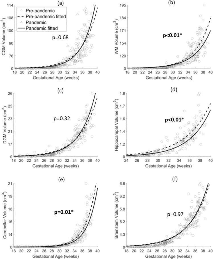
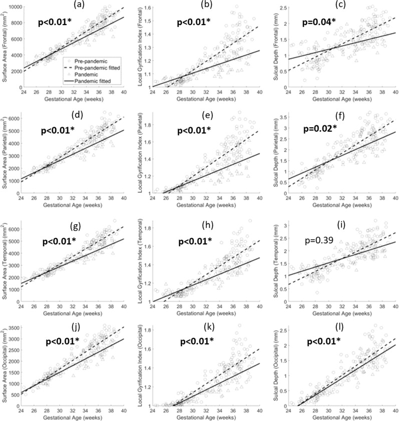
Results: Stress and depression scores are significantly higher in the pandemic cohort, compared to the pre-pandemic cohort. Fetal white matter, hippocampal, and cerebellar volumes are decreased in the pandemic cohort. Cortical surface area and local gyrification index are also decreased in all four lobes, while sulcal depth is lower in the frontal, parietal, and occipital lobes in the pandemic cohort, indicating delayed brain gyrification.
Conclusions: We report impaired fetal brain growth and delayed cerebral cortical gyrification in COVID-19 pandemic era pregnancies, in the setting of heightened maternal psychological distress. The potential long-term neurodevelopmental consequences of altered fetal brain development in COVID-era pregnancies merit further study.
Keywords: Brain imaging; Epidemiology; Paediatric research; Stress and resilience.
© The Author(s) 2022.
Conflict of interest statement
Competing interestsThe authors declare no competing interests.
Figures
References
-
- Amgalan A., Andescavage N., Limperopoulos C. Prenatal origins of neuropsychiatric diseases. Acta Paediatr. 10.1111/apa.15766 (2021). - PubMed
Grants and funding
LinkOut - more resources
Full Text Sources
