Case Reports
. 2022 Jun 29;10(3):e0045722.
doi: 10.1128/spectrum.00457-22. Epub 2022 Jun 1.

Compartmentalized into Bacteriocytes but Highly Invasive: the Puzzling Case of the Co-Obligate Symbiont Serratia symbiotica in the Aphid Periphyllus lyropictus

Affiliations
Case Reports

Compartmentalized into Bacteriocytes but Highly Invasive: the Puzzling Case of the Co-Obligate Symbiont Serratia symbiotica in the Aphid Periphyllus lyropictus

François Renoz et al. Microbiol Spectr. .

Abstract

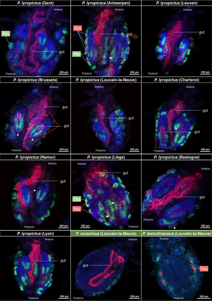
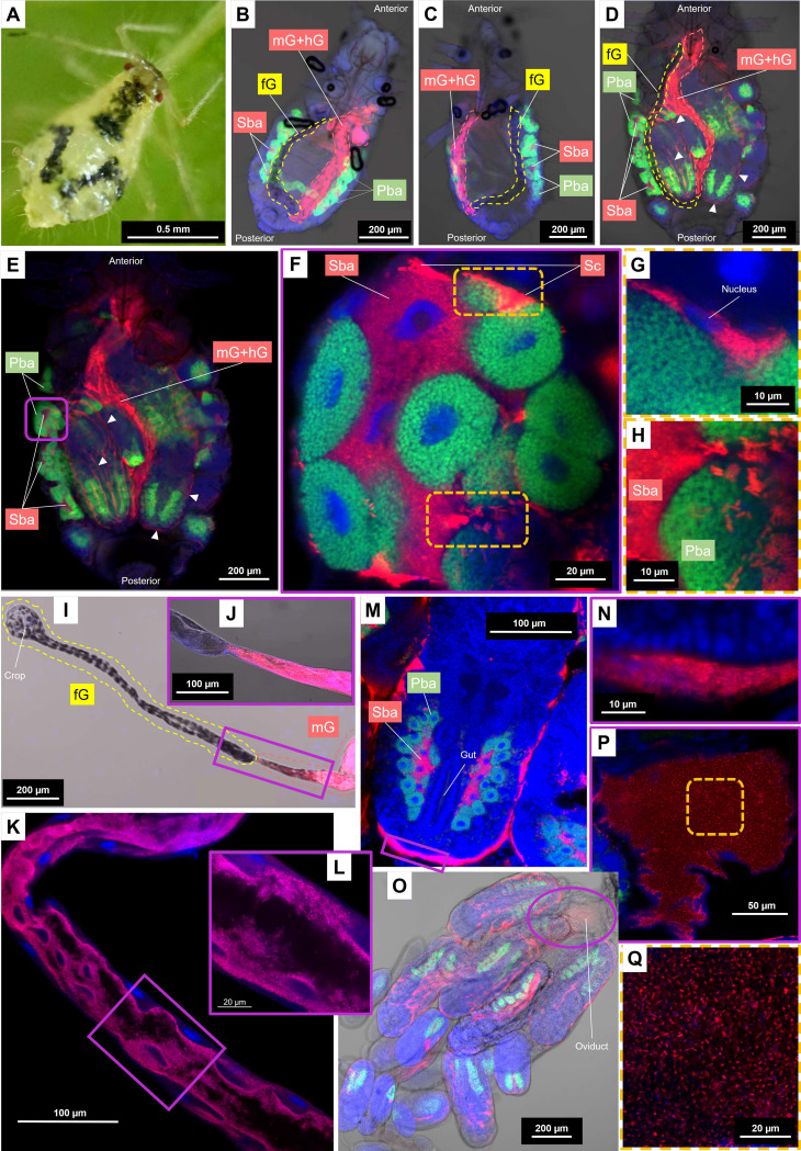
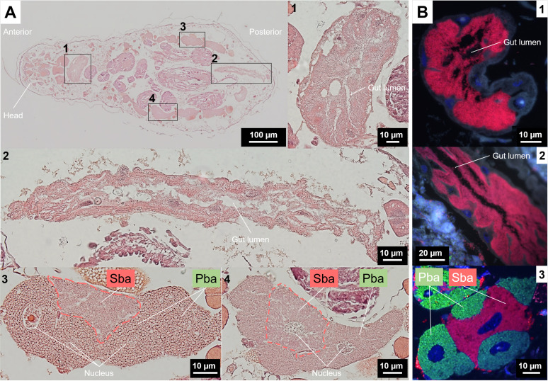
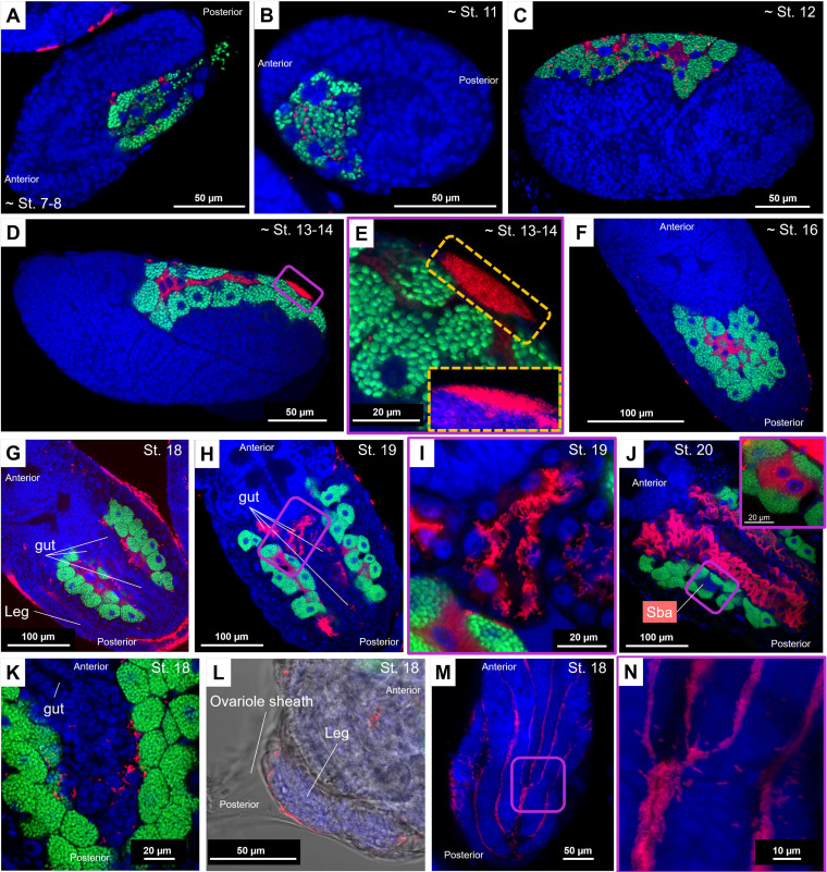
Dependence on multiple nutritional symbionts that form a metabolic unit has evolved many times in insects. Although it has been postulated that host dependence on these metabolically interconnected symbionts is sustained by their high degree of anatomical integration (these symbionts are often housed in distinct symbiotic cells, the bacteriocytes, assembled into a common symbiotic organ, the bacteriome), the developmental aspects of such multipartner systems have received little attention. Aphids of the subfamilies Chaitophorinae and Lachninae typically harbor disymbiotic systems in which the metabolic capabilities of the ancient obligate symbiont Buchnera aphidicola are complemented by those of a more recently acquired nutritional symbiont, often belonging to the species Serratia symbiotica. Here, we used microscopy approaches to finely characterize the tissue tropism and infection dynamics of the disymbiotic system formed by B. aphidicola and S. symbiotica in the Norway maple aphid Periphyllus lyropictus (Chaitophorinae). Our observations show that, in this aphid, the co-obligate symbiont S. symbiotica exhibits a dual lifestyle: intracellular by being housed in large syncytial bacteriocytes embedded between B. aphidicola-containing bacteriocytes in a well-organized compartmentalization pattern, and extracellular by massively invading the digestive tract and other tissues during embryogenesis. This is the first reported case of an obligate aphid symbiont that is internalized in bacteriocytes but simultaneously adopts an extracellular lifestyle. This unusual infection pattern for an obligate insect symbiont suggests that some bacteriocyte-associated obligate symbionts, despite their integration into a cooperative partnership, still exhibit invasive behavior and escape strict compartmentalization in bacteriocytes. IMPORTANCE Multipartner nutritional endosymbioses have evolved many times in insects. In Chaitophorinae aphids, the eroded metabolic capabilities of the ancient obligate symbiont B. aphidicola are complemented by those of more recently acquired symbionts. Here, we report the atypical case of the co-obligate S. symbiotica symbiont associated with P. lyropictus. This bacterium is compartmentalized into bacteriocytes nested into the ones harboring the more ancient symbiont B. aphidicola, reflecting metabolic convergences between the two symbionts. At the same time, S. symbiotica exhibits highly invasive behavior by colonizing various host tissues, including the digestive tract during embryogenesis. The discovery of this unusual phenotype for a co-obligate symbiont reveals a new face of multipartner nutritional endosymbiosis in insects. In particular, it shows that co-obligate symbionts can retain highly invasive traits and suggests that host dependence on these bacterial partners may evolve prior to their strict compartmentalization into specialized host structures.

Keywords: aphids; bacterial mutualism; bacteriocytes; co-obligate Serratia symbiotica; embryo invasion; gut symbiont.


Conflict of interest statement

The authors declare no conflict of interest.

Figures

FIG 1
FIG 1
Tissue tropism of S. symbiotica in field-collected adult P. lyropictus aphids. Green, red, and blue signals indicate Buchnera cells, Serratia cells, and host insect nuclei, respectively. S. symbiotica was consistently observed in the gut of P. lyropictus adults for all 10 sites examined. S. symbiotica was also found in the gut of certain P. lyropictus embryos (see embryos surrounded by a red frame) and in secondary bacteriocytes (Sba) nested between primary bacteriocytes (Pba) harboring Buchnera. White arrows indicate secondary bacteriocytes in the embryos. S. symbiotica is also visible at the periphery of the embryos (arrowheads). S. symbiotica was also observed in the gut of P. coracinus, while it is present only in secondary bacteriocytes in P. testudinaceus.
FIG 2
FIG 2
Detailed tissue tropism of the co-obligate S. symbiotica symbiont associated with P. lyropictus. Green, red, and blue signals indicate Buchnera cells, Serratia cells, and host insect nuclei, respectively. (A) A young adult of P. lyropictus feeding on Acer platanoides. (B) A second-stage nymph (3 days old; bright field) in which the horseshoe-shaped architecture of the bacteriome is well visible. S. symbiotica is present in secondary bacteriocytes and in the posterior part of the digestive tract (midgut and hindgut) but appears to be absent from the foregut. (C) A third instar nymph (5 days old; bright field) where S. symbiotica exhibits the same pattern of tissue tropism as that observed in the second instar. (D) A young adult (12 days old; bright field) where the developing embryos are more visible. S. symbiotica can be found in the secondary bacteriocytes, in the posterior part of the digestive tract, but also at the periphery of the embryos (arrowheads). (E) An enlarged dark field view of the adult stage from which the enlarged shots of the bacteriocyte structures that follow are derived. (F) One of the bacteriocyte clusters forming the bacteriome (Airyscan) composed of syncytial secondary bacteriocytes hosting S. symbiotica, embedded between the uninucleated primary bacteriocytes hosting Buchnera. Sheath cells that also house the co-obligate symbiont sparsely cover the periphery of the bacteriocytes. (G) Close-up view (Airyscan) of a sheath cell hosting S. symbiotica. (H) Close-up view (Airyscan) suggesting the occurrence of symbionts trafficking between secondary bacteriocytes and hemolymph. (I) Dissected digestive tract (bright field) demonstrating the absence of S. symbiotica in the foregut. (J) Close-up view (bright field) of the intersection between the foregut and the midgut confirming the presence S. symbiotica in the latter. (K) Midgut segment (Airyscan) showing S. symbiotica densely colonizing this part of the digestive tract. (L) Close-up view (Airyscan) showing S. symbiotica densely distributed at the inner periphery of the midgut. (M) An embryo of developmental stage 18 (Airyscan) where S. symbiotica is well visible in secondary bacteriocytes but also in the periphery of the embryos. (N) Close-up view (Airyscan) showing the presence of S. symbiotica around the embryos. (O) Dissected embryonic chain (bright field) revealing the presence of S. symbiotica the presence of S. symbiotica in the oviduct and around the embryos in the ovarioles. (P) Close-up view (Airyscan) confirming the presence of S. symbiotica in the oviduct. (Q) S. symbiotica cells (Airyscan) with their typical rod shape in the oviduct. fG, foregut; mG, midgut; hG, hindgut; Sba, secondary bacteriocyte; Pba, primary bacteriocyte; Sc, sheath cell.
FIG 3
FIG 3
Bacteriocyte structures and tissue tropism of S. symbiotica in P. lyropictus (third instar nymph). (A) Representative images of H&E-stained whole-aphid sections. (1 and 2) Enlarged sections of the digestive tract. (3 and 4) Magnified images of H&E-stained bacteriocyte clusters showing the nested pattern of secondary bacteriocytes between primary bacteriocytes. (B) FISH on tissue sections. Green, red, and blue signals indicate Buchnera cells, Serratia cells and host insect nuclei, respectively. (1 and 2) Enlarged sections of the digestive tract showing the dense presence of S. symbiotica. (3) A bacteriocyte cluster consisting of primary bacteriocytes wrapping a secondary bacteriocyte. Pba, primary bacteriocyte; Sba, secondary bacteriocyte.
FIG 4
FIG 4
Infection dynamics of Buchnera and S. symbiotica during embryonic development in P. lyropictus. Green, red, and blue signals indicate Buchnera cells, Serratia cells, and host insect nuclei, respectively. (A) Buchnera and S. symbiotica infect a stage 7 to 8 embryo by transiting via the posterior zone. (B) Cellularization symbiont-infected syncytium until the achievement of stage 11. (C) Individualization of the uninucleate bacteriocytes harboring Buchnera during stage 12. (D) Completion of the syncytial bacteriocytes harboring S. symbiotica. The nuclei of the syncytial bacteriocytes are clearly visible around stages 13 to 14. (E) Local accumulation of S. symbiotica on the embryo surface during stages 13 to 14. Inset: z-stack sections confirming the presence of S. symbiotica on the surface of the embryo where they form a dense bacterial mass. (F) Establishment of a compact bacteriome in which the bacteriocytes sheltering S. symbiotica are surrounded by those containing Buchnera. S. symbiotica remains sporadically visible on the surface of the embryo. (G) Establishment of the horseshoe-shaped architecture of the bacteriome at stage 18. The periphery of the embryo is densely colonized by the symbiont which can even be seen between the legs. (H and I) Beginning of the colonization of the digestive tract by S. symbiotica at stage 19. (J) Stage 20 is marked by a massive colonization of the digestive tract by the symbiont. Inset: zoom on a secondary bacteriocyte. The fluorescent signal associated with S. symbiotica is so intense in the digestive tract that the visibility of the symbiont in secondary bacteriocytes is impeded. This difference in signal intensity suggests that the gut is colonized much more densely by the symbiont than by the bacteriocytes. (K) An image showing S. symbiotica freely circulating in the hemolymph. (L) A bright-field image showing S. symbiotica in the ovariole. (M) The symbiont is established in the surface invaginations of the embryos. (N) Zoom-in on these invaginations showing the concentration of the symbiont in these cavities and suggesting that S. symbiotica could form biofilms on the surface of the embryos.

Similar articles

Cited by

References

    1. Wu D, Daugherty SC, Aken SEV, Pai GH, Watkins KL, Khouri H, Tallon LJ, Zaborsky JM, Dunbar HE, Tran PL, Moran NA, Eisen JA. 2006. Metabolic complementarity and genomics of the dual bacterial symbiosis of sharpshooters. PLoS Biol 4:e188. doi:10.1371/journal.pbio.0040188. - DOI - PMC - PubMed
    1. McCutcheon JP, Moran NA. 2010. Functional convergence in reduced genomes of bacterial symbionts spanning 200 My of evolution. Genome Biol Evol 2:708–718. doi:10.1093/gbe/evq055. - DOI - PMC - PubMed
    1. Koga R, Moran NA. 2014. Swapping symbionts in spittlebugs: evolutionary replacement of a reduced genome symbiont. ISME J 8:1237–1246. doi:10.1038/ismej.2013.235. - DOI - PMC - PubMed
    1. Manzano-Marín A, Latorre A. 2016. Snapshots of a shrinking partner: genome reduction in Serratia symbiotica. Sci Rep 6:32590. doi:10.1038/srep32590. - DOI - PMC - PubMed
    1. Mao M, Yang X, Poff K, Bennett G. 2017. Comparative genomics of the dual-obligate symbionts from the treehopper, Entylia carinata (Hemiptera: Membracidae), provide insight into the origins and evolution of an ancient symbiosis. Genome Biol Evol 9:1803–1815. doi:10.1093/gbe/evx134. - DOI - PMC - PubMed

Publication types

Supplementary concepts

LinkOut - more resources