Review
. 2022 Jun;70(6):1931-1944.
doi: 10.4103/ijo.IJO_2363_21.

Sympathetic ophthalmia: A comprehensive update

Affiliations
Review

Sympathetic ophthalmia: A comprehensive update

Swapnil Parchand et al. Indian J Ophthalmol. 2022 Jun.

Abstract

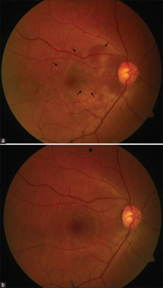
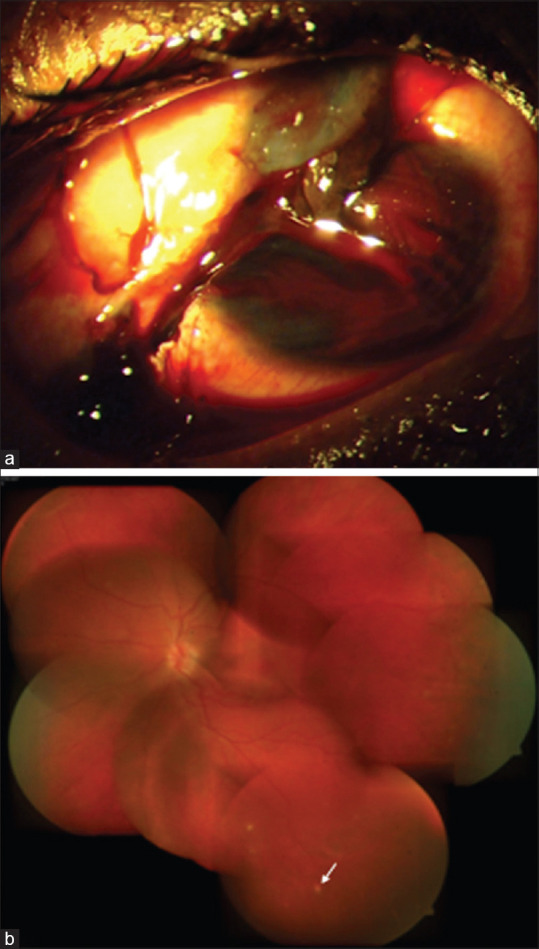
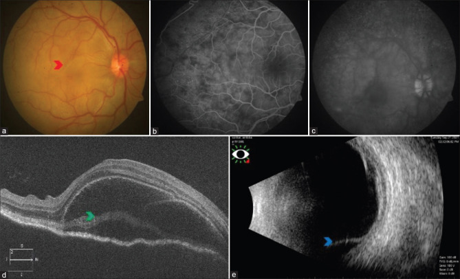
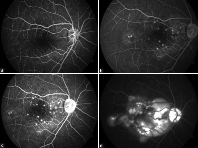
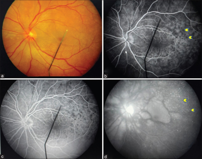
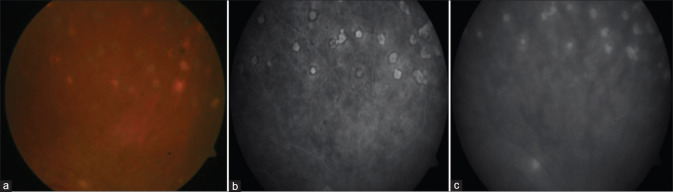
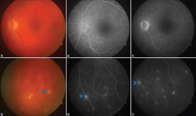
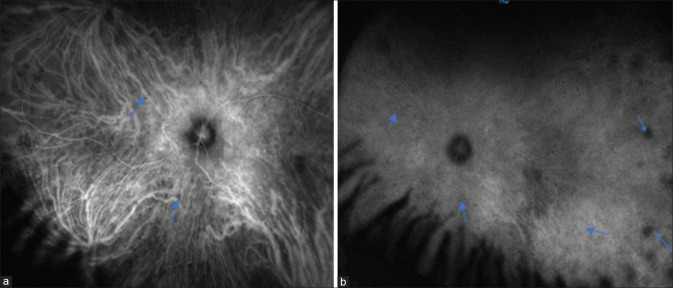
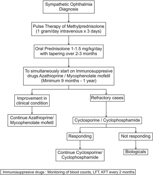
Sympathetic ophthalmia is a rare, bilateral, granulomatous, panuveitis following penetrating trauma or surgery to one eye. Clinical presentation commonly occurs within the first year of trauma occurrence but can be delayed by several years. It manifests as acute/chronic granulomatous uveitis with yellowish-white choroidal lesions or Dalen-Fuchs nodules. Initially, patients respond rapidly to corticosteroid therapy, but a majority require long-term use of corticosteroid-sparing agents to prevent recurrences. The purpose of this review is to elaborate on the current understanding of the pathophysiology, the importance of multimodal imaging in early diagnosis, and the role of newer immunomodulatory and biological agents in recalcitrant cases.

Keywords: Dalen–Fuchs nodule; granulomatous uveitis; immunosuppression; ocular trauma; panuveitis; sympathetic ophthalmia.


Conflict of interest statement

None

Figures

Figure 1
Figure 1
Color fundus photograph of a case of sympathetic ophthalmia with multiple neurosensory detachments (black arrows) (a), which resolved after systemic steroid and immunomodulators (b)
Figure 2
Figure 2
A 32-year-old male presented with corneoscleral tear and uveal tissue prolapse (a). He underwent primary wound repair. Four months after injury he developed sympathetic ophthalmia in the left eye. He was managed on systemic steroids and azathioprine. At 1 year follow-up, the left eye had sunset glow fundus with nummular scars in the periphery (white arrow) (b)
Figure 3
Figure 3
Right eye color fundus photograph of a case of sympathetic ophthalmia with disc hyperemia and multiple pockets of exudative retinal detachment (red arrowhead) (a). FFA showing pinpoint hyperfluorescence in the early phase (b), followed by the pooling of dye within the area of neurosensory detachment in the late phase (c). SD-OCT showing neurosensory detachment with the split of photoreceptors at the level of myloid (green arrowhead) (d). B-scan showing retinochoroid thickening and exudative retinal detachment inferiorly (blue arrowhead) (e)
Figure 4
Figure 4
FFA showing multiple hyperfluorescent leaks in the early phase (a, b, and c), followed by the pooling of dye within the area of neurosensory detachment in the late phase (d)
Figure 5
Figure 5
Fundus picture showing exudative retinal detachment in the peripapillary area and macula (a). FFA showing hypofluorescent spots (yellow arrows) along with multiple hyperfluorescent leaks in the early phase (b and c), followed by leakage and pooling in the late phase (d) (yellow arrows)
Figure 6
Figure 6
Color fundus photograph showing healed nummular scars (a), which appear hyperfluorescent (window defects) on FFA in early phase (b), and minimal increase in hyperflourescence (c) in the late phase
Figure 7
Figure 7
Left eye fundus picture showing sunset glow fundus (a) and Dalen–Fuchs spots (blue arrowhead) in inferior retina (d). FFA showing disc staining ( b and c) with Dalen–Fuchs spots appearing hyperfluorescent spots (blue arrowhead) both in the early and late phases (window defect) (e and f)
Figure 8
Figure 8
Widefield ICGA showing hypocyanescent spots (blue arrows) in the early phase (a) which persists in the late phase (b)
Figure 9
Figure 9
ICGA showing hypocyanescent spots (yellow arrows) (a) that become isocyanescent during the late phase (b)
Figure 10
Figure 10
SS-OCT showing neurosensory detachment associated with the split of photoreceptors at the level of myloid (white arrow) resulting in bacillary layer detachment
Figure 11
Figure 11
EDI-SD-OCT showing multiple neurosensory retinal detachments associated with diffuse choroidal thickening and loss of choroidal architecture. Subsequent scans show a reduction in choroidal thickness by restoring choroidal architecture (white arrows) after starting immunomodulatory treatment
Figure 12
Figure 12
OCTA showing flow voids (yellow arrows) in choriocapillaris slab, which reduce and disappear with treatment on subsequent follow-up
Figure 13
Figure 13
Flowchart for the management of sympathetic ophthalmia

References

    1. Albert D, Diaz-Rohena R. A historical review of sympathetic ophthalmia and its epidemiology. Surv Ophthalmol. 1989;34:1–14. - PubMed
    1. Mackenzie W. 3rd edition. London: Longmans; 1840. A Practical Treatise on the Diseases of the Eye; pp. 523–34.
    1. Dalen A. Zur Kenntnis der sogenannten Choroiditis sympathetica. Mitt Augen Klin Carolin Med. Chirurg Inst Stockholm. 1904;6:1.
    1. Fuchs E. On sympathetic inflammation (initially remarks on serous iritis) Graefes Arch Clinical Exp Ophthalmology. 1905;61:365–456.
    1. Elschnig A. Studies on sympathetic ophthalmia, II:The antigenic effects of eye pigments. Graefes Arch Clin Exp Ophthalmol. 1910;76:509–46.

MeSH terms

Substances