. 2022 Jun 1;79(6):332.
doi: 10.1007/s00018-022-04293-3.

Consequences of the Hsp110DE9 mutation in tumorigenesis and the 5-fluorouracil-based chemotherapy response in Msh2-deficient mice

Affiliations

Consequences of the Hsp110DE9 mutation in tumorigenesis and the 5-fluorouracil-based chemotherapy response in Msh2-deficient mice

Kathleen Noel et al. Cell Mol Life Sci. .

Abstract

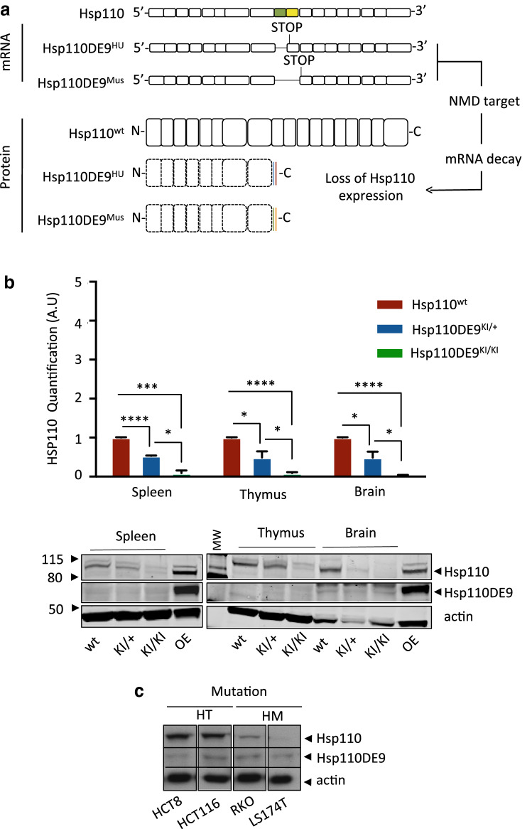
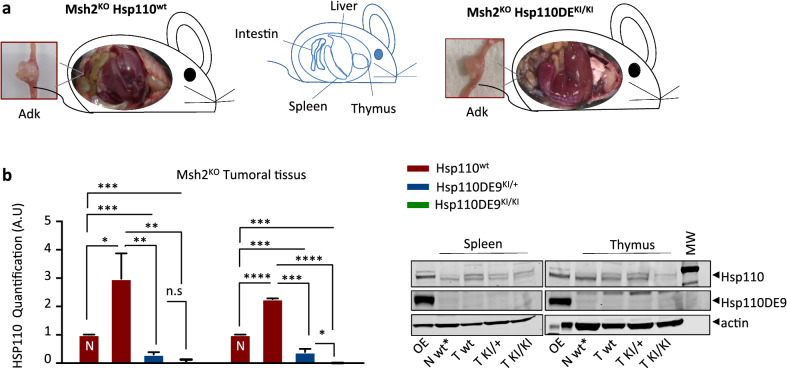
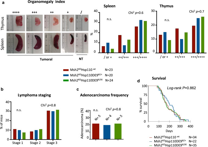
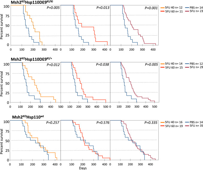
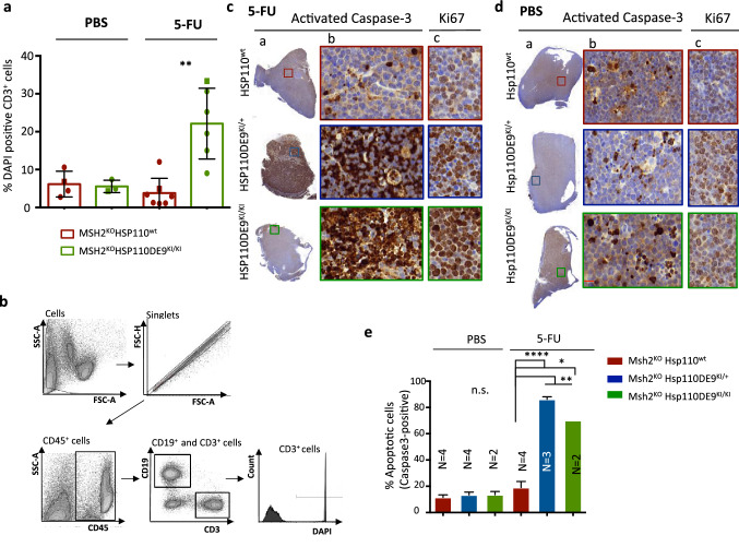
Heat shock proteins (HSPs) play oncogenic roles in human tumours. We reported a somatic inactivating mutation of HSP110 (HSP110DE9) in mismatch repair-deficient (dMMR) cancers displaying microsatellite instability (MSI) but did not assess its impact. We evaluated the impact of the Hsp110DE9 mutation on tumour development and the chemotherapy response in a dMMR knock-in mouse model (Hsp110DE9KIMsh2KO mice). The effect of the Hsp110DE9 mutation on tumorigenesis and survival was evaluated in Msh2KO mice that were null (Hsp110wt), heterozygous (Hsp110DE9KI/+), or homozygous (Hsp110DE9KI/KI) for the Hsp110DE9 mutation by assessing tumoral syndrome (organomegaly index, tumour staging) and survival (Kaplan-Meier curves). 5-Fluorouracil (5-FU), which is the backbone of chemotherapy regimens in gastrointestinal cancers and is commonly used in other tumour types but is not effective against dMMR cells in vivo, was administered to Hsp110DE9KI/KI, Hsp110DE9KI/+, and Hsp110wtMsh2KO mice. Hsp110, Ki67 (proliferation marker) and activated caspase-3 (apoptosis marker) expression were assessed in normal and tumour tissue samples by western blotting, immunophenotyping and cell sorting. Hsp110wt expression was drastically reduced or totally lost in tumours from Msh2KOHsp110DE9KI/+ and Msh2KOHsp110DE9KI/KI mice. The Hsp110DE9 mutation did not affect overall survival or tumoral syndrome in Msh2KOHsp110DE9KI/+ and Msh2KOHsp110DE9KI/KI mice but drastically improved the 5-FU response in all cohorts (Msh2KOHsp110DE9KI/KI: P5fu = 0.001; Msh2KOHsp110DE9KI/+: P5fu = 0.005; Msh2KOHsp110wt: P5fu = 0.335). Histopathological examination and cell sorting analyses confirmed major hypersensitization to 5-FU-induced death of both Hsp110DE9KI/KI and Hsp110DE9KI/+ dMMR cancer cells. This study highlights how dMMR tumour cells adapt to HSP110 inactivation but become hypersensitive to 5-FU, suggesting Hsp110DE9 as a predictive factor of 5-FU efficacy.

Keywords: 5-FU chemotherapy; Hsp110; MMR deficiency; Microsatellite instability; Mouse model.


Conflict of interest statement

The authors have stated explicitly that there are no conflicts of interest in connection with this article.

Figures

Fig. 1
Fig. 1
Hsp110DE9 KI model induces the loss of wild-type HSP110 expression in mouse tissues. a Schematic drawing of Hsp110 mRNA and the consequence of the Hsp110DE9 mutation (exon 9 skipping and stop codon formation in humans (HU) (Hsp110DE9HU)). In the Hsp110DE9 KI mouse allele, the endogenous HSP110 locus was modified to produce the same effect as the human Hsp110DE9 mutation and the putative Hsp110DE9 mutant protein. The mutated pre-mRNA is a target of the NMD system, as we described previously [11], with consecutive loss of the expression of the putative mutant Hsp110DE9 protein. b Upper panel: Hsp110 protein quantification analysis in non-tumoral tissues (spleen, thymus and brain) from wild-type, Hsp110DE9KI/+ and Hsp110DE9KI/KI mice. Bottom panel: The western blot shown is representative of 3 independent experiments. c Western blot analysis of 4 MSI CRC cell lines: HCT8 and HCT116 cells heterozygous for the Hsp110DE9 mutation; RKO and LS174T cells homozygous for the Hsp110DE9 mutation. Actin was used as a loading control. An unpaired t test was performed to assess significance. *P < 0.05, **P < 0.01, ***P < 0.001, ****P < 0.0001
Fig. 2
Fig. 2
Hsp110 depletion does not suppress or change the tumour phenotype in the MSI mouse model (I). a The pictures show representative images of the two mouse genotypes evaluated in this study (i.e., Msh2KOHsp110wt, left panel, and Msh2KOHsp110DE9KI/KI, right panel). Each mouse exhibited signs suggestive of organomegaly in the three organs (spleen, thymus and liver), consistent with lymphoma development. The boxed images represent an enlargement of the intestinal tract with representative adenocarcinoma. b Left panel: Hsp110 protein quantification analysis of T cells infiltrating spleen and thymus tissues from Msh2KOHsp110wt, Msh2KOHsp110DE9KI/+ and Msh2KOHsp110DE9KI/KI mice; (N) indicates extracts from nontumoral tissues. Right panel: Representative western blot membranes from 3 independent experiments. Actin was used as a loading control. An unpaired t test was performed to determine significance. *P < 0.05, **P < 0.01, ***P < 0.001, ****P < 0.0001
Fig. 3
Fig. 3
Hsp110 depletion does not suppress or change the tumour phenotype in the MSI mouse model (II). a Left panel: Representative pictures of organomegaly in the two localizations (spleen and thymus) and the associated index depending on the size of the organ at the macroanalysis after sacrifice. Right panel: Analysis of the organomegaly index of the three groups of mice. No significant differences were observed among the three mouse genotypes in terms of the development of splenomegaly (Chi2 P = 0.6) or thymomegaly (Chi2 P = 0.7). b Lymphoma staging analysis of the three study cohorts (for more details, see the Materials and Methods section and Supplementary Fig. S2c). c Adenocarcinoma formation rate in the three groups of this study. No significant differences were observed among the three mouse genotype groups in terms of adenocarcinoma development (Chi2 P = 0.8). d Kaplan–Meier survival analysis of the Msh2KOHsp110wt (red line), Msh2KOHsp110DE9KI/+ (blue line) and Msh2KOHsp110DE9KI/KI (green line) mouse groups. The log-rank test was used for statistical analysis. An unpaired t test was performed to determine significance
Fig. 4
Fig. 4
Hsp110 depletion improved the response to 5-FU chemotherapy in the MSI mouse model. Kaplan–Meier survival analysis of Msh2KOHsp110DE9KI/KI (upper panel), Msh2KOHsp110DE9KI/+ (middle panel), Msh2KOHsp110wt (bottom panel) mice treated with 5-FU (5-FU 40 mg/kg orange line; 5-FU 60 mg/kg red line; 5-FU all doses burgundy line) and/or placebo (PBS; blue line). The log-rank test was used for statistical analysis
Fig. 5
Fig. 5
Hsp110 protein depletion increases the susceptibility of MSI T-lymphoma cells to apoptosis after 5-FU treatment. a Quantification of cell viability based on a comparison of the percentage of DAPI-stained T-lymphoma cells (CD3+) in tumoral organs (spleen and thymus) determined by cytometry analysis with the organomegaly status of Msh2KOHsh110wt and Msh2KOHsp110DE9KI/KI mice treated with 5-FU or with placebo. The square points correspond to representative flow cytometry data shown in Supplementary Fig. S8 b Representative FACS dot plots showing the gating of cells used to detect T-lymphoma cells positive for DAPI staining. An unpaired t test was performed to determine significance. *P < 0.05, **P < 0.01, ***P < 0.001, ****P < 0.0001. (c and d) Representative activated caspase-3 immunostaining of the entire field of view (a) or a magnified region (b) corresponding to the coloured boxed area after 5-FU (c) and/or placebo (PBS) (d) treatment for the three groups (Msh2KOHsp110wt, Msh2KOHsp110DE9KI/+ and Msh2KO Hsp110DE9KI/KI). The image in (c) is an enlargement of the coloured boxes for Ki67 immunostaining. e Apoptotic index calculated as the percentage of activated caspase-3-positive cells in at least 2000 cells of the area of the slide with the most marked staining

Similar articles

Cited by

References

    1. Svrcek M, Lascols O, Cohen R, Collura A, Jonchere V, Flejou JF, Buhard O, Duval A. MSI/MMR-deficient tumor diagnosis: Which standard for screening and for diagnosis? Diagnostic modalities for the colon and other sites: Differences between tumors. Bull Cancer. 2019;106:119–128. doi: 10.1016/j.bulcan.2018.12.008. - DOI - PubMed
    1. Dorard C, de Thonel A, Collura A, et al. Expression of a mutant HSP110 sensitizes colorectal cancer cells to chemotherapy and improves disease prognosis. Nat Med. 2011;17:1283–1289. doi: 10.1038/nm.2457. - DOI - PubMed
    1. Boudesco C, Verhoeyen E, Martin L, et al. HSP110 sustains chronic NF-κB signaling in activated B-cell diffuse large B-cell lymphoma through MyD88 stabilization. Blood J Am Soc Hematol. 2018;132(5):510–520. - PubMed
    1. Zappasodi R, Bongarzone I, Ghedini GC, et al. Serological identification of HSP105 as a novel non-Hodgkin lymphoma therapeutic target. Blood. 2011;118:4421–4430. doi: 10.1182/blood-2011-06-364570. - DOI - PubMed
    1. Chatterjee S, Burns TF. Targeting heat shock proteins in cancer: a promising therapeutic approach. Int J Mol Sci. 2017;18:1978. doi: 10.3390/ijms18091978. - DOI - PMC - PubMed