. 2022 Jun 1;79(6):331.
doi: 10.1007/s00018-022-04347-6.

Impact of IL-21-associated peripheral and brain crosstalk on the Alzheimer's disease neuropathology

Affiliations

Impact of IL-21-associated peripheral and brain crosstalk on the Alzheimer's disease neuropathology

Sudhanshu Agrawal et al. Cell Mol Life Sci. .

Abstract

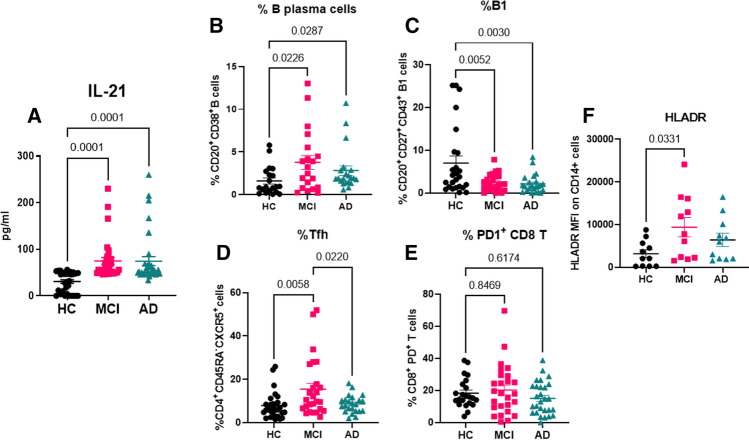
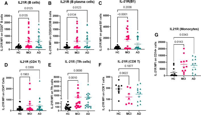
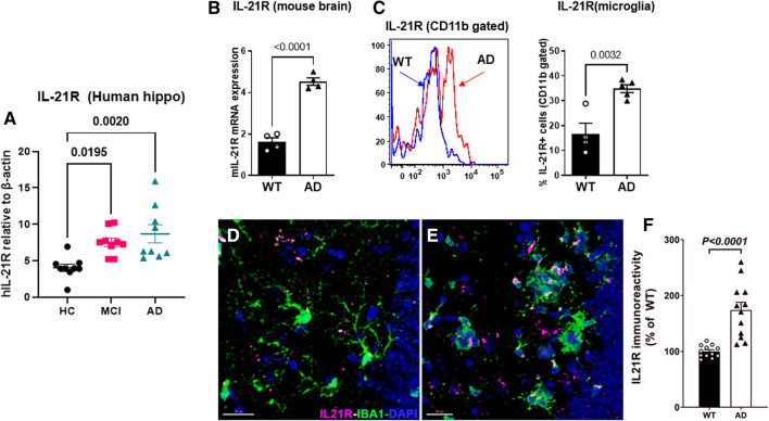
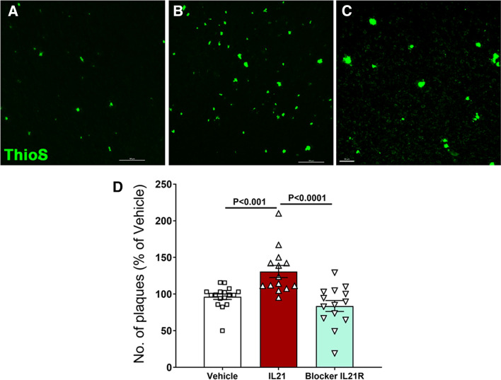
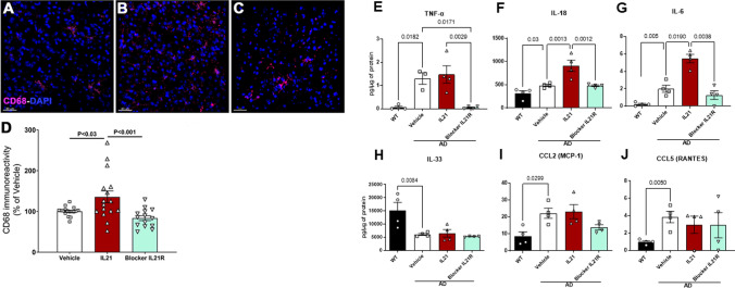
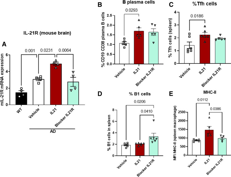
Alzheimer's disease (AD) is associated with dysregulated immune and inflammatory responses. Emerging evidence indicates that peripheral immune activation is linked to neuroinflammation and AD pathogenesis. The present study focuses on determining the role of IL-21 in the pathogenesis of AD using human samples and the 5xFAD mice model. We find that the levels of IL-21 are increased in the periphery of both humans and mice in AD. In addition, the proportions of IL-21 target cells, Tfh and B plasma cells as well as activation of monocytes is increased in PBMCs from AD and mild cognitively impaired (MCI) subjects as compared to age-matched controls, indicating immune activation. In contrast, the percentage of B1 cells that control inflammation is decreased. These changes are due to IL-21 as the expression of IL-21 receptor (IL-21R) is higher on all these cells in AD. Furthermore, treatment with recombinant IL-21 in AD mice also leads to similar alterations in Tfh, B, B1, and macrophages. The effect of IL-21 is not confined to the periphery since increased expression of IL-21R is also observed in both humans and mice hippocampus derived from the AD brains. In addition, mice injected with IL-21 display increased deposition of amyloid beta (Aβ) plaques in the brain which is reduced following anti-IL-21R antibody that blocks the IL-21 signaling. Moreover, activation of microglia was enhanced in IL-21-injected mice. In keeping with enhanced microglial activation, we also observed increased production of pro-inflammatory cytokines, IL-18 and IL-6 in IL-21-injected mice. The microglial activation and cytokines were both inhibited following IL-21R blockage. Altogether, IL-21 escalates AD pathology by enhancing peripheral and brain immune and inflammatory responses leading to increased Aβ plaque deposition. IL-21 impacts AD neuropathology by enhancing peripheral and neuronal immune activation, inflammation, and Aβ plaque deposition. Increased levels of IL-21 in the circulation of AD and MCI subjects enhances the proportions of Tfh and B plasma cells indicative of peripheral immune activation. On the other hand, the proportions of B1 cells that help reduce inflammation and clear Aβ are reduced. In addition to the periphery, IL-21 also acts on the brain via IL-21 receptor, IL-21R that displays increased expression in the hippocampi of AD and MCI subjects. IL-21 enhances the activation of microglia, induces the secretion of pro-inflammatory cytokines and deposition of Aβ plaques in the brain in AD.

Keywords: Alzheimer’s disease; B1 cells; IL-21; Neuroinflammation; Tfh.


Conflict of interest statement

The authors declare no conflict of interest.

Figures

Fig. 1
Fig. 1
Comparison of the IL-21 plasma levels and percentages of B and T cells and activation of monocytes in AD, MCI and healthy controls. Plasma samples from AD, MCI and age-matched healthy controls (HC) were analyzed for IL-21 using specific ELISA. N = 34; M = 17; F = 17. A Dot plot depict the level of IL-21 in the three groups. PBMCs from AD, MCI and HC were subjected to flow cytometry and live gated cells were analyzed for the following—B B plasma cells (CD20+CD38+); C B1 cells (CD20+, CD27+, CD43+); D Tfh cells (CD4+CD45RACXCR5+); E exhausted CD8T cells (CD8+, PD1+); F activated monocytes (CD14+, HLADR+). N = 20; M = 10; F = 10. One way ANOVA followed by Tukey’s test was used for analysis
Fig. 2
Fig. 2
IL-21R expression on cells in PBMCs of AD MCI and healthy controls. IL-21R expression on immune cells was determined on PBMCs from AD, MCI and HC using flow cytometry. Dot plots depict the mean fluorescence intensity (MFI) of IL-21R on live gated cells. A Total B cells; B plasma B cells; C B1 cells; D total CD4 T cells; E Tfh cells; F CD8 T cells; G monocytes. N = 20; M = 10; F = 10. One way ANOVA followed by Tukey’s test was used for analysis
Fig. 3
Fig. 3
IL-21R expression in humans and mice AD brain. IL-21R expression was determined in the hippocampi of AD, MCI and HC using q-PCR. A Dot plot depicts the IL-21R gene expression; N = 9; B graph depicts the IL-21R expression in the hippocampi of 5xFAD mice and WT littermates using q-PCR; C IL-21R detection in the microglia of 5xFAD and WT mice by flow cytometry; IHC staining of IL-21R and microglia (IBA-1) was performed in the hippocampus of WT (D) and AD (E) mice; F plot depicts the volumetric quantification quantitation of IL-21R immunoreactivity
Fig. 4
Fig. 4
Administration of recombinant IL-21 enhances Aβ deposition in the brains of 5xFAD mice. Mice were given five injections of IL-21 or four injections of anti-IL-21R antibody (IL-21R blocker) and Aβ plaques were stained in the brain using thioflavin-S. Accumulation of plaques in—A vehicle-injected control; B IL-21 injected; C IL-21R blocker injected; D bar graph depicts the quantitation of the same in both males and females. N = 14
Fig. 5
Fig. 5
Effect of IL-21 administration on neuroinflammation in 5xFAD mice. Mice were given five injections of IL-21 or four injections of anti-IL-21R antibody (IL-21R blocker) and microglia were stained with CD68. CD68 staining in—A vehicle injected; B IL-21 injected; C IL-21R blocker injected; D bar graph depicts the quantitation of the same. The level of cytokines and chemokines were determined in the brain using multiplex. Bar graphs depict the pg/µg levels of—E TNF-α; F IL-18; G IL-6; H IL-33; I CCL-2; J CCL-5
Fig. 6
Fig. 6
Effect of IL-21 administration on IL-21R expression and peripheral immune cells. Mice were given five injections of IL-21 or four injections of anti-IL-21R antibody (IL-21R blocker) and the following were determined. A IL-21R expression in the brain by qPCR; dot plot depict the percentages of cells in the spleen as determined by flow cytometry. B B plasma cells; C Tfh cells; D B1 cells; E MHC-II expression on macrophages

Similar articles

Cited by

References

    1. Acharya MM, Green KN, Allen BD, Najafi AR, Syage A, Minasyan H, Le MT, Kawashita T, Giedzinski E, Parihar VK, et al. Elimination of microglia improves cognitive function following cranial irradiation. Sci Rep. 2016;6:31545. doi: 10.1038/srep31545. - DOI - PMC - PubMed
    1. Agrawal S, Abud EM, Snigdha S, Agrawal A. IgM response against amyloid-beta in aging: a potential peripheral protective mechanism. Alzheimers Res Ther. 2018;10:81. doi: 10.1186/s13195-018-0412-9. - DOI - PMC - PubMed
    1. Apodaca LA, Baddour AAD, Garcia C, Jr, Alikhani L, Giedzinski E, Ru N, Agrawal A, Acharya MM, Baulch JE. Human neural stem cell-derived extracellular vesicles mitigate hallmarks of Alzheimer’s disease. Alzheimers Res Ther. 2021;13:57. doi: 10.1186/s13195-021-00791-x. - DOI - PMC - PubMed
    1. Baglio F, Saresella M, Preti MG, Cabinio M, Griffanti L, Marventano I, Piancone F, Calabrese E, Nemni R, Clerici M. Neuroinflammation and brain functional disconnection in Alzheimer’s disease. Front Aging Neurosci. 2013;5:81. doi: 10.3389/fnagi.2013.00081. - DOI - PMC - PubMed
    1. Baulch JE, Acharya MM, Agrawal S, Apodaca LA, Monteiro C, Agrawal A. Immune and inflammatory determinants underlying Alzheimer’s disease pathology. J Neuroimmune Pharmacol. 2020 doi: 10.1007/s11481-020-09908-9. - DOI - PMC - PubMed