. 2022 Jul;36(7):e22318.
doi: 10.1096/fj.202002588RRR.

Laminin N-terminus α31 expression during development is lethal and causes widespread tissue-specific defects in a transgenic mouse model

Affiliations

Laminin N-terminus α31 expression during development is lethal and causes widespread tissue-specific defects in a transgenic mouse model

Conor J Sugden et al. FASEB J. 2022 Jul.

Abstract

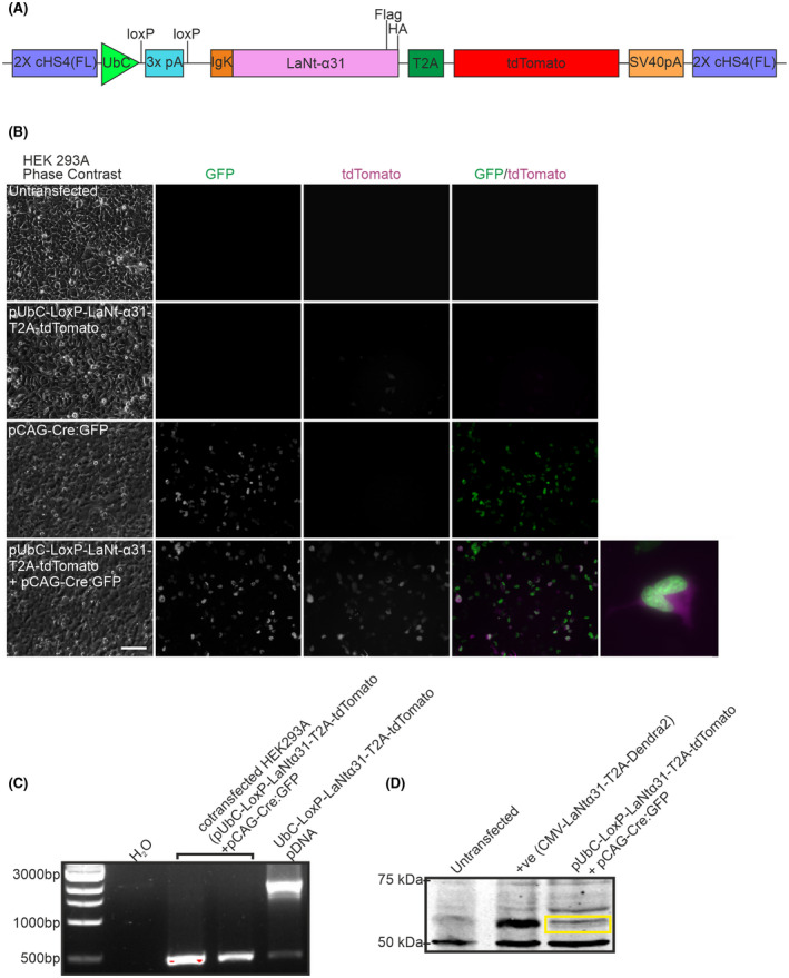
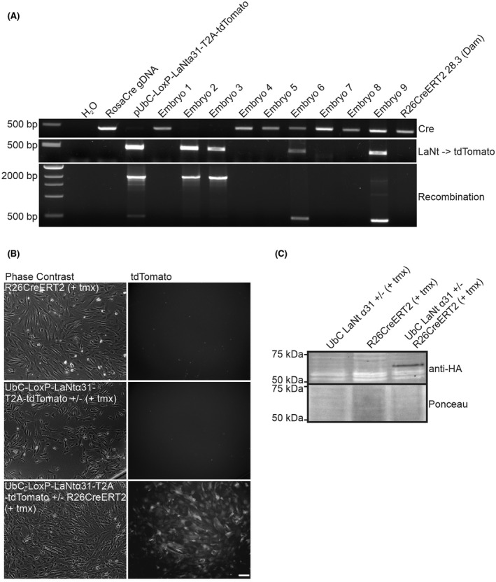
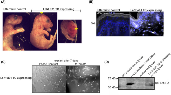
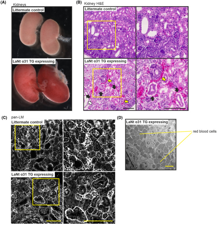
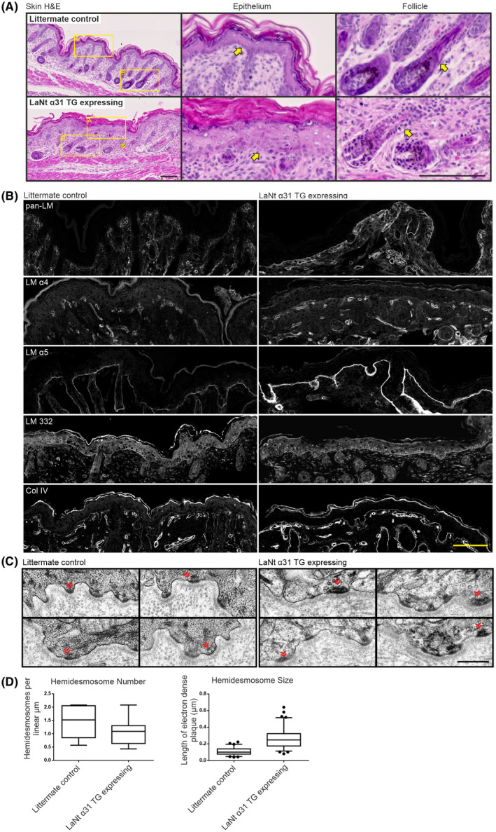
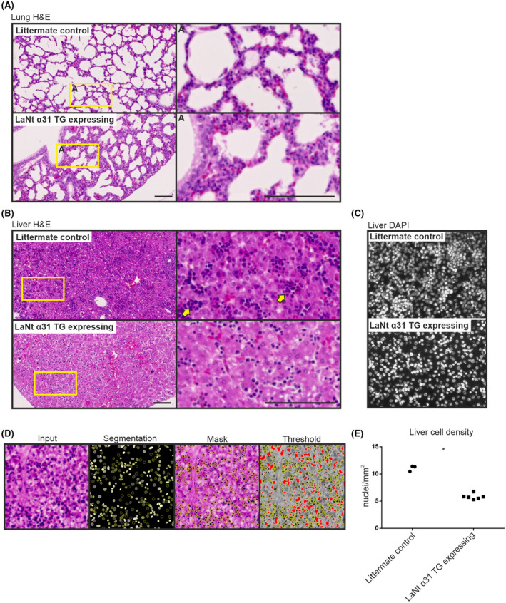
Laminins (LMs) are essential components of all basement membranes where they regulate an extensive array of tissue functions. Alternative splicing from the laminin α3 gene produces a non-laminin but netrin-like protein, Laminin N terminus α31 (LaNt α31). LaNt α31 is widely expressed in intact tissue and is upregulated in epithelial cancers and during wound healing. In vitro functional studies have shown that LaNt α31 can influence numerous aspects of epithelial cell behavior via modifying matrix organization, suggesting a new model of laminin auto-regulation. However, the function of this protein has not been established in vivo. Here, a mouse transgenic line was generated using the ubiquitin C promoter to drive inducible expression of LaNt α31. When expression was induced at embryonic day 15.5, LaNt α31 transgenic animals were not viable at birth, exhibiting localized regions of erythema. Histologically, the most striking defect was widespread evidence of extravascular bleeding across multiple tissues. Additionally, LaNt α31 transgene expressing animals exhibited kidney epithelial detachment, tubular dilation, disruption of the epidermal basal cell layer and of the hair follicle outer root sheath, and ~50% reduction of cell numbers in the liver, associated with depletion of hematopoietic erythrocytic foci. These findings provide the first in vivo evidence that LaNt α31 can influence tissue morphogenesis.

Keywords: basement membrane; development; laminin; netrin.


Figures

FIGURE 1
FIGURE 1
Validation of UbCLaNt Cre‐inducible construct in vitro. (A) Diagram of the pUbC‐LoxP‐LaNt‐α31‐T2A‐tdTomato construct. (B) HEK 293A cells were transfected with pUbC‐LoxP‐LaNt‐α31‐T2A‐tdTomato, pCAG‐Cre:GFP, or pUbC‐LoxP‐LaNt‐α31‐T2A‐tdTomato and pCAG‐Cre:GFP and imaged 48 h after transfection. Scale bar 100 µm. (C) PCR products using primers flanking the stop cassette on DNA extracted from HEK293A cells co‐transfected with pUbC‐LoxP‐LaNt‐α31‐T2A‐tdTomato and pCAG‐Cre:GFP. (D) Western blot of lysates from HEK293 cells either untransfected or transfected with CMV‐ LaNt‐α31‐T2A‐Dendra2 (positive control), or pUbC‐LoxP‐LaNt‐α31‐T2A‐tdTomato and pCAG‐Cre:GFP then probed with anti‐Flag antibodies
FIGURE 2
FIGURE 2
UbCLaNtα31 x R26CreERT2 ER transgenic mice express the UbC‐LaNtα31 transgene following exposure to tamoxifen. (A) PCR products on DNA extracted from transgenic mouse from UbCLaNtα31 x R26CreERT2 mating embryos using primers flanking the stop cassette. (B) Phase contrast and fluorescence microscopy images of explanted cells from UbCLaNtα31::R26CreERT2 embryos. Scale bar =100 µm. (C) Western blot of lysates from UbCLaNtα31::R26CreERT2 embryo explants processed with anti‐HA antibodies
FIGURE 3
FIGURE 3
Transgenic mice overexpressing LaNtα31 display localized regions of erythema. (A) Representative images of UbCLaNtα31::R26CreERT2 embryos. Animals subsequently confirmed as expressing the LaNt α31 transgene are labeled as LaNt α31 TG expressing. *Indicates regions of visible erythema. (B) Representative fluorescence microscopy UbCLaNtα31::R26CreERT2 OCT sections tdTomato fluorescence. Scale bar =100 µm. (C) Fluorescence microscopy images of explanted cells from LaNt α31 TG expressing. Scale bar =100 µm. (D) Western blot of tissue lysates from WT, UbCLaNtα31::R26CreERT2 embryos or explanted cells processed with anti‐HA antibodies. HEK293A cells cotransfected with the LaNt α31 transgene expression construct and Cre‐GFP expression construct are included as a positive control
FIGURE 4
FIGURE 4
LaNt α31 overexpression leads to epithelial detachment, tubular dilation and interstitial bleeding in the kidney and disruption of capillary basement membrane integrity. (A) Representative images of whole kidneys of newborn UbCLaNtα31::R26CreERT2 mouse kidneys from non‐expressing littermate controls (top) or LaNt α31 TG expressing animals (bottom). (B) Representative images of H&E stained FFPE sections (5 μm) of newborn littermate controls of LaNt α31 TG expressing mouse kidneys. Right column shows areas of increased magnification. Black arrows point to areas of epithelial detachment. White arrows point to tubular dilation. Yellow arrows point to areas of interstitial bleeding. (C) FFPE sections (5 μm) from littermate controls or LaNt α31 TG expressing animals processed for immunohistochemistry with pan‐laminin polyclonal antibodies. Right column shows areas of increased magnification. Scale bars =100 μm
FIGURE 5
FIGURE 5
LaNt α31 overexpression disrupts epidermal‐dermal cell organization. (A) H&E staining of FFPE sections (5 μm) of newborn UbCLaNtα31::R26CreERT2 transgenic mice dorsal skin. Upper panel non‐expressing littermate controls, lower panels LaNt α31TG expressing animals. Yellow chevrons indicate areas of extravascular erythrocytes. Middle and right columns show increased magnification of the epithelium or hair follicles respectively. Yellow arrows indicate basal layer of epithelial cells. Scale bar =100 μm. (B) Littermate controls (left) or LaNt α31 TG expressing OCT sections (10 μm) processed for immunohistochemistry with anti‐laminin 111 (pan‐LM), anti‐laminin α4 (LM α4), anti‐laminin α5 (LM α5), anti‐laminin 332 (LM 332) and anti‐Type IV collagen (Col IV). Scale bar =50 μm. (C) Transmission electron micrographs of littermate control or LaNt α31 TG expressing skin sections imaged at the dermal‐epidermal junctions. Chevrons indicate hemidesmosomes. Scale bar =0.5 µm. (D) Box and whisker graphs of quantification of hemidesmosome number per µm of basement membrane (n = 12 and 17 images), and of size of the hemidesmome measured as the length of the electron dense plaque at the cell membrane (n = 67 and 81 hemidesmosomes). Boxes represent 25th–75th percentile with line at median, whiskers 5th and 95th percentile. Dots represent outliers
FIGURE 6
FIGURE 6
Mice expressing the LaNtα31 transgene display structural differences in the lung and a reduction of hematopoietic colonies in the liver. (A) H&E staining of FFPE sections (5 μm) of newborn UbCLaNtα31::R26CreERT2 transgenic mice lungs (A) and liver (B). Upper panel non‐expressing littermate controls, lower panels LaNt α31TG expressing animals. Right columns show increased magnification. Yellow arrowheads highlight areas of increased cell density. Scale bars =100 μm. (C) DAPI staining of littermate controls or LaNt α31 TG expressing mouse livers. (D) Representative image analysis method of determining nuclei count. (E) Quantification of nuclei. Each point represents the mean of the quantification of nuclei/mm2 from 2 separate microscope slides at different sectioning depths per mouse

Similar articles

Cited by

References

    1. Yamada KM, Collins JW, Cruz Walma DA, et al. Extracellular matrix dynamics in cell migration, invasion and tissue morphogenesis. Int J Exp Pathol. 2019;100:144‐152. - PMC - PubMed
    1. Sekiguchi R, Yamada KM. Basement membranes in development and disease. Curr Top Dev Biol. 2018;130:143‐191. - PMC - PubMed
    1. Yurchenco PD. Basement membranes: cell scaffoldings and signaling platforms. Cold Spring Harb Perspect Biol. 2011;3(2):a004911. - PMC - PubMed
    1. Pozzi A, Yurchenco PD, Iozzo RV. The nature and biology of basement membranes. Matrix Biol. 2017;57‐58:1‐11. - PMC - PubMed
    1. Walma DAC, Yamada KM. The extracellular matrix in development. Development. 2020;147. 10.1242/dev.175596 - DOI - PMC - PubMed

LinkOut - more resources